Non-destructive supersonic induced-luminessing imaging method and equipment

A technology of induced luminescence and imaging method, applied in measurement devices, ultrasonic/sonic/infrasonic diagnosis, material analysis using sonic/ultrasonic/infrasonic waves, etc., can solve the problems of low cost, poor contrast, single imaging contrast, etc. Easy operation, easy production and assembly, and improved contrast

Inactive Publication Date: 2004-04-14
SOUTH CHINA NORMAL UNIVERSITY
View PDF0 Cites 0 Cited by
  • Summary
  • Abstract
  • Description
  • Claims
  • Application Information

AI Technical Summary

Problems solved by technology

[0002] Among the imaging technologies commonly used in medical imaging diagnosis at present, X-ray transmission imaging, X-ray tomography (CT), and nuclear magnetic resonance imaging all have a large degree of damage to human tissues, and X-ray tomography, nuclear magnetic resonance imaging, etc. The cost of the resonance imager is relatively expensive; the B-type ultrasonic imaging has basically no damage to the human body, and the cost of the B-type ultrasonic imager is also low, but because it is tissue interface reflection imaging, it cannot achieve tomography, and the resolution is low ( For in vivo tissues with simple structures, the best resolution is a few millimeters), and the contrast is poor
Optical imaging technology (including time-resolved optical imaging, frequency-domain optical imaging, optical coherence tomography, etc.) has the characteristics of no damage to the human body and high resolution. With strong scattering and absorption, the current technical level can only achieve imaging of shallow biological tissues with a depth of millimeters, and it is difficult to achieve accurate measurement of deep tissues. At the same time, these methods of imaging are realized by using the optical properties of tissues However, its imaging contrast is solely derived from the difference in the optical properties of the tissue

Method used


Image

  • Non-destructive supersonic induced-luminessing imaging method and equipment
  • Non-destructive supersonic induced-luminessing imaging method and equipment
  • Non-destructive supersonic induced-luminessing imaging method and equipment

Examples


Embodiment 1

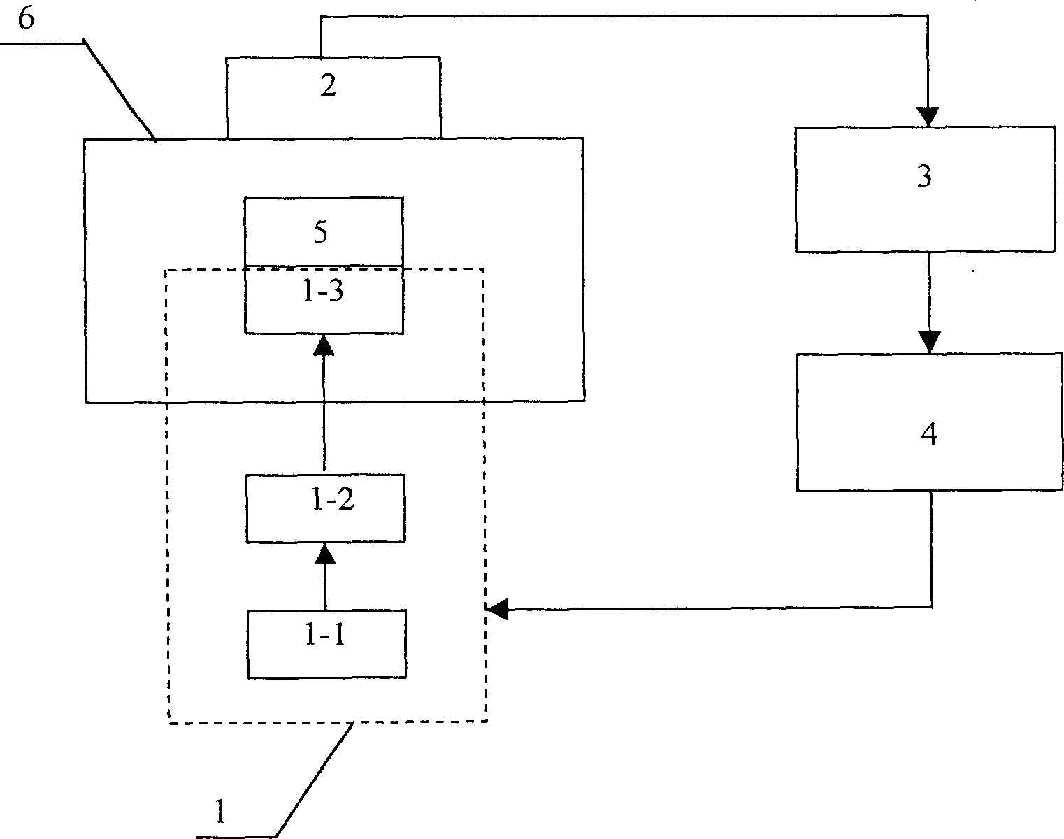
[0027] figure 1 shows the structure of the ultrasound-induced luminescence non-destructive imaging device of the present invention, by figure 1 It can be seen that the device is composed of an ultrasonic generating component 1, a light receiving component 2, an analog-to-digital converter 3, and a computer 4, wherein the ultrasonic generating component 1 is composed of a function generator 1-1, a power amplifier 1-2, an ultrasonic transducer 1- 3 are electrically connected in sequence; the light receiving component 2 is formed by connecting the camera lens 2-1 and the detector 2-2; Electrical connection in turn; 5 is the measured organism (or tissue), 6 is the darkroom. The device is composed of various components connected, among which: the function generator 1-1 selects the AFG320 arbitrary function generator manufactured by Tektronix Company; the power amplifier 1-2 selects the 2100L broadband power amplifier manufactured by ENI Company; the ultrasonic transducer 1- 3 is ...

Embodiment 2

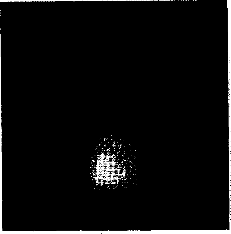
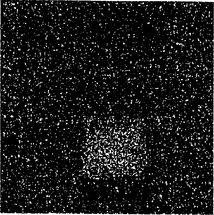
[0030] Apply the device described in Example 1 to the imaging of living organisms, take a nude mouse for experimentation, place it on the ultrasonic transducer 1-3 in the darkroom 6 after anesthesia, and record under weak light as follows: Figure 4 As shown in the outline drawing, close the dark room; adjust the function generator 1-1 by the computer 4 to generate a sinusoidal signal with a frequency of 40KHz, and drive the ultrasonic transducer 1-3 after being amplified by the power amplifier 1-2 to 50W to generate weak ultrasonic waves. The power density of weak ultrasound reaching the rat body is about 0.18W / cm 2 (far lower than the US FDA's ultrasound safety standard for human body 23bar, equivalent to 19.32W / cm 2), under the induction of weak ultrasound, the nude mouse body produces its own endogenous luminescence, the detector 2-2 records the luminescence signal for 15 minutes through the camera lens 2-1 and converts the luminescence signal into an electrical signal, an...


PUM

PropertyMeasurementUnit
diameteraaaaaaaaaa

Abstract

The imaging method includes applying weak supersonic wave directly onto organism or tissue to induce endogenous luminescence; receiving the light by an optical receptor and converting the light into electrical signal; A/D converting the electrical signal into digital signal; and digital signal treatment to obtain the structure image of the organism or tissue. The imaging equipment consists of a supersonic wave generator assembly, an optical receptor assembly, an A/D converter and a computer. Combining the strong permeability of supersonic wave in organism and the high resolution and no damage of optical imaging, the present invention can obtain high-resolution image for biomedical imaging diagnosis.

Description

technical field [0001] The present invention relates to a medical imaging diagnosis technology, in particular to an ultrasound-induced ultrasound-induced luminescence that uses harmless weak ultrasound to induce organisms or tissues to produce their own internal luminescence, and uses the endogenous luminescence to perform non-damaging structural imaging. Luminescent non-destructive imaging method and device thereof. Background technique [0002] Among the imaging technologies commonly used in medical imaging diagnosis at present, X-ray transmission imaging, X-ray tomography (CT), and nuclear magnetic resonance imaging all have a large degree of damage to human tissues, and X-ray tomography, nuclear magnetic resonance imaging, etc. The cost of the resonance imager is relatively expensive; the B-type ultrasonic imaging has basically no damage to the human body, and the cost of the B-type ultrasonic imager is also low, but because it is tissue interfa...

Claims


Application Information

Patent Timeline
Patent Type & Authority Patents(China)
IPC IPC(8): A61B8/00G01N21/62G01N29/00
Inventor 邢达何永红
Owner SOUTH CHINA NORMAL UNIVERSITY
Why Eureka
Try Eureka