Computed tomography system

a tomography system and computed tomography technology, applied in the field of computed tomography system, can solve problems such as image artifacts in the finally reconstructed image, and achieve the effect of improving image quality

Active Publication Date: 2019-09-24
KONINKLJIJKE PHILIPS NV
View PDF13 Cites 0 Cited by
  • Summary
  • Abstract
  • Description
  • Claims
  • Application Information

AI Technical Summary

Benefits of technology

[0006]It is an object of the present invention to provide a computed tomography system, method and computer program for generating an image of a subject comprising a contrast agent, wherein the quality of the image can be improved.
[0011]By the scaling procedure, which may be performed in the projection domain and / or in the image domain, different amounts of the contrast agent, which may be present at different times, can be balanced, which allows for an improved quality of the finally reconstructed image.
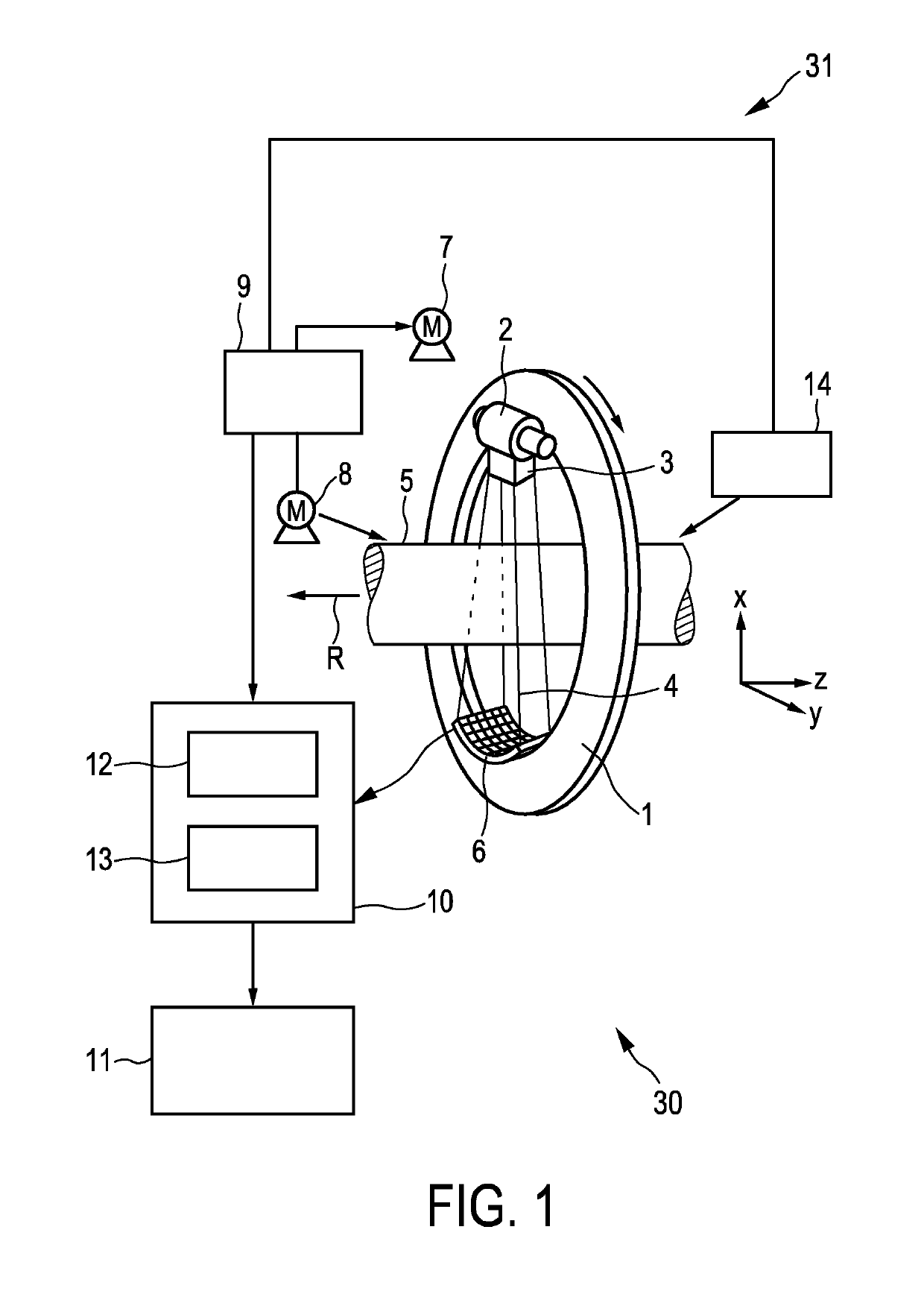
[0017]The subject is preferentially a living being and the provided sets of spectral projections are preferentially cardiac gated, especially prospective cardiac gated. For performing the prospective cardiac gated acquisition of the spectral projections an electrocardiography signal generated by an electrocardiography unit connected to the subject may be used. By using cardiac gated spectral projections, image artifacts, which may be caused by a cardiac movement, may be reduced, thereby further improving the quality of the reconstructed image. Moreover, by performing a prospective cardiac acquisition of the spectral projections, the radiation dose applied to the subject can be significantly reduced.
[0018]The predefined similarity measure preferentially depends on an average of the intensity values of the respective first image in the respective overlap region. The average may be, for instance, the arithmetic mean or the median or another average. Using this average as a similarity measure leads to an improved image quality, wherein the average of the intensity values of the respective first image in the respective overlap region can be calculated with low computational effort. First images may be regarded as having a same intensity in an overlap region, if a difference between a) a similarity value obtained by applying the similarity measure to the intensities of one of the first images in the overlap region and b) a similarity value obtained by applying the similarity measure to the intensities of the other of the first images in the overlap region is smaller than a predefined threshold. The predefined threshold may be predetermined by calibration or in another way such that the image quality is improved. In an embodiment first images may be regarded as having a same intensity in an overlap region, if a difference between a) a similarity value obtained by applying the similarity measure to the intensities of one of the first images in the overlap region and b) a similarity value obtained by applying the similarity measure to the intensities of the other of the first images in the overlap region are equal.
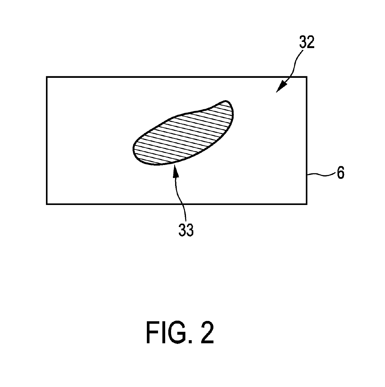
[0020]In another embodiment the respective region of interest on the detection surface corresponds to a virtual projection of a predefined object of interest within the subject on the detection surface. Preferentially, the projections providing unit is adapted to provide the respective acquisition geometry used for acquiring the respective projection, wherein the image generation unit is adapted to provide an object of interest image showing the object of interest within the subject and to determine the respective region of interest on the detection surface by virtually projecting the object of interest onto the detection surface under consideration of the provided respective acquisition geometry and the provided object of interest image. For instance, if the object of interest is the heart, the region of interest on the detection surface may correspond to a virtual projection of the heart onto the detection surface. Thus, for determining the contrast value being indicative of the total amount of the contrast agent based on projection values of a respective first projection only projection values may be used, which are within the region of interest on the detection surface, wherein the region of interest corresponds to the virtual projection of the predefined object of interest onto the detection surface. The contrast value may therefore be indicative of the total amount of contrast agent in the object of interest only. This can lead to an improved scaling regarding the object of interest and hence finally to an improved quality of an image of the object of interest.
[0021]The image generation unit may be adapted to parallel rebin the first projections and optionally also the second projections before using the first projections and optionally also the second projections for reconstruction, especially before determining contrast values for the first projections. The parallel rebinning can lead to further reduced computational efforts for generating the image.

Problems solved by technology

Detection values, which have been generated at different times, relate therefore to different concentrations of the contrast agent within the subject, which can lead to image artifacts in the finally reconstructed image.

Method used


Image

  • Computed tomography system
  • Computed tomography system
  • Computed tomography system

Examples


Embodiment Construction

[0034]FIG. 1 shows schematically and exemplarily a computed tomography system 30 for generating an image of a subject. The computed tomography system 30 comprises a projections acquisition unit 31 for acquiring several sets of spectral projections of the subject, a processing unit 10 for processing the provided sets of spectral projections, especially for generating an image, and a display 11 for showing the generated image. In this embodiment the subject is a person and the projections acquisition unit 31 is adapted to perform a prospective cardiac gated acquisition of the spectral projections.

[0035]The projections acquisition unit 31 includes a gantry 1 which is capable of rotation around a rotation axis R which extends parallel to a z direction. A polychromatic radiation source 2, which is, in this embodiment, an x-ray tube, is mounted on the gantry 1. The radiation source 2 is provided with the collimator 3 for forming, in this embodiment, a conical radiation beam 4 from the rad...


PUM


Abstract

The invention relates to a computed tomography system (30). Several sets of spectral projections, which correspond to different positions of a radiation source (2) along a rotation axis (R), are decomposed into first projections being indicative of a contrast agent and second projections being not indicative of the contrast agent. An image is generated by a) determining for each first projection a contrast value being indicative of a total amount of contrast agent and scaling the first projections such that for different first projections of a same set the same contrast value is determined, and reconstructing an image based on the scaled first projections, and / or b) reconstructing for the different sets first images, scaling the first images such that they have a same intensity in overlap regions and combining the scaled first images. Thus, different contrast agent amounts can be balanced, thereby allowing for an improved image quality.

Description

CROSS REFERENCE TO RELATED APPLICATIONS[0001]This application is the U.S. National Phase application under 35 U.S.C. § 371 of International Application No. PCT / EP2015 / 075650, filed Nov. 4, 2015, published as WO 2016 / 071374 on May 12, 2016, which claims the benefit of European Patent Application Number 14192039.7 filed Nov. 6, 2014. These applications are hereby incorporated by reference herein.FIELD OF THE INVENTION[0002]The invention relates to a computed tomography system, method and computer program for generating an image of a subject comprising a contrast agent.BACKGROUND OF THE INVENTION[0003]A computed tomography system comprises a radiation source emitting radiation traversing a subject to be imaged and a detector for generating detection values depending on the radiation having traversed the subject. The detection values are generated, while the radiation source and the subject move relative to each other, for instance, while the radiation source rotates around the subject....

Claims


Application Information

Patent Timeline
Patent Type & Authority Patents(United States)
IPC IPC(8): G06K9/00A61B6/03A61B6/02A61B6/00
CPCA61B6/027A61B6/5241A61B6/482A61B6/5205A61B6/481A61B6/4241A61B6/032
Inventor KOEHLER, THOMASPROKSA, ROLANDGRASS, MICHAEL
Owner KONINKLJIJKE PHILIPS NV
Why Eureka
Try Eureka