Method for assigning tissue normalization factors for digital image analysis
a tissue normalization factor and image analysis technology, applied in image analysis, image enhancement, instruments, etc., can solve the problems of inability to accurately distinguish between tissue objects (e.g., cells), and limited in their ability to exclude area
- Summary
- Abstract
- Description
- Claims
- Application Information
AI Technical Summary
Problems solved by technology
Method used
Image

Examples
Embodiment Construction
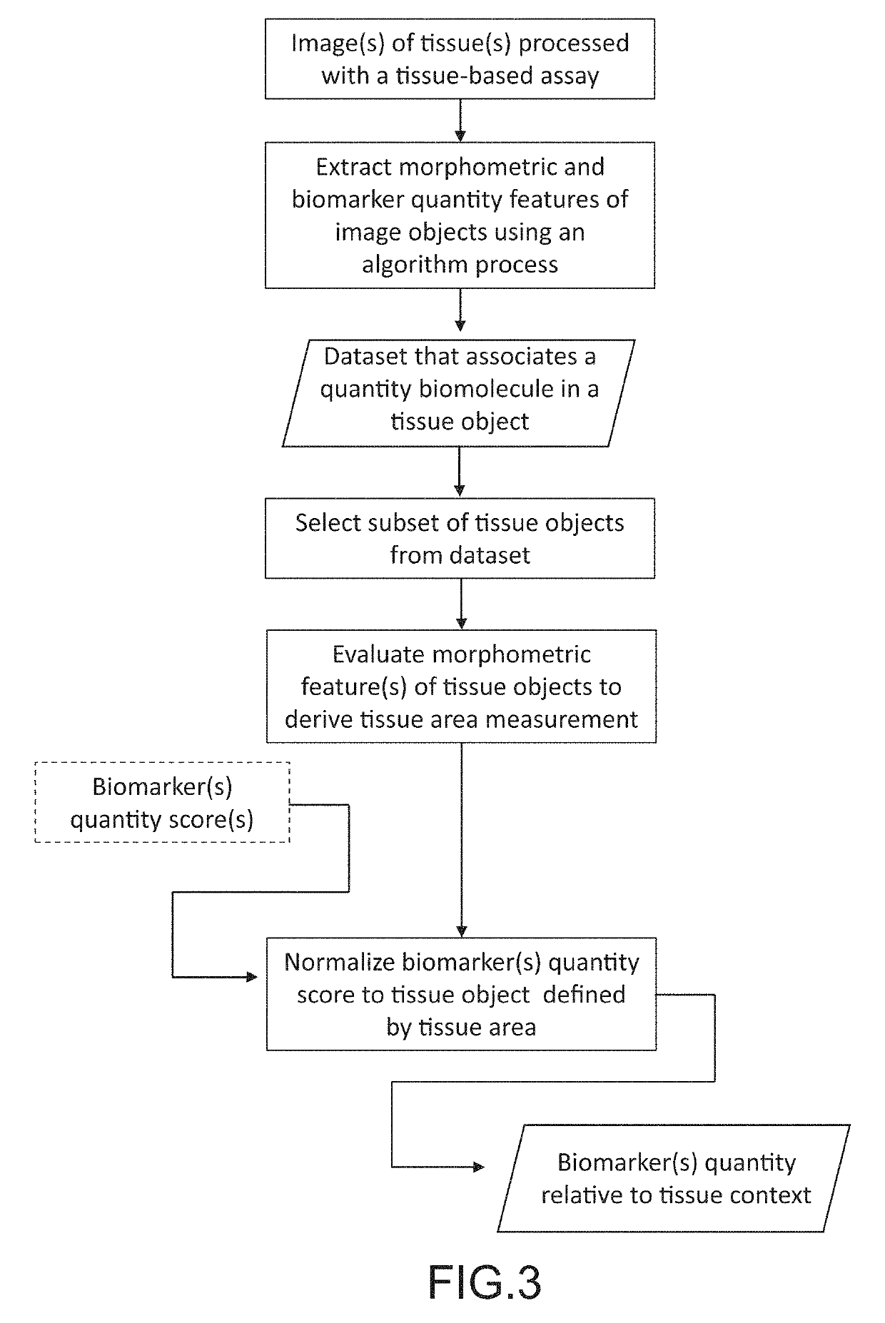
[0023]In the following description, for purposes of explanation and not limitation, details and descriptions are set forth in order to provide a thorough understanding of the present invention. However, it will be apparent to those skilled in the art that the present invention may be practiced in other embodiments that depart from these details and descriptions without departing from the spirit and scope of the invention.
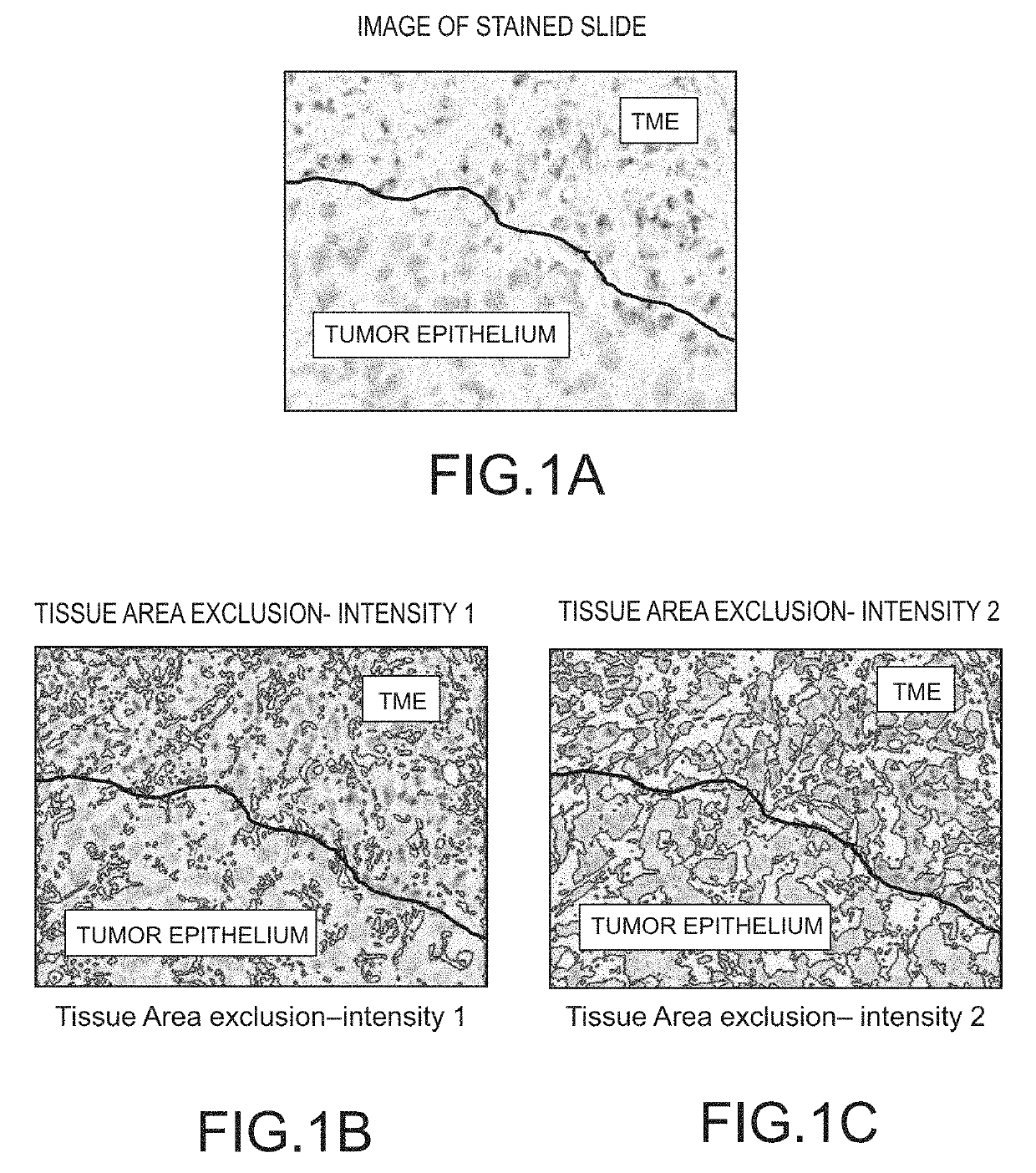
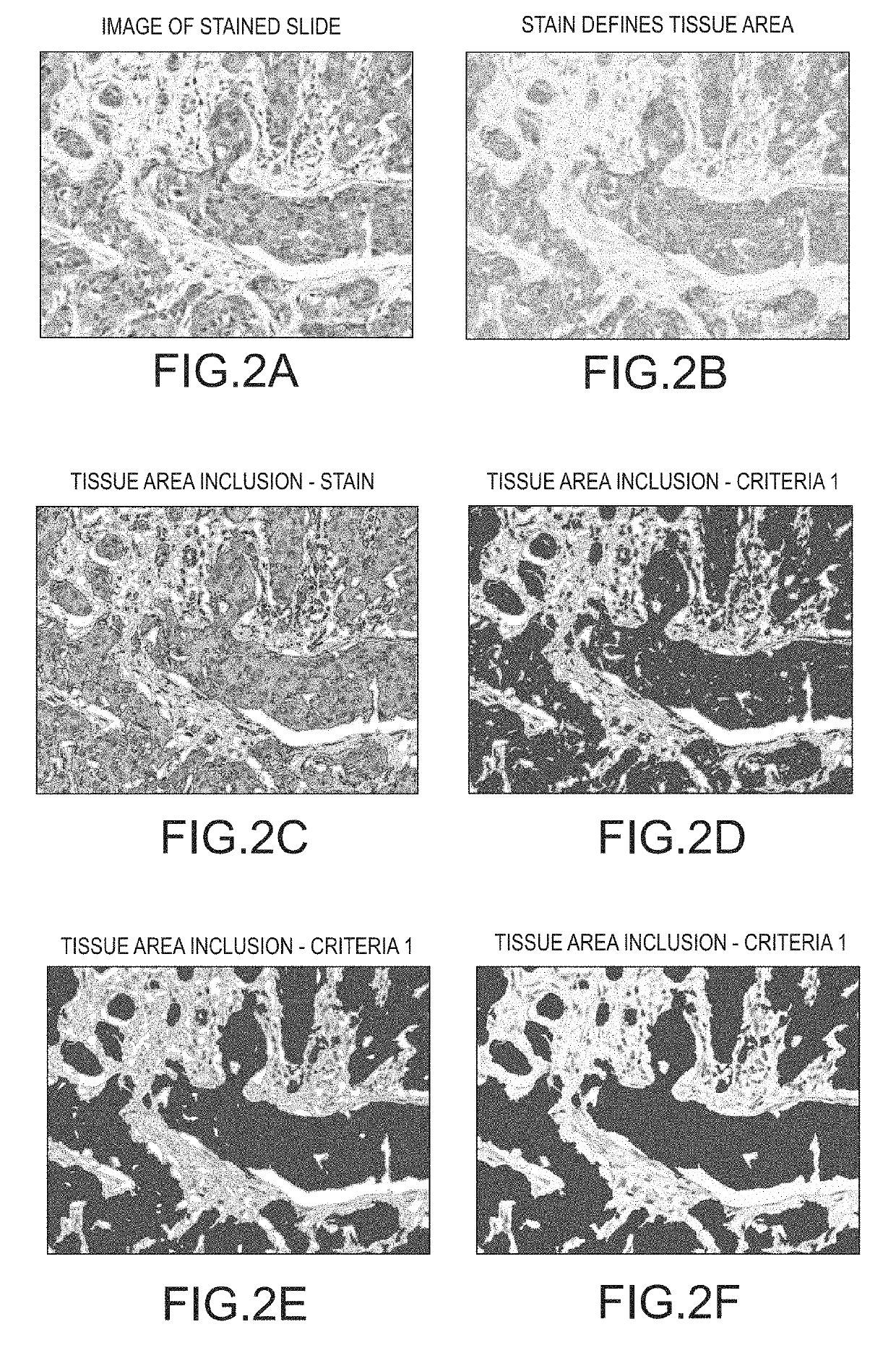
[0024]For purpose of definition, a tissue object is one or more of a cell (e.g., immune cell), cell sub-compartment (e.g., nucleus, cytoplasm, membrane, organelle), cell neighborhood, a tissue compartment (e.g., tumor, tumor microenvironment (TME), stroma, lymphoid follicle, healthy tissue), biomarker, blood vessel, and a lymphatic vessel. Tissue objects are visualized by histologic stains which highlight the presence and localization of a tissue object. Tissue objects can be identified directly by stains specifically applied to highlight said tissue object (e.g., h...
PUM
 Abstract
Description
Claims
Application Information
 - R&D Engineer
- R&D Manager
- IP Professional
- Industry Leading Data Capabilities
- Powerful AI technology
- Patent DNA Extraction
Browse by: Latest US Patents, China's latest patents, Technical Efficacy Thesaurus, Application Domain, Technology Topic.
© 2024 PatSnap. All rights reserved.Legal|Privacy policy|Modern Slavery Act Transparency Statement|Sitemap