Cutting device for endovascular surgery

a cutting device and endovascular technology, applied in the field of endovascular surgery, can solve the problems of reducing the reliability and precision of the operation of the patient, reducing the accuracy of the operation, so as to improve the reliability and precision of the operation.

Active Publication Date: 2020-02-04
ASSISTANCE PUBLIQUE HOPITAUX DE PARIS
View PDF7 Cites 0 Cited by
  • Summary
  • Abstract
  • Description
  • Claims
  • Application Information

AI Technical Summary

Benefits of technology

[0024]Thus, the invention is based on an innovative method and discloses an endovascular surgical device so that the practitioner can cut a piece of vascular wall commonly referred to as a flap, rather than tear it off in an approximate and uncontrolled gesture. It is important to specify that this cutting gesture is a clean action fully controlled by the practitioner. It does not involve tearing or ripping the flap using any type of instrument. The practitioner uses the device according to the invention to make a perfect cut of the flap, and he personally controls the force applied on the flap by the blades and also the speed at which the gesture is performed. Therefore, the practitioner's gesture is much safer and more precise, which is much more comfortable for the surgeon and much safer for the patient. Unlike the use of the introducer in the conventional backward fenestration technique, the device according to the invention can be used to operate on aortic dissections in the acute phase and also in the chronic phase when the flap has already begun to become rigid.
[0025]The flexibility or suppleness of the transmission means are such that the sliding device can be inserted into the arterial vascular system on two guide rods and can follow meanders without any risk of injury or perforation of a vessel. Although it is technically difficult to have remote control over the operation of the cutting blades, the movement of blades on the guide rods firstly directs the cutting blades reliably and precisely until they come into contact with the flap to be cut and secondly stabilises the cutting blades so that their remote operation can be controlled.

Problems solved by technology

Dissection of the thorax-abdominal aorta is a serious pathology, leading to a fatal prognostic of patients in the short and long term.
The main complication of the aortic dissection is the result of the fact that the blood pressure in the false channel is too high, consequently it compresses the blood circulation in the true channel.
This pressure gradient between the two channels is related to the fact that the resistance opposing the blood flow in the false channel is too high.
Consequently, blood circulates badly in the portion of the dissected aorta and the result is poor perfusion of the arteries irrigating the lower organs and limbs.
If compression of the true channel by the false channel is not quickly released, the organs for which perfusion depends on the aorta will quickly experience malfunctions, sometimes irreversible.
Although this is efficient, it is sometimes impossible to use endoprosthesis due to difficult anatomic conditions, or insufficient considering the fact that there are other reentry orifices.
However, the practitioner does not have very good control over it because he has to work “blind” despite radioscopic monitoring.
These aneurysms have a serious risk of rupture due to the high blood pressure inside them, and to degeneration of the remaining wall of the aorta.
Furthermore, it is rare that the dissection diagnosis is made in the acute phase.
Thus, the use of endoprostheses fitted with visceral branches is a choice that can only rarely be put into practice in this type of chronic aneurysmal dissection.
The use of endoprostheses with visceral branches can only be envisaged in the true aortic channel because very often rigidification of the flap makes catheterisation of the visceral arteries impossible.
Therefore, this solution is completely unacceptable.
The fenestration technique using an introducer as described above is also unacceptable; although it can give good results with a flexible flap during the first days after the dissection, tearing becomes impossible and even dangerous with a flap that has become rigid over time.
The surgeon would then be obliged to apply pressure on the flap that could break suddenly and weaken the aorta or even cause its rupture, which would be fatal for the patient.
Since these treatments are unacceptable, all that remains is open surgery, in other words the patient is opened up at the thorax-abdomen level so that the surgeon can operate on the aorta directly.
However, open surgery is very difficult, both for the patient and for the medical team.
There are many complications related to such an operation (death, paraplegia, terminal renal insufficiency, etc.), and post-operation recovery is long and difficult for the patient.
It is also very expensive for the health system.
These risks are particularly high when patients usually have subjacent pathologies such as high blood pressure, and a poor general condition.
Furthermore, it frequently happens that these patients cannot receive this type of surgery because the dangers are worse than the benefits.

Method used


Image

  • Cutting device for endovascular surgery
  • Cutting device for endovascular surgery
  • Cutting device for endovascular surgery

Examples


first embodiment

A) Example of a First Embodiment

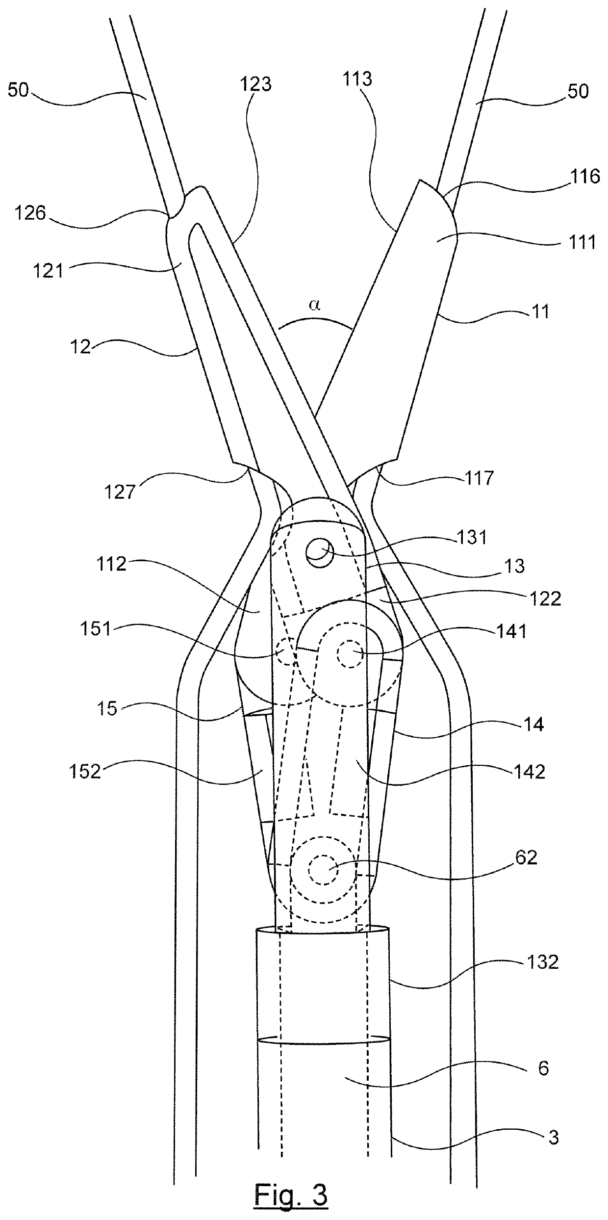
[0058]FIGS. 3 and 4 show a detailed view of the cutting blades of a first embodiment according to the invention, in the open and closed positions respectively. The hidden parts are shown in dashed lines. Each of the cutting blades 11, 12 has a rounded distal end 111, 121, and a proximal end 112, 122. This rounded form means that the cutting blades can be inserted in the closed position without creating any lesions in the inside wall of the vessels inside which the device passes. It also makes it possible to actuate the cutting blades safely within a vessel. This characteristic is particularly important when operating on a patient who already has intravascular lesions. Each blade has a cutting edge 113, 123 used to cut the vascular wall, even when a fibrous scar tissue has begun to develop. Blades are hinged to each other by a hinge pin 131, that also holds them to the attachment part 13. Each blade 11, 12 is hinged at its proximal part to a connecting...

second embodiment

b) Example of a Second Embodiment

[0074]According to this second embodiment shown in FIG. 6, the cutting blades 101, 102 are solid and slide along the guide rods 500 by means of the rings 191, 192. The thickness of the rings is about 2 mm, their outer diameter is approximately 2 mm.

[0075]Operation and articulation of the cutting blades with the connecting rods 14, 15, the attachment part 13, the cable 6, the catheter 3 and the control element 4 are identical in all respects with the example described in section 5a). The part dimensions are identical to the first embodiment.


PUM


Abstract

The invention relates to an endovascular cutting device for carrying out surgical or medical operations. According to the invention, such a device includes two cutting blades, a means for remotely actuating the two cutting blades, and at least two flexible guide rods, said cutting blades being slidably mounted onto said guide rods, and said remote actuation means including a flexible transmission means, wherein a channel, through which the flexible guide rods (50) are to be inserted, extends through said cutting blades (11, 12, 101, 102) over the entire outer length thereof.

Description

CROSS-REFERENCE TO RELATED APPLICATION[0001]This application is the 35 U.S.C. § 371 national stage of PCT Application No. PCT / EP2013 / 064842, filed Jul. 12, 2013, which is herein incorporated by reference in its entirety and which also claims priority to, and the benefit of, French Patent Application No. 1256751, filed Jul. 12, 2012, which is herein incorporated by reference in its entirety.1. DOMAIN OF THE INVENTION[0002]The invention relates to medical devices for making surgical or medical operations. More specifically, the invention relates to a cutting device for endovascular surgery operations, particularly for surgery of acute or chronic dissections of the aorta.2. PRIOR ART[0003]Dissection of the thorax-abdominal aorta is a serious pathology, leading to a fatal prognostic of patients in the short and long term. Risk factors of this type of cardiovascular accident are particularly high blood pressure or congenital pathologies such as Marfan's disease that affects the elastic f...

Claims


Application Information

Patent Timeline
Patent Type & Authority Patents(United States)
IPC IPC(8): A61B17/3201A61B17/32A61B17/00A61B17/22
CPCA61B17/320016A61B17/3201A61B2017/22039A61B2017/00778A61B2017/22095A61B2017/22044A61B2017/2905
Inventor ALSAC, JEAN-MARCDUFETELLE, EDOUARD
Owner ASSISTANCE PUBLIQUE HOPITAUX DE PARIS
Why Eureka
Try Eureka