Device for training tracheal suctioning

a technology for tracheal suction and simulator, which is applied in the field of medical training equipment for health workers, can solve the problems of inability to use cloth or other means designed to prevent droplets from entering the airway, insufficient suction training, and harm to patients, and achieve the effect of real-time training on tracheal suction

Active Publication Date: 2020-04-28
LAERDAL MEDICAL AS
View PDF10 Cites 0 Cited by
  • Summary
  • Abstract
  • Description
  • Claims
  • Application Information

AI Technical Summary

Benefits of technology

[0012]The present invention has as an object to provide a device that enables realistic training on tracheal suction and ventilation through the tracheostomy tube without the risk of simulated mucus entering the lungs of the manikin.

Problems solved by technology

However, such a cloth or other means that are designed to prevent droplets from entering the airways are not possible to use when training on tracheal suctioning where a catheter or thin tube is inserted through the tracheostomy tube to suck out any mucus in the airways.
This dry suction may not provide adequate training as the health worker will not know is if sufficient or any mucus would have been sucked out by the procedure.
Mucus in the lungs may cause harm to the patient and prevent air from properly entering the lungs.
This known device cannot be used for simulating tracheal suction.

Method used


Image

  • Device for training tracheal suctioning
  • Device for training tracheal suctioning
  • Device for training tracheal suctioning

Examples


Embodiment Construction

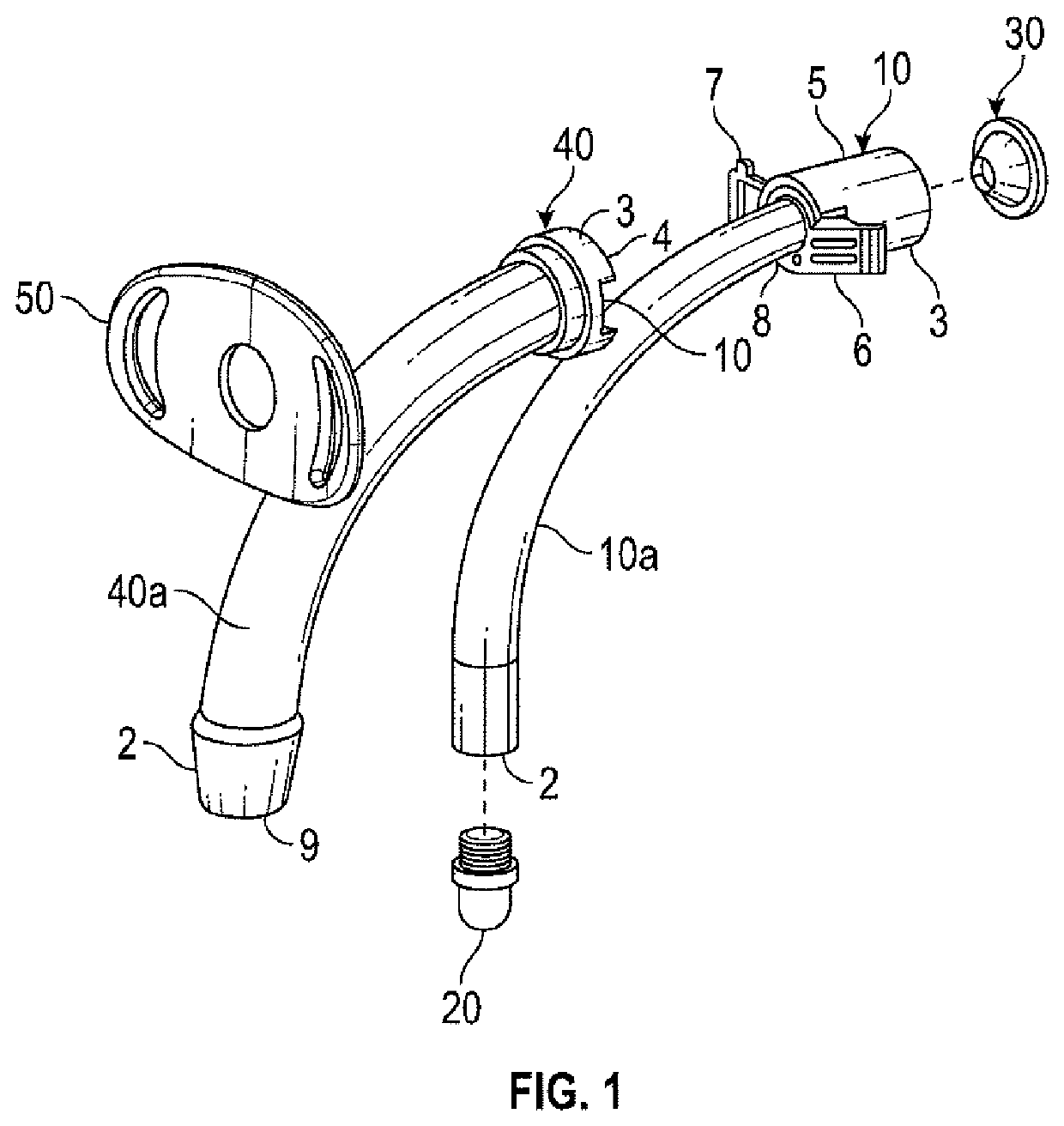
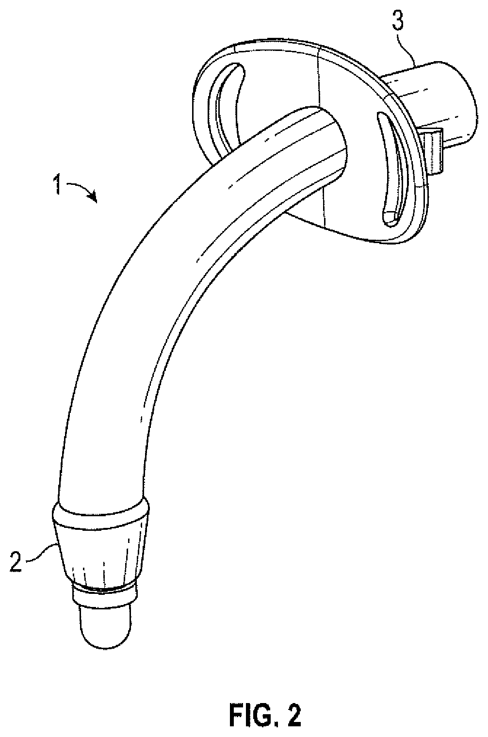
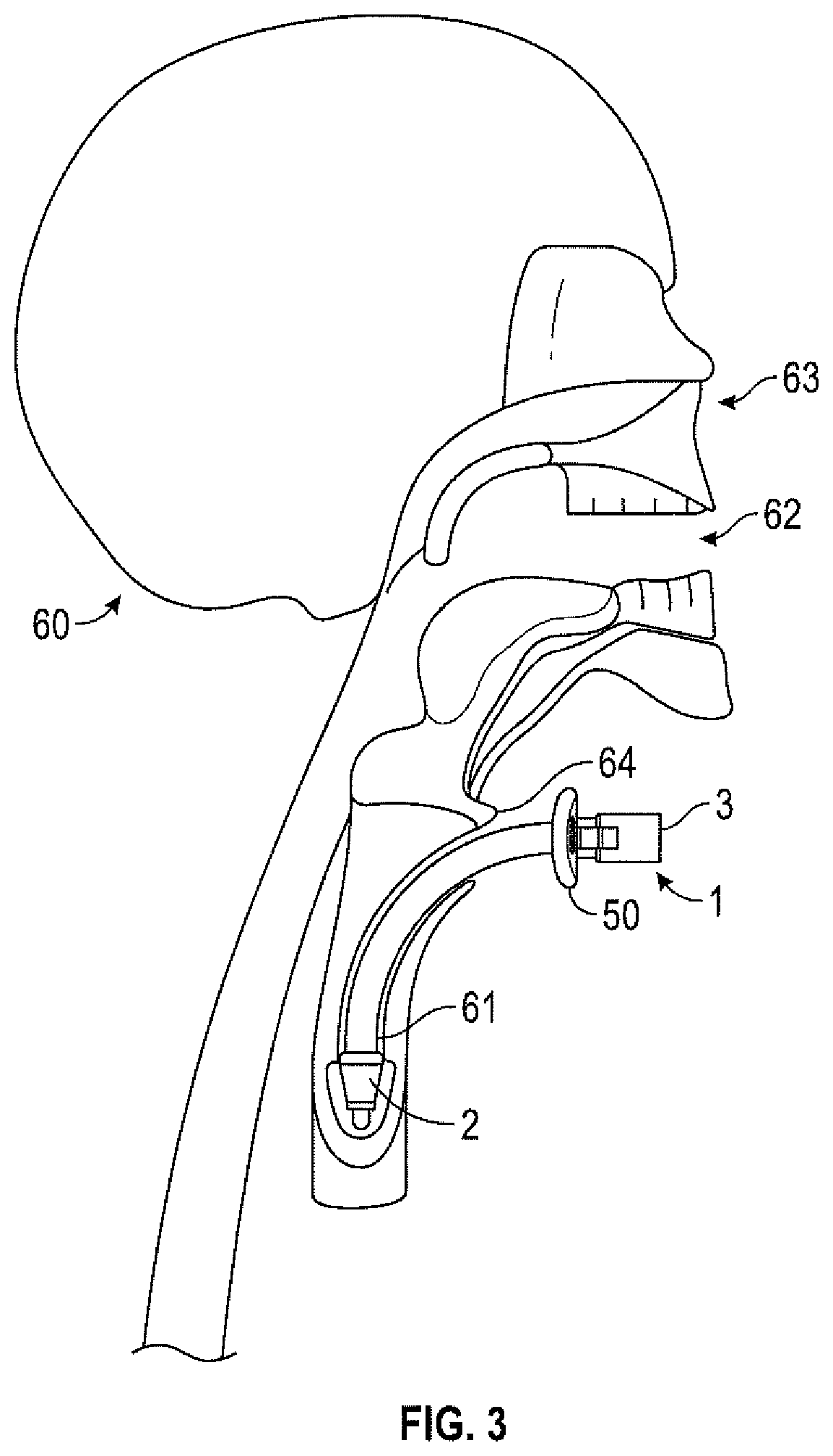
[0017]The training device 1 of the invention is primarily adapted to enter through a trachea opening or slit at the throat of a manikin 60 (FIG. 3), but may also be used through the mouth 62 (FIG. 3) or nose opening 63 (FIG. 3) of a manikin. It has a distal end 2, which is put through the trachea opening 64 (FIG. 3) and a proximal end 3, which is adapted to stay at the outside of the throat close to the trachea opening 64.

[0018]As shown in FIG. 1, the device 1 comprises a tracheal core 40, which has a tubular part 40a that at the distal end 2 is adapted to be arranged within the tracheal tube 61 (FIG. 3) of the manikin 60, which in turn is leading to the lower airways, including the lungs. At the proximal end 3, the tracheal core 40 has a flange 4, which will be explained further hereinafter.

[0019]The device also comprises a tracheal insert 10, which comprises a tubular part 10a. At the distal end 2 it may be equipped with a plug 20, but in a preferred embodiment, the distal end is ...


PUM


Abstract

Training device for training tracheal suctioning. The training device includes a tracheal core adapted to be arranged within a tracheal, nasal or oral cavity of a medical training simulator, and coupled to the airways of the simulator and an insert adapted to be inserted into the core, the insert forming a reservoir for simulated mucus.

Description

TECHNICAL FIELD[0001]The present invention relates to training equipment for medical training of health workers, such as paramedics or physicians or nurses. It specifically concerns a device for training tracheal suctioning on a medical simulator, such as a manikin.BACKGROUND ART[0002]A medical simulator, such as a manikin is commonly equipped with oral and nasal airways that are communicating with a simulated lung. The lung is fluidly coupled to the airways through a tube extending from the head portion, through a neck portion, and into a chest portion wherein the lung is arranged. In most manikins, there are two lungs that are each coupled via a simulated bronchus to a common simulated trachea. The bronchi and the trachea are constituted by tubes.[0003]Such manikins are frequently used by medical staff or the populace to train CPR or more advanced medical procedures, depending on the design of the manikin.[0004]The device is primarily intended for insertion through an opening made...

Claims


Application Information

Patent Timeline
Patent Type & Authority Patents(United States)
IPC IPC(8): G09B23/28G09B23/32G09B23/34G09B23/30A61M16/04
CPCA61M16/0475G09B23/32G09B23/285G09B23/34A61M16/0463G09B23/288G09B23/30A61M2209/02A61M16/0465A61M16/0461A61M16/0477A61M16/107
Inventor NECESSARY, GREGTEDESCHI, BENGRIFFITH, PAULTHANE, DAVID
Owner LAERDAL MEDICAL AS
Why Eureka
Try Eureka