Profiling microvesicle nucleic acids and uses thereof as signatures in diagnosis of renal transplant rejection

a technology of microvesicle nucleic acids and signatures, which is applied in the field of using microvesicle rna signatures, can solve the problems that urine cells are not able to reliably and successfully discriminate between patients

Active Publication Date: 2022-01-04
THE BRIGHAM & WOMEN S HOSPITAL INC +1
View PDF14 Cites 0 Cited by
  • Summary
  • Abstract
  • Description
  • Claims
  • Application Information

AI Technical Summary

Benefits of technology

[0009]Traditionally, biomarker discovery and development has required the use of material obtained from tissue biopsies. However, recent developments in the exosome field have allowed biomarker research in biofluids to evolve. Exosomes are highly stable microvesicles, approximately 30-200 nm in diameter, that are shed by cells into all biofluids, including blood, urine, and cerebrospinal fluid, carrying a rich source of intact protein and RNA. Exosomes and other vesicles can be released by multi-vesicular body pathway or through direct budding at the plasma membrane. RNA can be efficiently isolated and addressed using technologies such as RT-qPCR and NGS (see e.g., Brock, G. et al. (2015) Liquid biopsy for cancer screening, patient stratification and monitoring. Translational Cancer Research, 4(3), 280-290; and Enderle, D. et al. (2015) Characterization of RNA from Exosomes and Other Extracellular Vesicles Isolated by a Novel Spin Column-Based Method. PLoS ONE, 10(8): e0136133. doi:10.1371 / journal.pone.0136133).
[0020]In some embodiments, the extracted nucleic acids are subject to further analysis. Various nucleic acid sequencing techniques are used to detect and analyze nucleic acids such as cell free DNA and / or RNA extracted from the microvesicle fraction from biological samples. Analysis of nucleic acids such as cell free DNA and / or nucleic acids extracted from microvesicles for diagnostic purposes has wide-ranging implications due to the non-invasive nature in which microvesicles can be easily collected.

Problems solved by technology

Interestingly, the cells in urine were not able to reliably and successfully discriminate between patients who experienced kidney transplant rejection, and those who did not experience any rejection symptoms or other indications.

Method used


Image

  • Profiling microvesicle nucleic acids and uses thereof as signatures in diagnosis of renal transplant rejection
  • Profiling microvesicle nucleic acids and uses thereof as signatures in diagnosis of renal transplant rejection
  • Profiling microvesicle nucleic acids and uses thereof as signatures in diagnosis of renal transplant rejection

Examples


example 1

dy of Urinary Microvesicle Signature for the Diagnosis of Human Kidney Transplant Rejection

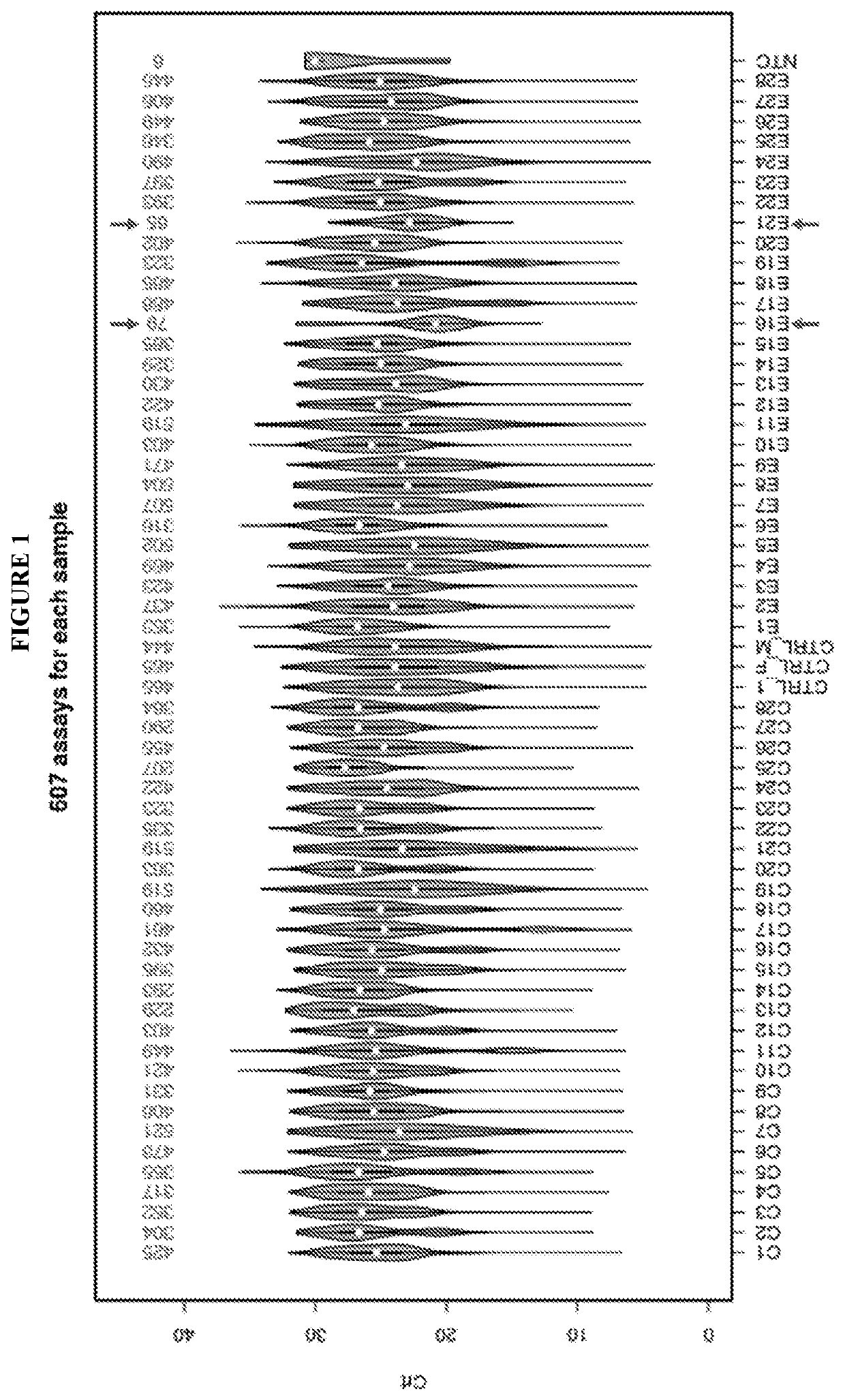
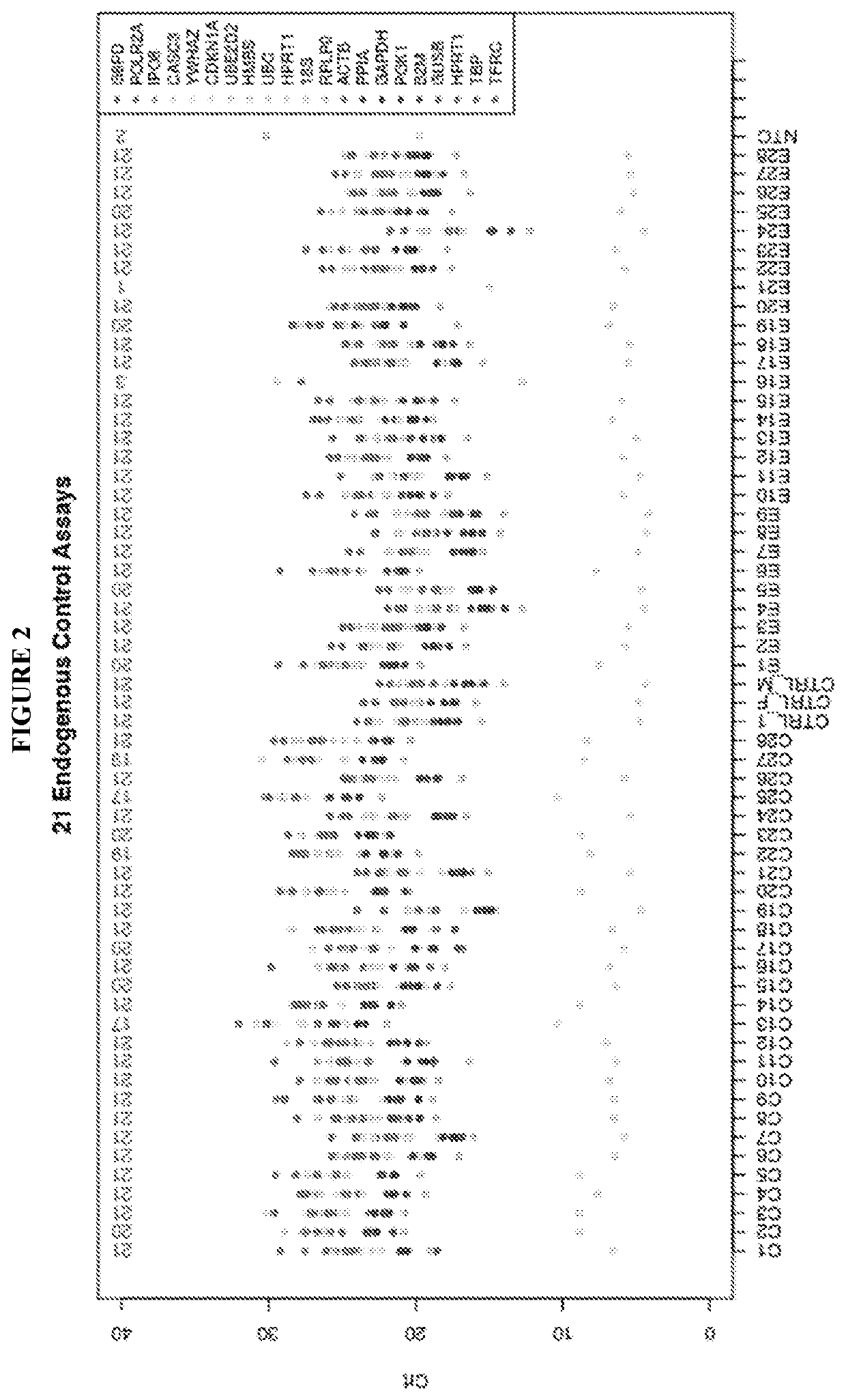
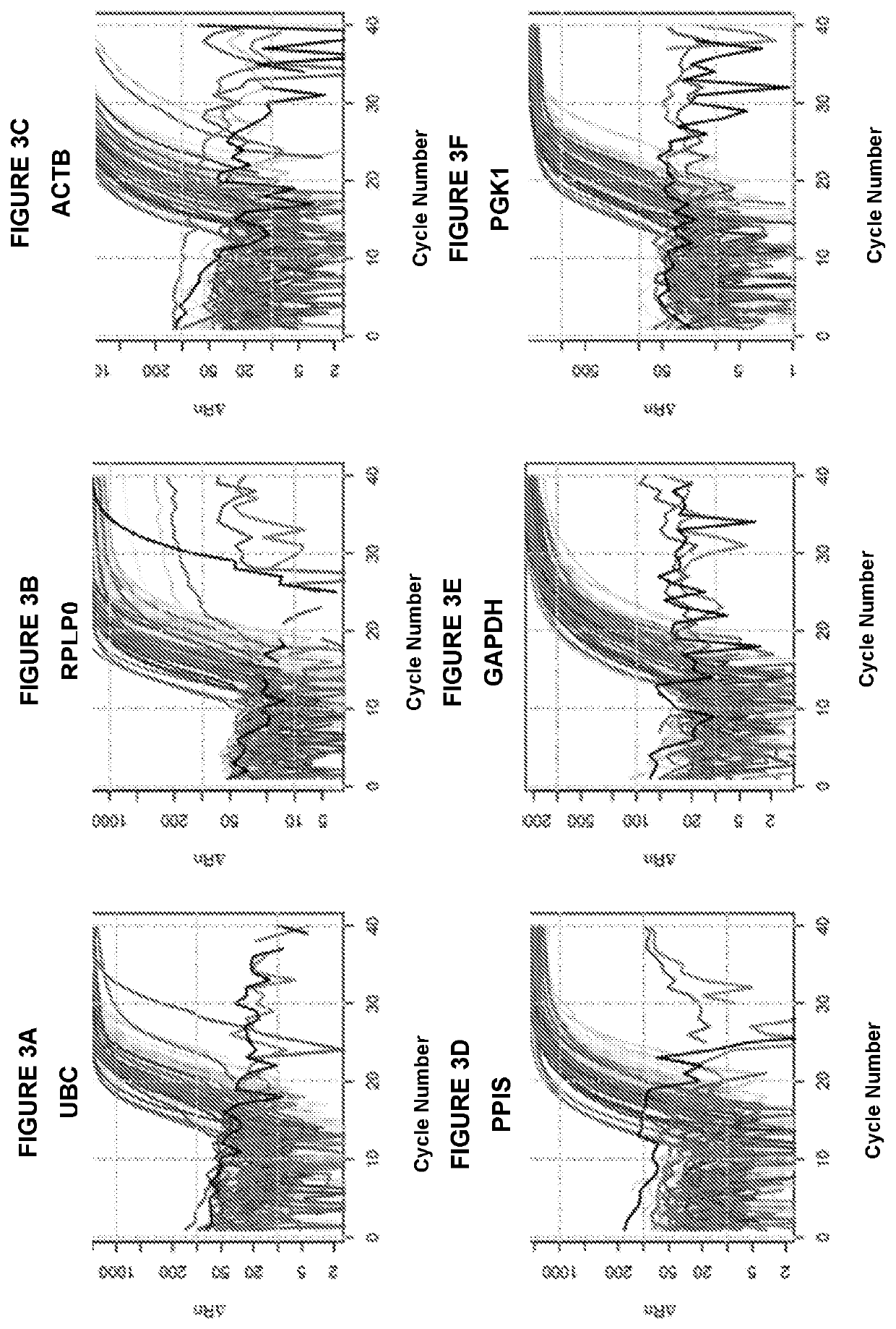
[0087]The studies presented herein were run on a pilot of 28 patients, and as detailed herein, the microvesicle RNA signatures, also referred to herein as the urinary microvesicle RNA signature, the exosome RNA signature, and / or the urinary exosome RNA signature, perfectly clustered the patients with rejection. In the studies presented herein, the microvesicle RNA signature is a microvesicle mRNA signature.

[0088]In the studies presented herein, 28 samples from 24 patients were used. Four patients with two samples each for two visits were used. Each sample was processed to extract RNA from cellular fraction or microvesicle fraction. The demographics of the samples were as follows: eight female samples and 20 male samples. Fourteen patients had transplant rejection, and fourteen patients had no symptoms or other indication of transplant rejection. In addition, three in-house control samples were...

example 2

and Validation of a Urinary Exosomes mRNA Signature for the Diagnosis of Human Kidney Transplant Rejection

[0101]Patients with end stage renal disease usually undergo transplantation, however, as many as 10-15% of these patients develop acute kidney rejection. Methods for monitoring clinical rejection include increase in serum creatinine and urinary protein secretion. These methods are not very accurate and may not reflect subclinical rejection. Currently, the patients are often monitored by repeat biopsies that may result in increased complications and cost. An accurate, non-invasive method would allow for earlier diagnosis and minimize the amount of immunosuppression needed to manage these patients. Extracellular vesicles such as exosomes (also referred to herein as microvesicles or the microvesicle fraction) are a promising new platform for biomarkers and can be used to monitor RNA and protein expression. Exosomes shed from the rejected kidney into the urine are likely originating...


PUM

PropertyMeasurementUnit
diameteraaaaaaaaaa
volumeaaaaaaaaaa
volumeaaaaaaaaaa

Abstract

The invention relates generally to the use of microvesicle RNA signatures for diagnosis, predicting, and / or to monitor treatment efficacy, including patients who are candidates for renal transplant and / or who have received a renal transplant.

Description

RELATED APPLICATIONS[0001]This application is a U.S. National Phase Application, filed under 35 U.S.C. 371, of International Application No. PCT / US2017 / 031212, filed on May 5, 2017, which claims priority to, and the benefit of, U.S. Provisional Application No. 62 / 332,279, filed May 5, 2016; the contents of each of which are incorporated herein by reference in their entirety.FIELD OF THE INVENTION[0002]The invention relates generally to the use of microvesicle RNA signatures for diagnosis, predicting, and / or to monitor treatment efficacy in patients, e.g., patients who are candidates for renal transplant and / or who have received a renal transplant.BACKGROUND[0003]Increasing knowledge of the genetic and epigenetic changes occurring in cells provides an opportunity to detect, characterize, and monitor diseases and disorders by analyzing disease-specific nucleic acid sequences and profiles. These changes can be observed by detecting any of a variety of disease-related biomarkers. Variou...

Claims


Application Information

Patent Timeline
Patent Type & Authority Patents(United States)
IPC IPC(8): C12Q1/68C12P19/34C12Q1/6876C12Q1/686
CPCC12Q1/6876C12Q1/686C12Q2600/112C12Q2600/118C12Q2600/158
Inventor SKOG, JOHAN KARL OLOVAZZI, JAMIL
Owner THE BRIGHAM & WOMEN S HOSPITAL INC
Why Eureka
Try Eureka