Isolated placental stem cell recruiting factors
a technology placental stem cell, which is applied in the direction of prosthesis, pharmaceutical delivery mechanism, peptide/protein ingredient, etc., can solve the problems of limited use and exceptionally difficult in vitro recruitment, and achieve the effect of prolonging the release of stem cell recruiting factor
- Summary
- Abstract
- Description
- Claims
- Application Information
AI Technical Summary
Benefits of technology
Problems solved by technology
Method used
Image

Examples
example 1
ation in the Presence of EpiFix®
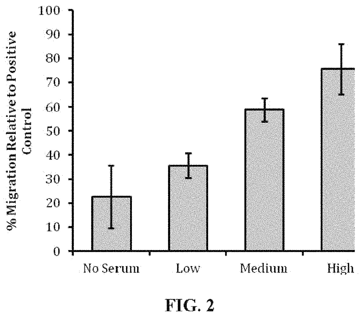
[0067]Human mesenchymal stem cells (human MSC) were evaluated in cell culture in the presence of samples of EpiFix® to determine whether the EpiFix® would induce migration of the human MSC. EpiFix® is a layer of amnion and chorion with the epithelial layer intact.
[0068]Materials and Methods
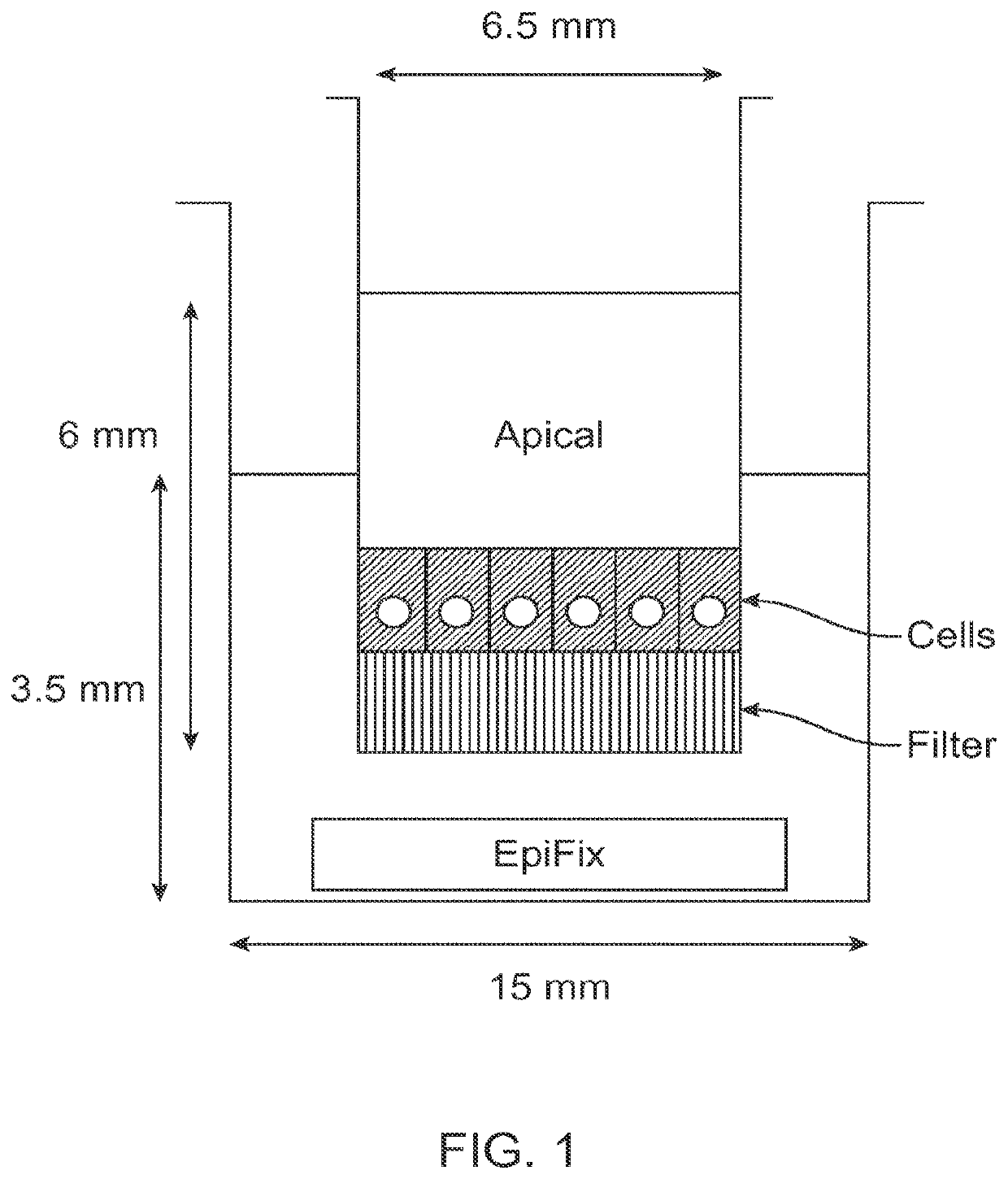
[0069]Standard migration assays were performed in 24-well cell culture inserts with 8-μm pore membrane filters at the bottom of the insert (see FIG. 1; BD Biosciences). 24 hours prior to the start of the experiment, human MSCs (one donor, passage 3) were cultured in serum free media, and 300 μL of 5 μg / mL fibronectin in PBS was placed into each cell culture insert to enable adsorption of fibronectin to the cell culture insert surface overnight.
[0070]On the day of the experiment, 700 μL of serum-free culture medium was loaded into the bottom wells of the plate, followed by the addition of differently sized portions of sterilized EpiFix® (Low: 1.5-mm diameter disk; Me...
example 2
Recruitment in Mice Receiving EpiFix® Implants
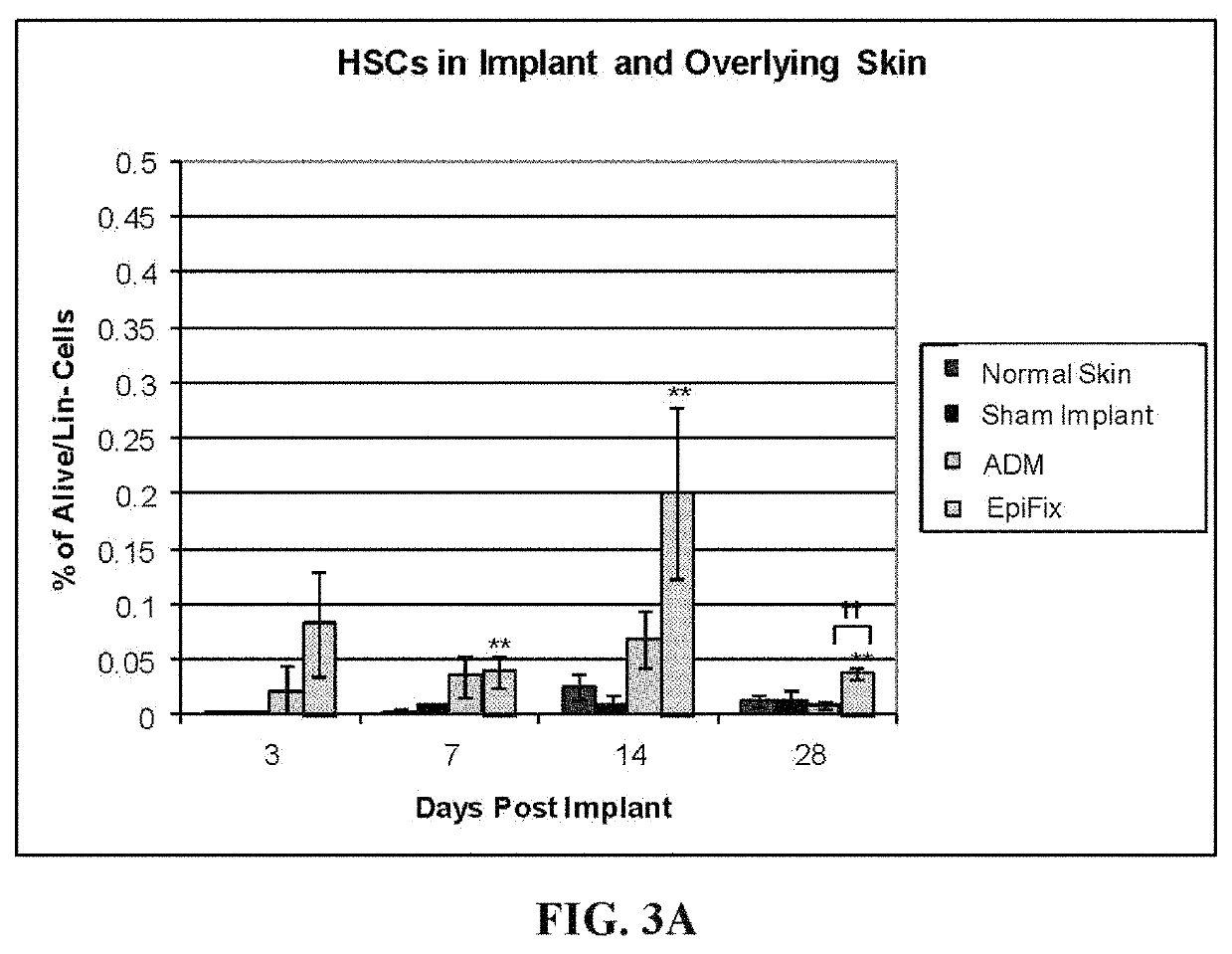
[0074]A study was undertaken to determine whether EpiFix® implanted in normal mice caused recruitment of stem / progenitor cells, focusing on mouse hematopoietic stem cells (HSCs) and mouse mesenchymal stem cells (mouse MSCs).
[0075]Materials and Methods
[0076]EpiFix® products from six donors were used for implantation in normal mice. A 5×5 mm square of EpiFix® was surgically placed subcutaneously in 4 month old FVB / NJ mice (weighing between about 23.50 g and about 30 g). Four mice were implanted per sample per time point. The time points were 3, 7, 14 and 28 days. The negative controls were normal skin and sham operated mice (surgical incision but no implant). Decellularized dermal matrix (acellular dermal matrix; ADM) was used as the comparative implant (Type I collagen, no cytokines). The implant and overlying skin was harvested for fluorescence-activated cell sorting (FACS).
[0077]Implants and overlying skin were harvested, cut into 1 mm2...
example 3
Characterization in Mice Receiving EpiFix® Implants
[0083]A study was undertaken to characterize stem cells recruited to EpiFix® implantation sites in mice, using flow cytometry and immunohistochemistry.
[0084]Materials and Methods
[0085]Sterile, Purion® processed EpiFix® in a 5×5 mm square patch was implanted subcutaneously through a skin incision on the backs of sixteen 4 month old FVB / NJ mice. Identical skin incisions were made in another sixteen mice to function as a control treatment (sham). For comparison with a collagen scaffold, a 5×5 mm square patch of decellularized human dermis (acellular dermal matrix; ADM) was implanted subcutaneously on the backs of sixteen mice. Un-operated mice were used as a source of “normal” back skin for the analyses.
[0086]The surgical site was removed at 3, 7, 14 and 28 days following implantation for analyses of stem cells. Four animals / group were used at each time point. Stem cells were identified with two distinct methods: Fluorescence-activated...
PUM
| Property | Measurement | Unit |
|---|---|---|
| distance | aaaaa | aaaaa |
| distance | aaaaa | aaaaa |
| distance | aaaaa | aaaaa |
Abstract
Description
Claims
Application Information
 - R&D Engineer
- R&D Manager
- IP Professional
- Industry Leading Data Capabilities
- Powerful AI technology
- Patent DNA Extraction
Browse by: Latest US Patents, China's latest patents, Technical Efficacy Thesaurus, Application Domain, Technology Topic.
© 2024 PatSnap. All rights reserved.Legal|Privacy policy|Modern Slavery Act Transparency Statement|Sitemap