
Automatic detection of growing nodules

Inactive Publication Date: 2005-01-06
SIEMENS MEDICAL SOLUTIONS USA INC
View PDF10 Cites 24 Cited by
  • Summary
  • Abstract
  • Description
  • Claims
  • Application Information

AI Technical Summary

Benefits of technology

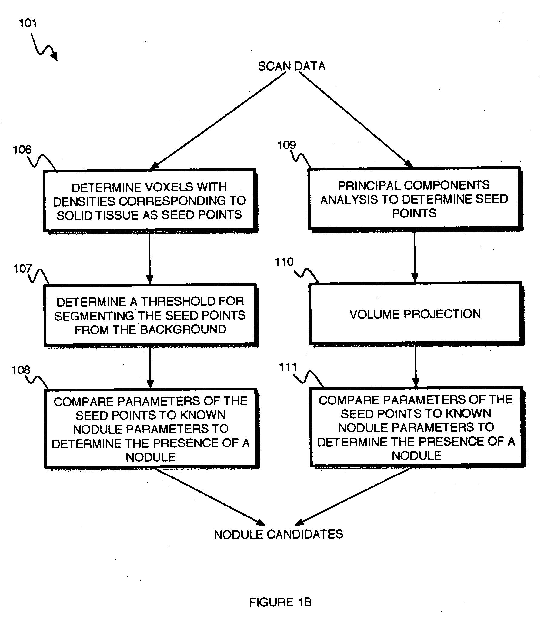
The nodule detection module includes a solitary module detecting solitary nodules, a pleura-attached module detecting pleura-attached nodules, and a vessel attached module detecting vessel-attached nodules. The system includes a false-positive module for removing false positive results from the list of nodule candidates as determined by one or more of the solitary module, the pleura-attached module and the vessel attached module.
The system includes a classification module for classifying nodule candidates of the segmentation module. The classification module determines a density of each nodule candidate, and removes nodule candidates from the list of growing nodule candidates determined to have a density above a predetermined density threshold. The classification module determines a size varian

Problems solved by technology

Despite decades of research into cancer treatment, the prognosis for patients diagnosed with lung cancer is very dismal, with an average 5-year survival rate of just 14%.
However the large datasets associated with multi-slice CT represent an increasing workload for radiologists.
However the vast majority of small nodules detected by radiologists during screening are benign.

Method used


Image

  • Automatic detection of growing nodules
  • Automatic detection of growing nodules
  • Automatic detection of growing nodules

Examples


Embodiment Construction

A method for detecting growing lung nodules uses the availability of prior scans to target the detection of precisely those nodules that are at highest likelihood of malignancy due to demonstrated growth.

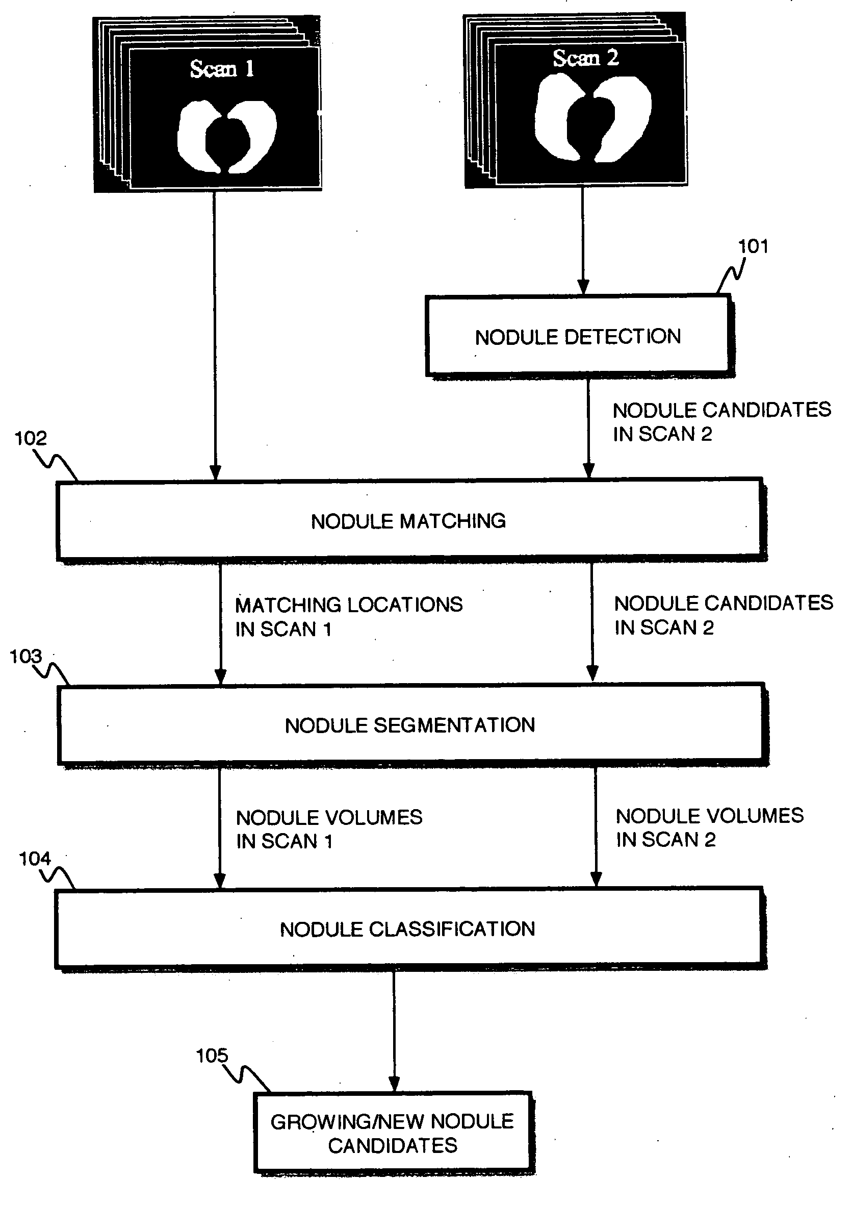
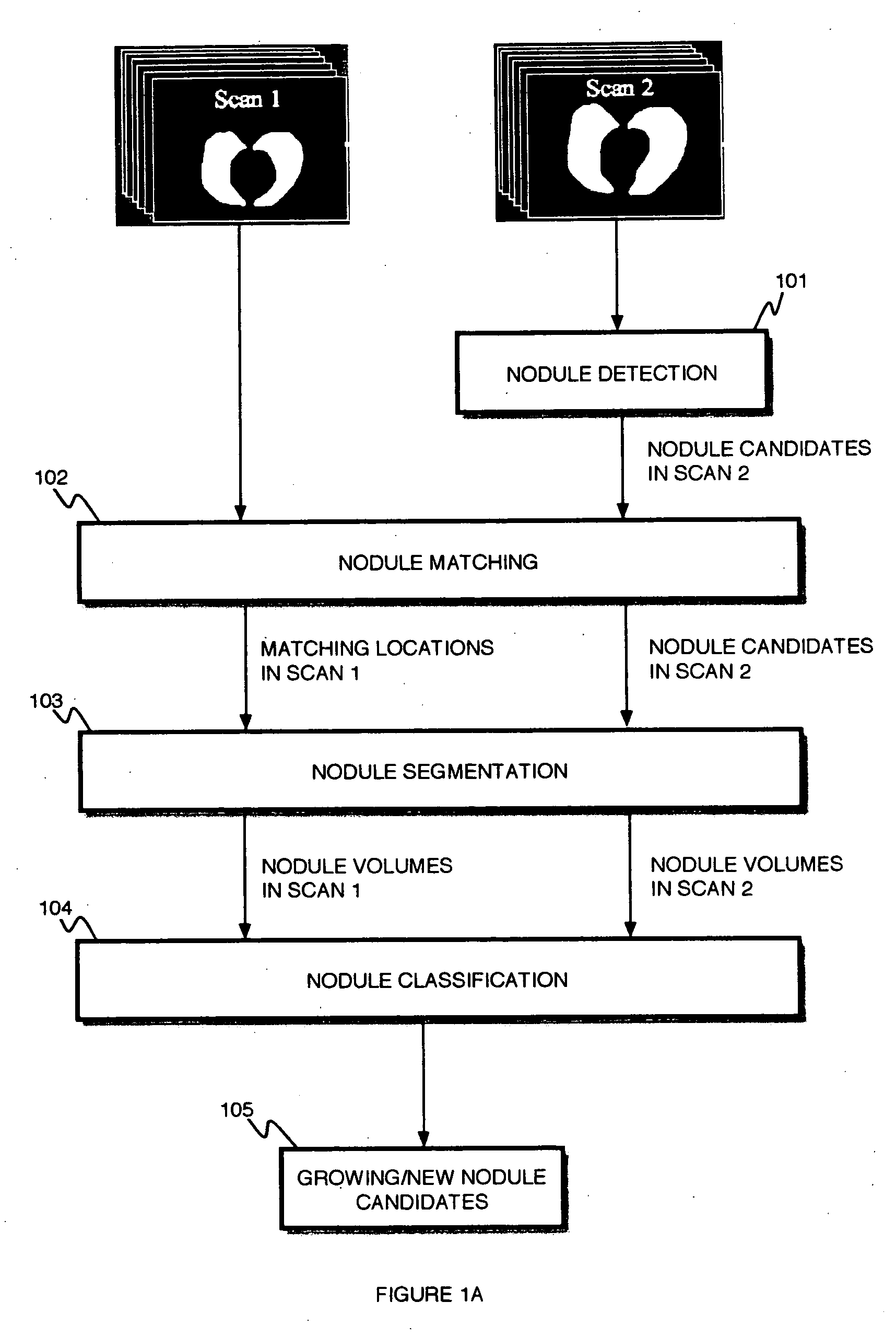
The method detects nodule candidates in the later of two scans of a patient 101. Locations in one scan are matched with the corresponding locations in another scan of the same patient 102. Once the location for the candidate in each of the two scans has been determined, an automatic method for nodule segmentation is applied to the voxels around each location 103. The volumes from each segmentation result are compared 104. A list of candidate nodules is generated where the nodule is determined to be larger or newly appeared since the previous scan 105.

The system and method operate on two multi-slice scans of the same patient taken at different times.

For each of patient an automatic detection program is applied to the later study. This gives a set of candidate nodules P. The foll...


PUM


Abstract

A system and method for detecting a growing nodule in multi-slice data detects a nodule candidate in a later scan, and matches a location of the nodule candidate in the later scan to a location in an earlier scan, wherein the earlier and later scans are of the same patient. The system and method segments the nodule candidate in the earlier and later scans, compares volumes from each segmentation, and determines a nodule, wherein the nodule is determined to be larger or newly appeared in the later scan as compared to the earlier scan.

Description

BACKGROUND OF THE INVENTION 1. Field of the Invention The present invention relates to volumetric medical image data, and more particularly to a system and method for automatic detection of growing or new nodules. 2. Discussion of Related Art Lung cancer is the leading cause of cancer death in the United States and around the world. Despite decades of research into cancer treatment, the prognosis for patients diagnosed with lung cancer is very dismal, with an average 5-year survival rate of just 14%. Early stages of lung cancer do not usually cause specific symptoms, and most patients are diagnosed at advanced stages. However for those patients who are diagnosed in stage I, the prognosis is much better, with average 5-year survival rates of 60-70%. Lung cancer screening offers the most promising option for detecting cancer in the early stages when cure is most likely. Lung cancer screening by computed tomography (CT) has been shown to increase the percentage of cancers detected...

Claims


Application Information

Patent Timeline
IPC IPC(8): G06K9/00G06K9/68G06T7/00
CPCG06T2207/30061G06T7/0012
Inventor NOVAK, CAROL L.SHEN, HONGODRY, BENJAMIN
Owner SIEMENS MEDICAL SOLUTIONS USA INC
Learn More