Eureka AIR delivers breakthrough ideas for toughest innovation challenges, trusted by R&D personnel around the world.

Multi-slice double inversion-recovery black-blood imaging with simultaneous slice re-inversion

Active Publication Date: 2008-01-01
UNIV OF WASHINGTON
View PDF1 Cites 18 Cited by
  • Summary
  • Abstract
  • Description
  • Claims
  • Application Information

AI Technical Summary

Benefits of technology

[0009]The predefined TI and the predefined delay time are determined so that the sequence of RF pulses and magnetic field gradient pulses are applied to read out the signal for imaging each selected single slice at a time when the magnetization signal for the flowing blood is substantially zero. Since a benefit of the present invention is suppressing the contribution of flowing blood to the signals used for imaging the successive slices, the site of the imaging will normally include at least one artery conveying flowing blood.
[0013]Yet another aspect of the present invention is directed to a method for enabling visual evaluation of atherosclerotic plaque at a site in a patient. This method is similar to the method noted above, but in addition, repeats all of the steps for each of a plurality of transverse slices at the site being imaged. The plurality of transverse slices is within planes that are generally transverse relative to the plurality of slices initially imaged to provide a reference when acquiring the signals for imaging the transverse slices. In addition, the method includes the step of displaying selected transverse slices of the site, so that any atherosclerotic plaque formation in an artery of the patient at the site is clearly visible. A contribution due to flowing blood in each transverse slice is thus suppressed to more clearly visually display any atherosclerotic plaque formation in an artery at the site.

Problems solved by technology

Efficient suppression of the signal from flowing blood is especially important for cardiovascular MRI, where anatomic structures and pathologic tissues of interest are close to blood, and flow artifacts may cause severe problems for diagnostic interpretation.
The method of in-flow saturation usually does not enable complete elimination of the signal from flowing blood and may be ineffective in applications that require clear visualization of the interface between vessel wall and lumen, such as high-resolution imaging of atherosclerotic plaque.
However, DIR, as used in this manner, is essentially applicable to imaging only a single slice per repetition time (TR), and therefore, a long scan time is required if several slices need to be imaged using the DIR method.
A common problem of the above-noted multi-slice DIR techniques is the construction of a preparative module, which includes a train of slice-selective inversions, creating unequal conditions for the evolution of magnetization of different slices, since the delay between non-selective inversion and slice-selective re-inversion depends on the actual slice number.
Furthermore, the restrictions on specific absorption rate (SAR) also may preclude a further increase of slice quantity.

Method used


Image

  • Multi-slice double inversion-recovery black-blood imaging with simultaneous slice re-inversion
  • Multi-slice double inversion-recovery black-blood imaging with simultaneous slice re-inversion
  • Multi-slice double inversion-recovery black-blood imaging with simultaneous slice re-inversion

Examples


Embodiment Construction

Principle of Multi-Slice DIR Acquisition

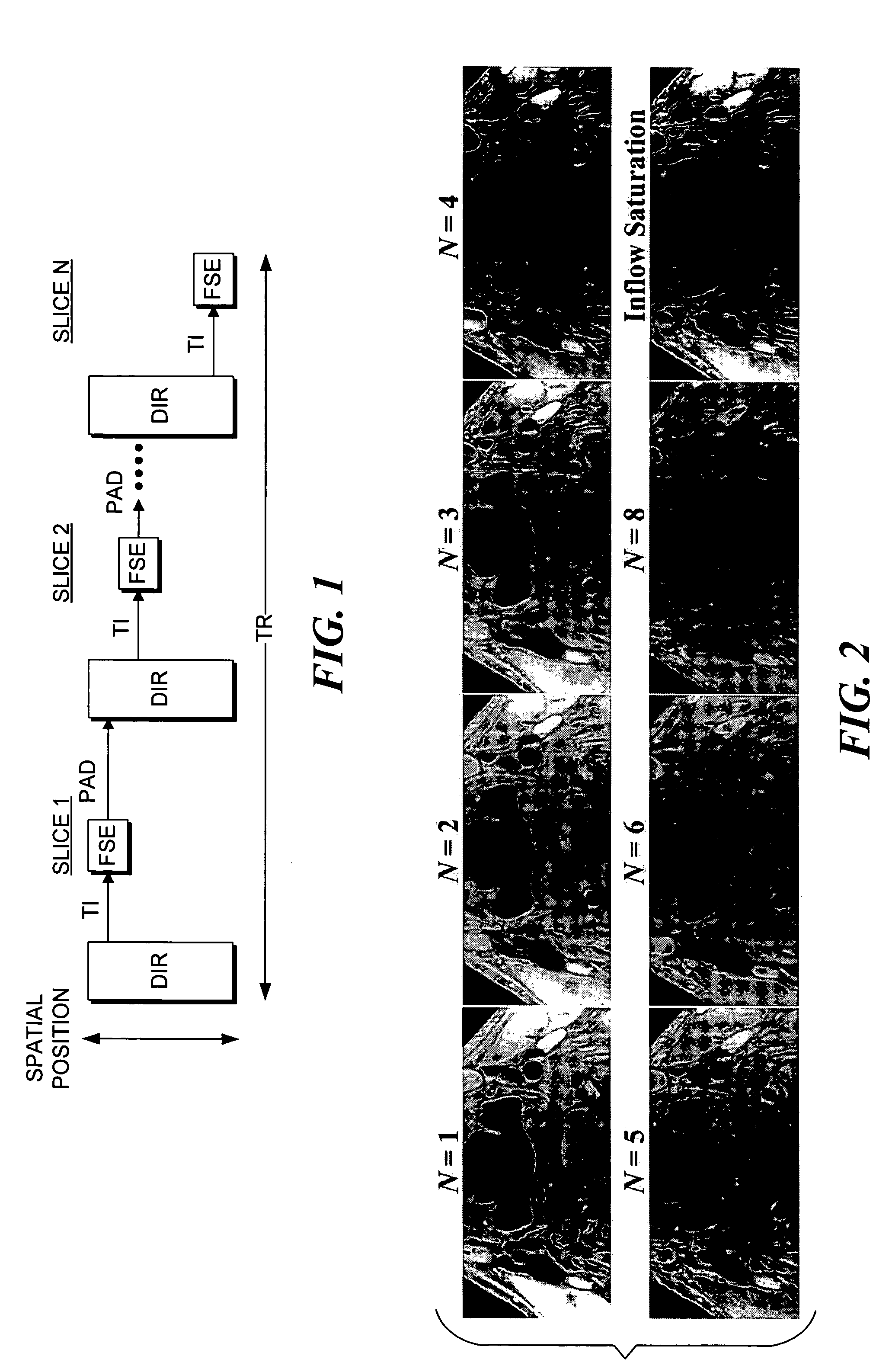
[0023]A schematic functional diagram of a multi-slice DIR-FSE sequence in accord with the novel method described herein is shown in FIG. 1. In FIG. 1, slices are imaged sequentially within the TR, as shown by FSE readout blocks. A DIR preparation is executed before read out of each slice. Each DIR block includes a slab-selective inversion applied to an entire slice pack. The proper timing of the sequence is controlled by a post-acquisition delay (PAD), which allows for coincidence of the zero-crossing point of blood (see Eq. [1] below) and the start of an FSE readout. The general idea of this method is to replace a slice-selective inversion in the DIR preparation by a slab-selective one, which is applied to an entire slab of slices to be imaged within a TR. The DIR sequence is repeated for each slice, while a shortened TI provides imaging of several slices per TR. A predefined post-acquisition delay (referred to as “PAD” in FIG. 1) is used aft...


PUM


Abstract

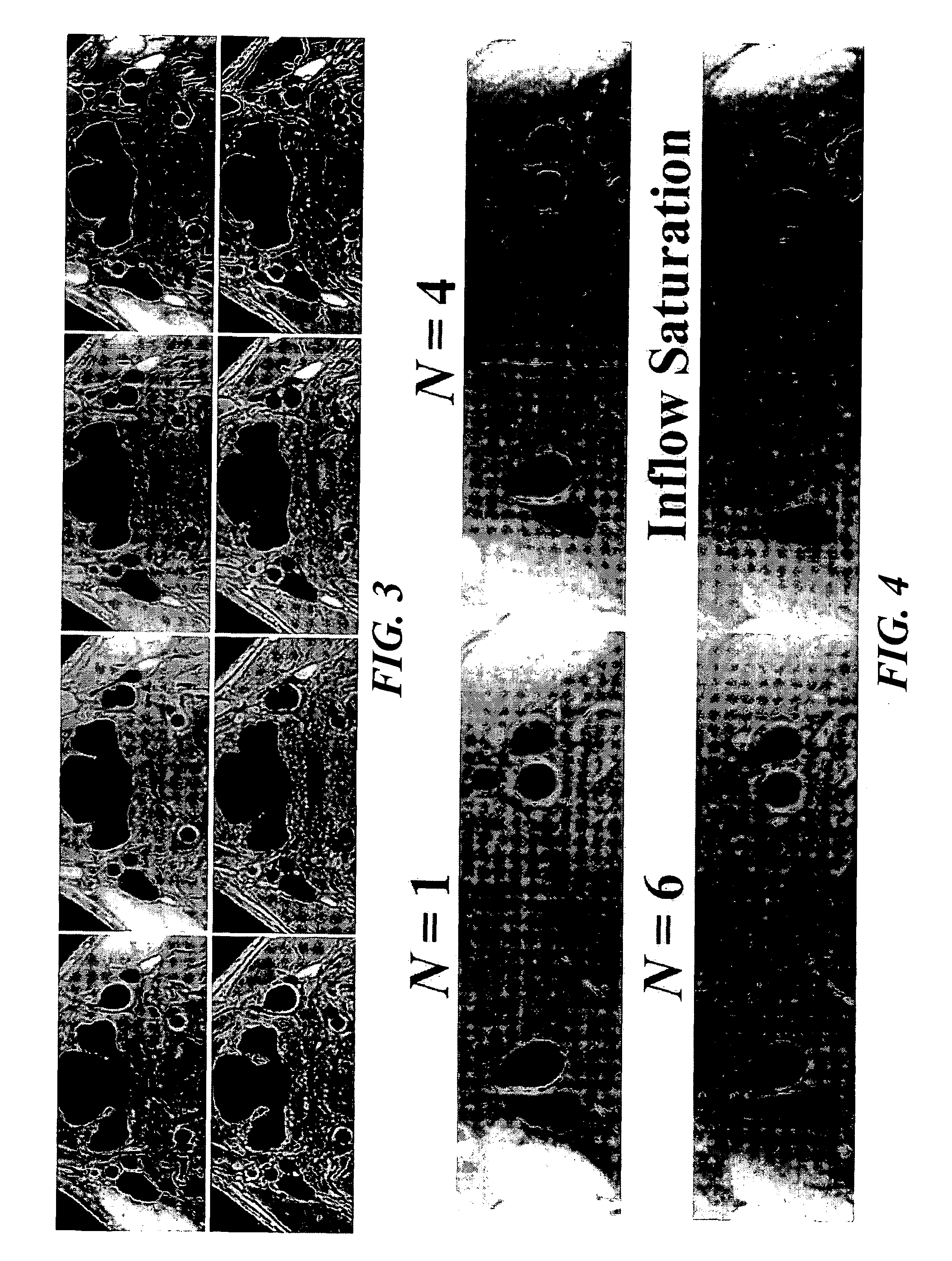
A multi-slice double inversion recovery (DIR) pulse sequence with read out of a signal for imaging successive slices implemented on a magnetic resonance image scanner. In the method, when the DIR pulse sequence is applied before imaging each slice, a slab-selective inversion re-inverts the entire slab that includes all of the slices. All slices are imaged within a predefined repetition time (TR). The number, N, of slices acquired per TR controls the inversion time to execute the read out of the signal for imaging each slice at a zero-crossing point of blood. In a test, multi-slice DIR images of carotid arteries were obtained with N ranging from 2-8, for four subjects. The results were compared with those for both standard single-slice DIR, and inflow saturation techniques. Multi-slice DIR with N=2-6 provided blood flow suppression in carotid arteries similar to that of single-slice DIR, and significantly better than inflow saturation.

Description

RELATED APPLICATIONS[0001]This application is based on copending provisional patent application Ser. No. 60 / 452,071, filed Mar. 3, 2003, the benefit of the filing date of which is hereby claimed under 35 U.S.C. § 119(e).GOVERNMENT RIGHTS[0002]This invention was made with government support under Grant No. RO1-HL56874, awarded by NIH-NIHBI, and the federal government may have certain rights in this invention.FIELD OF THE INVENTION[0003]This invention generally pertains to magnetic resonance imaging (MRI) and more specifically, pertains to a method and a system that use a plurality of radio frequency (RF) inversion pulses grouped into double-inversion procedures in order to suppress the signal from flowing blood when imaging a plurality of slices, while performing a simultaneous slab-selective re-inversion, wherein the slab includes the plurality of slices being imaged.BACKGROUND OF THE INVENTION[0004]Efficient suppression of the signal from flowing blood is especially important for c...

Claims


Application Information

Patent Timeline
IPC IPC(8): A61B5/05A61B5/055G01R33/54G01R33/563
CPCA61B5/055G01R33/563A61B5/7285G01R33/4835
Inventor YARNYKH, VASILY L.YUAN, CHUN
Owner UNIV OF WASHINGTON
Why Eureka
Eureka Blog
Learn More