Multi-Plane/Multi-Slice Processing For 2-D Flow Imaging in Medical Diagnostic Ultrasound

a technology of flow imaging and multi-plane processing, applied in the field of medical diagnostic ultrasound, can solve the problems of insufficient sensitivity, long examination, and images acquired with proper settings may have flow holes or other problems

Active Publication Date: 2009-12-10
SIEMENS MEDICAL SOLUTIONS USA INC
View PDF12 Cites 17 Cited by
  • Summary
  • Abstract
  • Description
  • Claims
  • Application Information

AI Technical Summary

Benefits of technology

[0006]By way of introduction, the preferred embodiments described below include a method, system, computer readable medium, and instructions for 2-D flow imaging in medical diagnostic ultrasound. Flow data for a volume is acquired. For more rapid acquisition, broad beam transmission and simultaneous reception along many scan lines distributed in the volume is used. The flow data for the volume is filtered, such as by calculating statistical information. The statistical information is used to determine the display values for the flow imaging. The display value for each location in the image may be based on data in a surrounding three-dimensional region.

Problems solved by technology

However, this repositioning may result in a longer examination.
However, even images acquired with proper settings may have flow holes or insufficient sensitivity.

Method used


Image

  • Multi-Plane/Multi-Slice Processing For 2-D Flow Imaging in Medical Diagnostic Ultrasound
  • Multi-Plane/Multi-Slice Processing For 2-D Flow Imaging in Medical Diagnostic Ultrasound
  • Multi-Plane/Multi-Slice Processing For 2-D Flow Imaging in Medical Diagnostic Ultrasound

Examples


Embodiment Construction

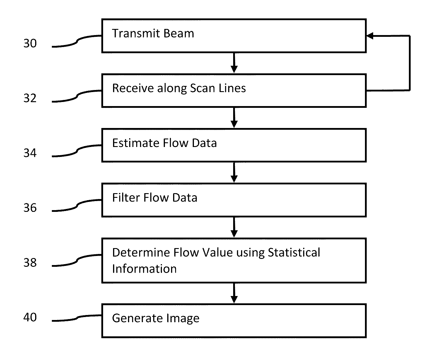
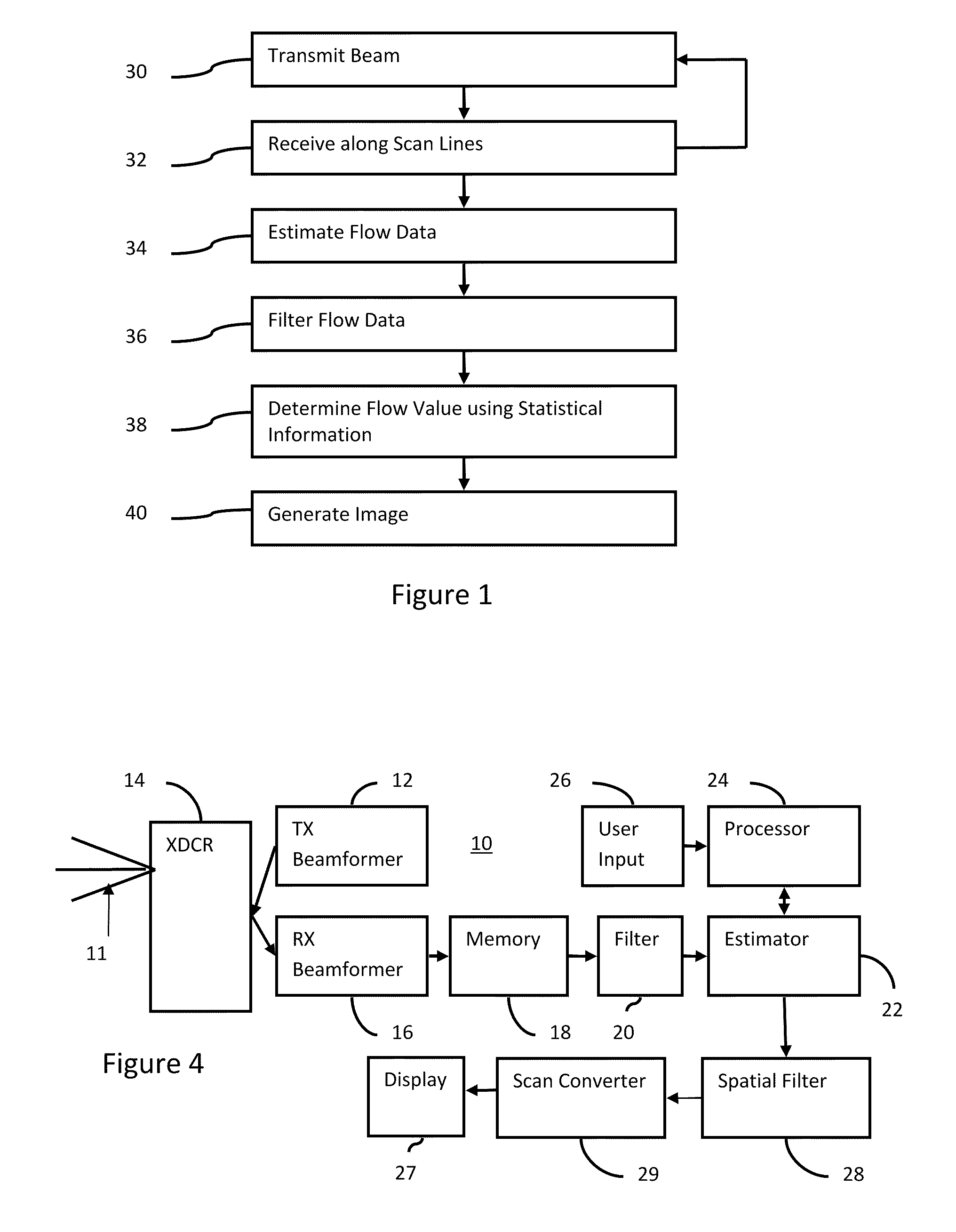
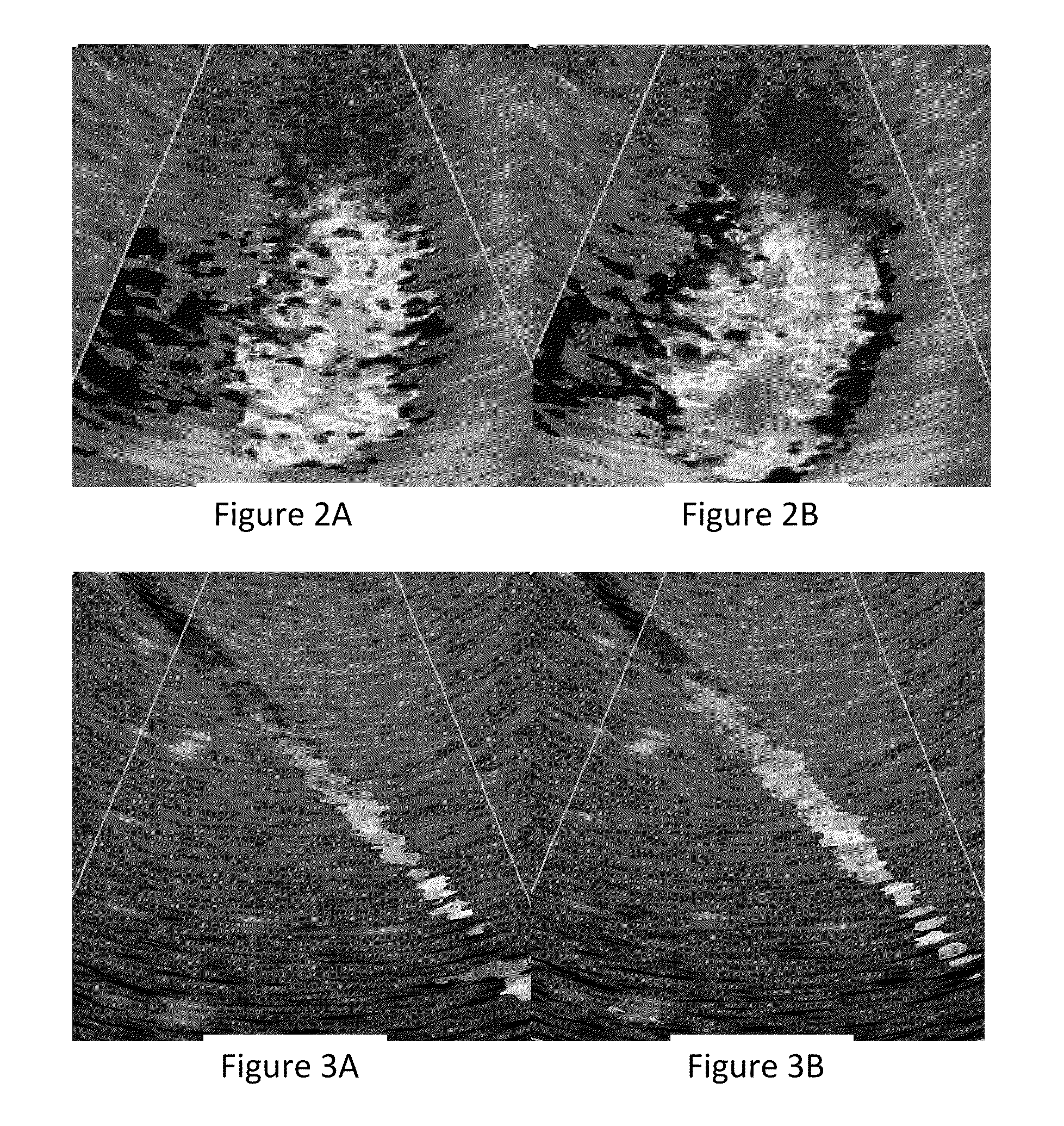
[0016]The color Doppler fill-in, sensitivity, and signal-to-noise ratio (SNR) for two-dimensional (2D) flow imaging may be improved by using simultaneous acquisition of multiple thin-slices or volume data. For two-dimensional imaging, the image may be displayed as a 2D slice through some combination of the data in the slices. Acquisition of thin slices may be performed using multi-dimensional array probes (1.5, 1.75D or 2D) or 1D arrays with mechanical steering. Volumetric processing of the thin slices through spatial filtering improves SNR and signal sensitivity. For 2D imaging, combination of the color information in the thin-slices may use the local and / or regional image statistics, such as mean, mix, max, median, and spatial variance. Using volume information may more likely provide a sufficient image, minimizing the time spent repositioning the transducer and improving workflow.

[0017]In one two-dimensional imaging example embodiment, color Doppler imaging is performed with simu...


PUM


Abstract

A volumetric method for 2-D flow imaging is provided in medical diagnostic ultrasound. Flow data for a volume is acquired. For more rapid acquisition, broad beam transmission and reception along many scan lines distributed in the volume is used. The volumetric flow data is filtered, such as by calculating statistical information, to generate a planar/2-D flow image. The statistical information from the three-dimensional flow data is used to determine the display values for the flow imaging.

Description

CROSS-REFERENCE TO RELATED APPLICATIONS[0001]The present patent document claims the benefit of the filing date under 35 U.S.C. §119(e) of Provisional U.S. Patent Application Ser. No. 61 / 059,681, filed Jun. 6, 2008, which is hereby incorporated by reference.BACKGROUND[0002]This present invention relates to medical diagnostic ultrasound. In particular, two dimensional flow imaging is provided.[0003]Flow imaging is performed with color Doppler processes. The Doppler effect is used to estimate velocity, energy and / or variance of flow. The flow information is mapped to a color for imaging.[0004]To image flow in a patient, the user positions the transducer at a desired location. Due to the sensitivity of the flow to the angle of insonification, the transducer is repositioned to identify the location with the best image. However, this repositioning may result in a longer examination.[0005]Flow imaging includes other variables. The user adjusts the overall gain and depth gains and velocity ...

Claims


Application Information

Patent Timeline
Patent Type & Authority Applications(United States)
IPC IPC(8): A61B8/06
CPCA61B8/488A61B8/06
Inventor SRINIVASAN, SESHADRIUSTUNER, KUTAY F.GURACAR, ISMAYIL M.
Owner SIEMENS MEDICAL SOLUTIONS USA INC
Why Eureka
Try Eureka