Polypeptide variants with altered effector function
a technology of effector function and polypeptide, which is applied in the field of polypeptide variants with altered effector function, can solve the problems of not removing foreign antigens, uneven distribution of variable domains of antibodies,
- Summary
- Abstract
- Description
- Claims
- Application Information
AI Technical Summary
Benefits of technology
Problems solved by technology
Method used
Image
Examples
example 1
Low Affinity Receptor Binding Assay
[0303] This assay determines binding of an IgG Fc region to recombinant FcγRIIA, FcγRIIB and FcγRIIIA α subunits expressed as His6-glutathione S transferase (GST)-tagged fusion proteins. Since the affinity of the Fc region of IgG1 for the FcγRI is in the nanomolar range, the binding of IgG1 Fc variants can be measured by titrating monomeric IgG and measuring bound IgG with a polyclonal anti-IgG in a standard ELISA format (Example 2 below). The affinity of the other members of the FcγR family, i.e. FcγRIIA, FcγRIIB and FcγRIIIA for IgG is however in the micromolar range and binding of monomeric IgG1 for these receptors can not be reliably measured in an ELISA format.
[0304] The following assay utilizes Fc variants of recombinant anti-IgE E27 (FIGS. 4A and 4B) which, when mixed with human IgE at a 1:1 molar ratio, forms a stable hexamer consisting of three anti-IgE molecules and three IgE molecules. A recombinant chimeric form of IgE (chimeric IgE) ...
example 2
Identification of Unique C1q Binding Sites in a Human IgG Antibody
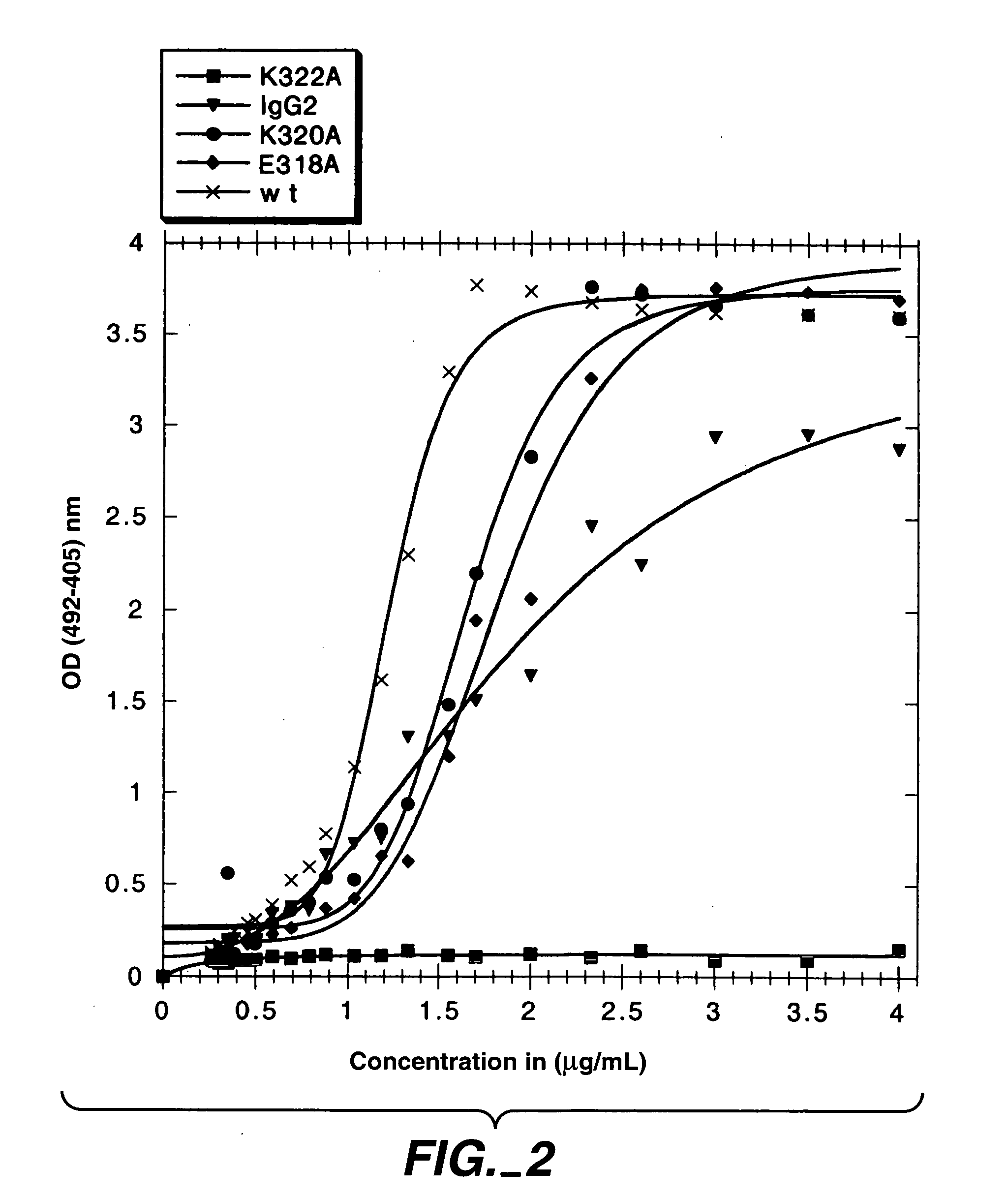
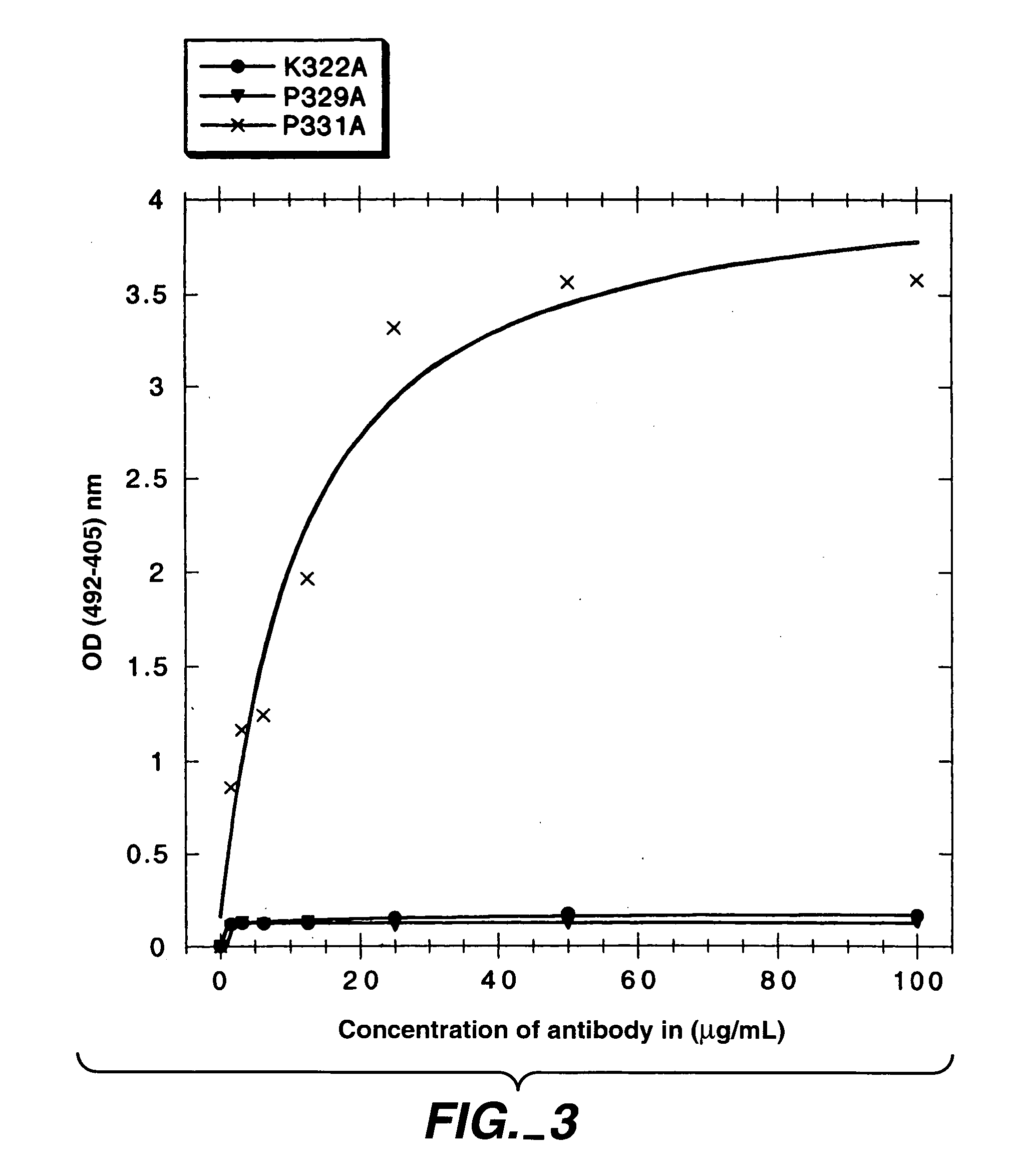
[0309] In the present study, mutations were identified in the CH2 domain of a human IgG1 antibody, “C2B8” (Reff et al., Blood 83:435 (1994)), that ablated binding of the antibody to C1q but did not alter the conformation of the antibody nor affect binding to each of the FcγRs. By alanine scanning mutagenesis, five variants in human IgG1 were identified, D270K, D270V, K322A P329A, and P331, that were non-lytic and had decreased binding to C1q. The data suggested that the core C1q binding sites in human IgG1 is different from that of murine IgG2b. In addition, K322A, P329A and P331A were found to bind normally to the CD20 antigen, and to four Fc receptors, FcγRI, FcγRII, FcγRIII and FcRn.
MATERIALS AND METHODS
[0310] Construction of C2B8 Variants: The chimeric light and heavy chains of anti-CD20 antibody C2B8 (Reff et al., Blood 83:435 (1994)) subcloned separately into previously described PRK vectors (Gorman et al., D...
example3
Variants with Improved C1q Binding
[0323] The following study shows that substitution of residues at positions K326, A327, E333 and K334 resulted in variants with at least about a 30% increase in binding to C1q when compared to the wild type antibody. This indicated K326, A327, E333 and K334 are potential sites for improving the efficacy of antibodies by way of the CDC pathway. The aim of this study was to improve CDC activity of an antibody by increasing binding to C1q. By site directed mutagenesis at K326 and E333, several variants with increased binding to C1q were constructed. The residues in order of increased binding at K326 are K<V<E<A<G<D<M<W, and the residues in order of increased binding at E333 are E<Q<D<V<G<A<S. Four variants, K326M, K326D, K326E and E333S were constructed with at least a two-fold increase in binding to C1q when compared to wild type. Variant K326W displayed about a five-fold increase in binding to C1q.
[0324] Variants of the wild type C2B8 antibody were...
PUM
| Property | Measurement | Unit |
|---|---|---|
| crystal structure | aaaaa | aaaaa |
| affinity | aaaaa | aaaaa |
| binding affinity | aaaaa | aaaaa |
Abstract
Description
Claims
Application Information
 - R&D
- Intellectual Property
- Life Sciences
- Materials
- Tech Scout
- Unparalleled Data Quality
- Higher Quality Content
- 60% Fewer Hallucinations
Browse by: Latest US Patents, China's latest patents, Technical Efficacy Thesaurus, Application Domain, Technology Topic, Popular Technical Reports.
© 2025 PatSnap. All rights reserved.Legal|Privacy policy|Modern Slavery Act Transparency Statement|Sitemap|About US| Contact US: help@patsnap.com


