Method and apparatus of image processing to detect and enhance edges

a technology of image processing and edge detection, applied in the field of methods and, can solve the problems of blurred edges or edges, noisy ultrasonic images, and fuzzy liver borders or edges, and achieve the effect of detecting and enhancing edges

Inactive Publication Date: 2006-01-26
HITACHI LTD
View PDF13 Cites 58 Cited by
  • Summary
  • Abstract
  • Description
  • Claims
  • Application Information

AI Technical Summary

Problems solved by technology

In such instances, the ultrasound image looks noisy as the true tissue is hidden behind this curtain of grain noises absent image processing to remove the noise.
If the same smoothing filter is applied to the liver borders or all areas in the image, the liver borders or edges become blurred or fuzzy.

Method used


Image

  • Method and apparatus of image processing to detect and enhance edges
  • Method and apparatus of image processing to detect and enhance edges
  • Method and apparatus of image processing to detect and enhance edges

Examples


Embodiment Construction

)

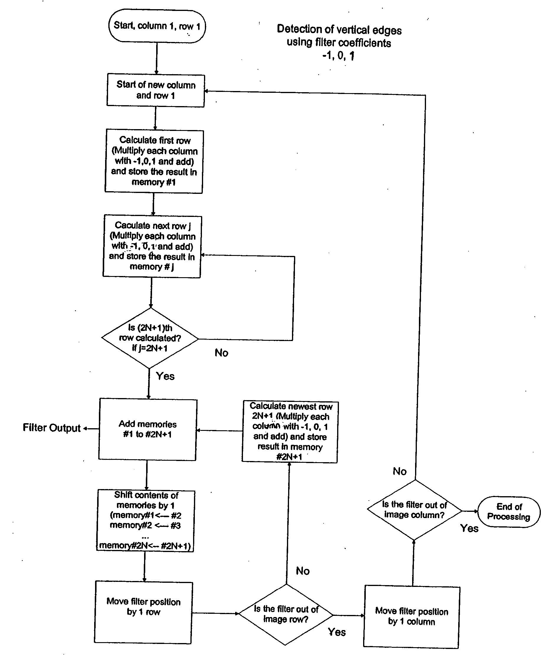
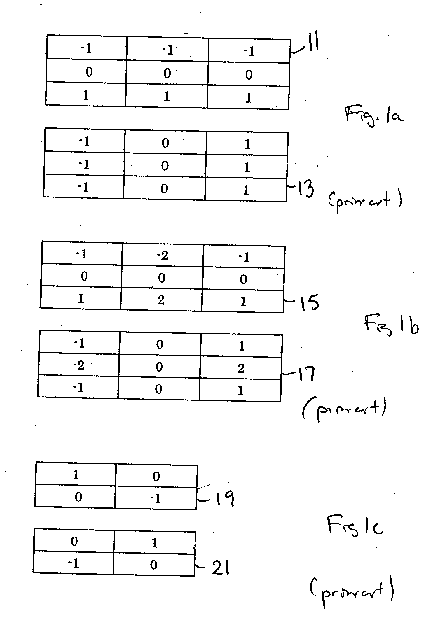
[0058] Accordingly, it is an object of the present invention to provide a method, and apparatus for performing such method, of image processing to detect and enhance edges and to reduce noise in ultrasound imaging. Specifically, the method relates to the detection of edges in noisy images and the enhancement the edges and the image.

[0059] It is therefore a teaching of the present invention to provide a method for determining if a smoothing filter should be applied to an ultrasound image depending on the output of an edge detection filter. The edge detection filter determines if an image pixel belongs to an edge or belongs in the middle of the homogeneous tissue area. The magnitude of the edge detection filter output is used to determine if the pixel belongs to an edge or a ‘strong’ or ‘steep’ edge. If the pixel belongs to a homogeneous tissue area, a smoothing filter is applied to the pixel. The ultrasound image may comprise one or more, or a combination of, any type of ultrasound...


PUM


Abstract

A method for detecting and enhancing edges in an ultrasound image includes the steps of applying an edge detection filter to at least one of a plurality of pixels of an ultrasound image to compute an edge detection value having a magnitude and a direction; and applying either a directional edge enhancement filter or a non-directional smoothing filter to at least one of the plurality of pixels of the ultrasound image.

Description

CROSS-REFERENCE TO RELATED APPLICATION [0001] This application claims the benefit of U.S. Provisional Application Ser. No. 60 / 586,589, filed Jul. 9, 2004.BACKGROUND OF THE INVENTION [0002] 1. Field of Invention [0003] The present invention relates to a method, and apparatus for performing such method, of image processing to detect and enhance edges and to reduce noise in ultrasound imaging. Specifically, the method relates to the detection of edges in noisy images and the enhancement the edges and the image. [0004] 2. Description of Related Art [0005] An ultrasound image consists of back-scattered signals from many scatterers in the sample volume (or image cell) Interference effects of these many signals may enhance signals or cancel each other, creating grainy images. These grains are called speckles and thus the ultrasound image is represented by these grains or speckles even if the imaged area is considered to be homogeneous tissue with monotone contrast. In such instances, the u...

Claims


Application Information

Patent Timeline
Patent Type & Authority Applications(United States)
IPC IPC(8): A61B8/00
CPCG06T5/20G06T2207/10132G06T2207/30004G06T2207/20192G06T5/003G06T7/0085G06T5/002G06T7/13
Inventor TAMURA, TADASHI
Owner HITACHI LTD
Why Eureka
Try Eureka