Method for the identification of extracellular domains of Epstein Barr Virus (EBV) tumor-associated latent membrane proteins and for the selection of antibody reagents reactive therewith

a technology latent membrane protein, which is applied in the field of antibody reagents reactive therewith and the selection of extracellular domains of epstein barr virus (ebv) tumor-associated latent membrane proteins, can solve the problems of lymphoproliferation disease, full-blown malignancy, and detection of epithelial replication of the virus

Inactive Publication Date: 2006-03-30
CYTO BARR
View PDF0 Cites 2 Cited by
  • Summary
  • Abstract
  • Description
  • Claims
  • Application Information

AI Technical Summary

Problems solved by technology

In the vast majority of cases EBV infection results in a lymphoproliferative disease that may be temporarily debilitating, but is always benign and self-limiting.
In certain immunosuppressed individuals, however, the result can be uncontrolled lymphoproliferation leading to full-blown malignancy.
In addition, in such patients uncontrolled epithelial replication of the virus is detectable in lesions of oral hairy leukoplakia.
In immunodeficient patients with defective cellular immunity directed against the dominant antigens as described above (esp.in XLP-patients, transplant recipients and HIV-infected individuals), the immune system fails to eradicate EBV+ B-cells expressing the latency-III phenotype, thus allowing their uncontrolled outgrowth frequently resulting in malignant oligoclonal and monoclonal B-cell tumours.
Thus, immunocompetent patients with EBV+ tumours are incapable of mounting effective immune responses directed against the "non-self" tumor-associated viral gene products.
Tumour-cell induced immunomodulation (suppression) and tumour cell apoptosis resistance may further render local T-cell immune responses ineffective.
Several EBV-encoded tumour cell-associated membrane proteins are described in the art; however, it has not yet been shown that such membrane proteins comprise extracellular domains, suitable for antibody interaction.
Alternatively, antibody reagents bound to the cell surface may bind and activate components of the complement cascade, causing lysis of the targets cells.
Although this is a very simple and consistent method, it fails to take into consideration that, for example, in an otherwise identical pair of sequences, one insertion or deletion will cause the following amino acid residues to be put out of alignment, thus potentially resulting in a large reduction in % homology when a global alignment is performed.
However, these more complex methods assign "gap penalties" to each gap that occurs in the alignment so that, for the same number of identical amino acids, a sequence alignment with as few gaps as possible--reflecting higher relatedness between the two compared sequences--will achieve a higher score than one with many gaps.

Method used


Image

  • Method for the identification of extracellular domains of Epstein Barr Virus (EBV) tumor-associated latent membrane proteins and for the selection of antibody reagents reactive therewith
  • Method for the identification of extracellular domains of Epstein Barr Virus (EBV) tumor-associated latent membrane proteins and for the selection of antibody reagents reactive therewith
  • Method for the identification of extracellular domains of Epstein Barr Virus (EBV) tumor-associated latent membrane proteins and for the selection of antibody reagents reactive therewith

Examples


example 1

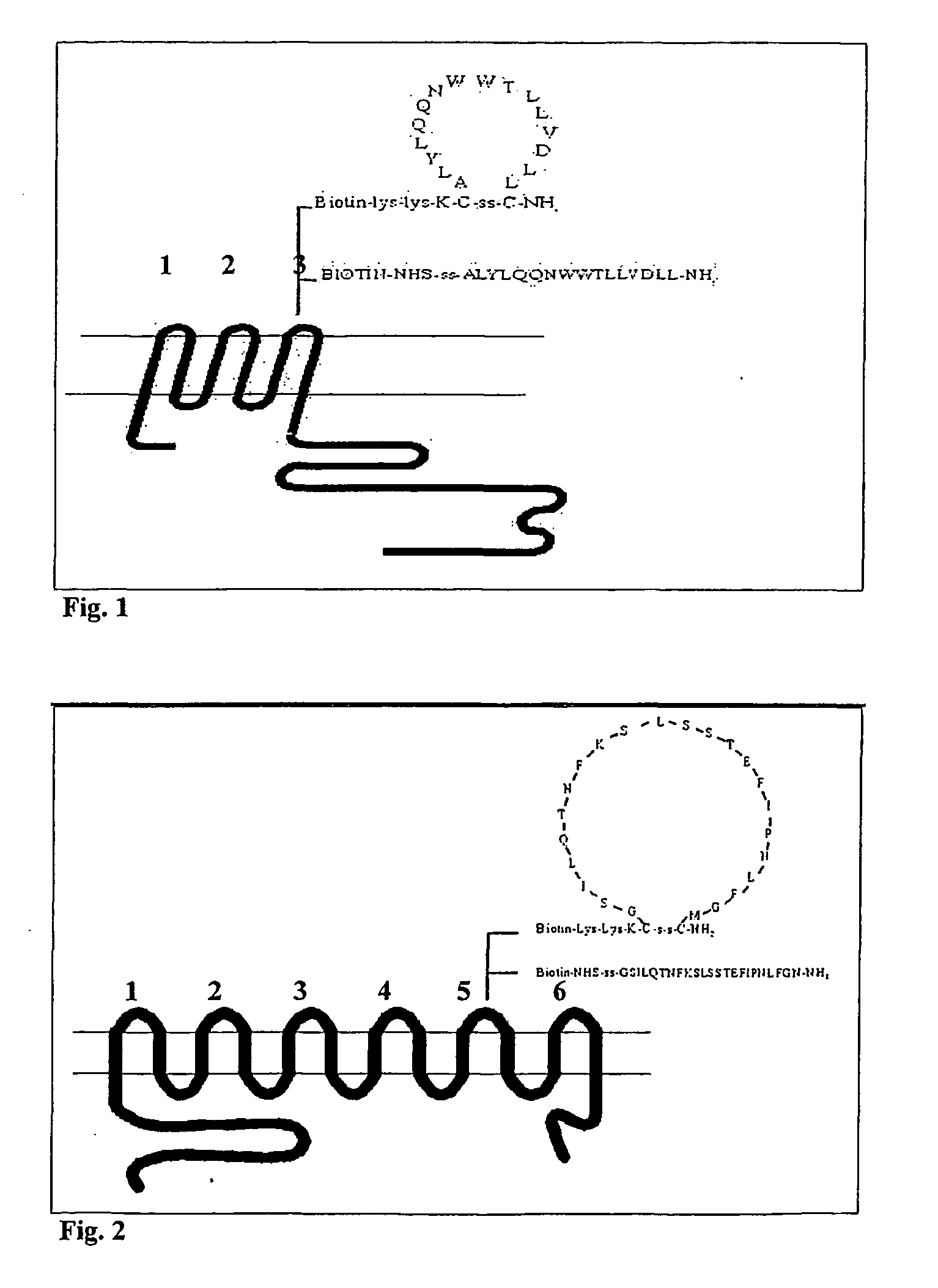
Design of LMP1, LMP2 and BARF1 Derived Peptides with a Putative Extracellular Localisation.

[0300] The amino acid sequences of LMP1, LMP2 and BARF1 were retrieved from the Swiss-Prot or Genbank database.

[0301] The aim was to define and demonstrate the existence of extracellular domains on these proteins as expressed in naturally infected and transformed (tumour) cells.

[0302] Thereto, as a first step the amino acid sequences derived from DNA encoding the LMP1, LMP2 and BARF1 proteins were used and the putative transmembrane domains were localised by hydrophobicity analysis using the GCG protein analysis program, supra.

[0303] This analysis revealed LMP1 to contain a hydrophilic N-terminus of about 20 amino acids followed by 6 subsequent hydrophobic domains, forming 6 repeated putative intra membrane helices connected by three short intracellular charged sequences and three putative extracellular domains, which are referred to as LMP1 loop -1, -2 and -3 (FIG. 1), then followed by a hydr...

example 2

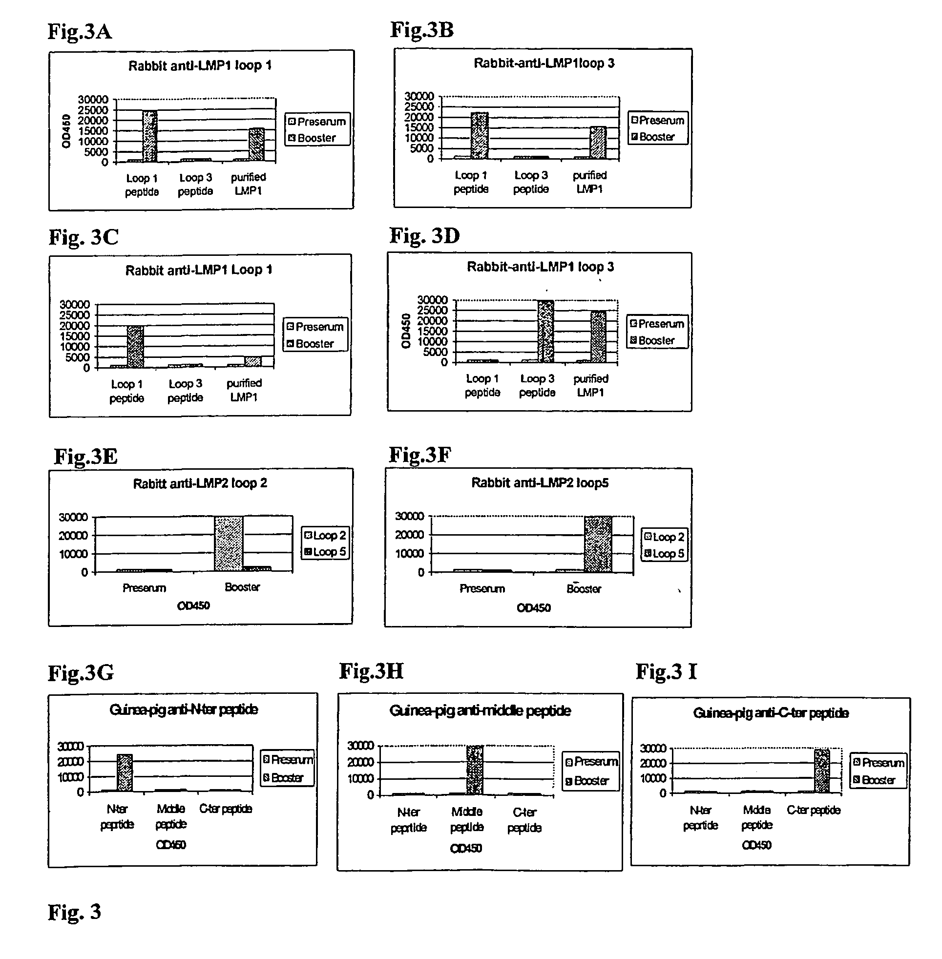
[0312] Generation of Antibody Reagents Reactive with Putative Extracellular Domains of LMP1, LMP2 and BARF1 Proteins by Immunisation with Designated Putative Extracellular Domain-Specific Peptides and Demonstration of the Existence of these Extracellular Domains in EBV-Transformed Cell Lines.

[0313] There is no direct proof for the existence and accessibility of LMP1, LMP2 and BARF1 domains on the outer surface of naturally EBV-infected transformed (tumour) cells.

[0314] Therefore, the first aim was to create specific reagents that would define such domains and would allow the demonstration of the functional accessibility of these domains.

Methods:

[0315] Peptides were made using fMoc chemistry using a solid phase peptide synthesizer (Applied Biosystems, USA). Following acid cleavage from the resin, peptides were washed repeatedly with diethylether and purified by RP-HPLC (Beckman System Gold). When relevant, peptides were circularised by oxidising the sulphydroxy-groups. Finally peptid...

example 3

Generation of Human Antibody Reagents Reactive with the Putative Extracellular Domains of LMP1, LMP2 or BARF1 from a Human Phage-Antibody Library by Phage Display Selection with Synthetic Loop-Domain Specific Peptides.

[0340] The generation of mouse and / or rat monoclonal antibodies by animal immunisation and the hybridoma cell-fusion technique of Kohler and Milstein is well known. However, this methodology does not readily allow generation of human monoclonal antibodies, which are the preferred antibody reagents for in vivo (ex vivo) therapeutic applications. Since The LMP1, LMP2 and BARF1 proteins are hardly immunogenic in man (Meij et al. 1999), the production of human antibodies with specificity for the extracellular domains of LMP1, LMP2 and BARF1 is not straight forward.

[0341] Therefore, an alternative approach was tried by using the novel and specifically constructed (now defined) extracellular domain peptides, described in example 1 and 2, to select for specific human antibody...


PUM

PropertyMeasurementUnit
concentrationaaaaaaaaaa
volumeaaaaaaaaaa
pHaaaaaaaaaa

Abstract

Described are a method for the identification of extracellular epitopes of Epstein Barr virus (EBV) encoded membrane proteins, expressed on the outer cell surface of EBV-transformed mammalian cells, methods fo rthe selection and preparation of antibody reagents specific for the said epitopes, as well as peptides, including extracellular domains of Epstein Barr Virus encoded tumour cell associated membrane proteins, the use of said peptide for immunization and therapeutic vaccination to induce antibodies and T-cells reactive with said domains, the use of said antibody reagents for the production of targeting cells, tumour cell purging and as diagnostic for and medicament against EBV-mediated malignant cell growth.

Description

[0001] The present application is a continuation of U.S. application Ser. No. 10 / 470,753 entitled, "METHOD FOR THE IDENTIFICATION OF EXTRACELLULAR DOMAINS OF EPSTEIN BARR VIRUS (EBV) TUMOR-ASSOCIATED LATENT MEMBRANE PROTEINS AND FOR THE SELECTION OF ANTIBODY REAGENTS REACTIVE THEREWITH," filed on Dec. 12, 2003, now abandoned, which itself claims priority from PCT / EP02 / 00997 having an international filing date of Jan. 30, 2002.[0002] The present invention relates to a method for identification of extracellular epitopes of Epstein Barr Virus (EBV) encoded membrane proteins, to a method for the selection of antibody reagents reactive with the said epitopes, to novel peptides, comprising extracellular domains of Epstein Barr Virus (EBV) encoded latency- and tumour-associated membrane proteins, to nucleotide sequences encoding said peptides, vectors comprising said nucleotide sequences, to antibody reagents reactive with said extracellular domains and to the use of the peptides, nucleoti...

Claims


Application Information

Patent Timeline
Patent Type & Authority Applications(United States)
IPC IPC(8): C12Q1/70C12Q1/68A61K38/00A61K48/00C07K14/05C07K16/08G01N33/569
CPCA61K38/00A61K48/00A61K2039/505C07K14/005C12N2710/16622C07K2317/21C07K2317/55C12N2710/16222C07K16/085A61P37/00
Inventor MIDDELDORP, JAAP MICHIEL
Owner CYTO BARR
Why Eureka
Try Eureka