Apparatus and method for fusion and in-operating-room presentation of volumetric data and 3-D angiographic data

Inactive Publication Date: 2006-04-06
PAIEON INC
View PDF51 Cites 241 Cited by
  • Summary
  • Abstract
  • Description
  • Claims
  • Application Information

AI Technical Summary

Benefits of technology

[0009] In accordance with a first aspect of the present invention, there is provided an apparatus for displaying a first image, said first image is a product of processing a second image taken by a medical imaging device prior to a medical operation, the first image comprising information about areas having sediments, wherein during the operation, said first image is presented to a user of the system. The first image is fused with a third image said third image is a product of processing a fourth image taken by a medical imaging device during the operation. The apparatus further comprises a computer program for registration of images of the medical imaging devices; and fusing information contained in and associated with products of processing images of the medical imaging devices; and presenting the first or third or a combination of the first and third images containing information obtained from the medical imaging devices or an image containing information obtained from the either one of the medical imaging devices. The apparatus also comprising a correction module to correct imaging errors in the second image, using the fourth image acquired during the operation. The imaging error is characterized by one or more calcified areas of one or more blood vessels depicted outsized on an image, said image is a product of processing of one or more images taken by one or more medical imaging device prior to an operation. In the preferred embodiment the first image and the third image, are presented on the same

Problems solved by technology

However, soft tissues are not visible in angiograms so a lot of information, and plaque information in particular is missing when coming to describe in details the current state of arteries.
However, CT scans lack up-to-date information, since they are taken prior to an operation.
In addition, the resolution of CTs is inferior to the resolu

Method used


Image

  • Apparatus and method for fusion and in-operating-room presentation of volumetric data and 3-D angiographic data
  • Apparatus and method for fusion and in-operating-room presentation of volumetric data and 3-D angiographic data
  • Apparatus and method for fusion and in-operating-room presentation of volumetric data and 3-D angiographic data

Examples


Embodiment Construction

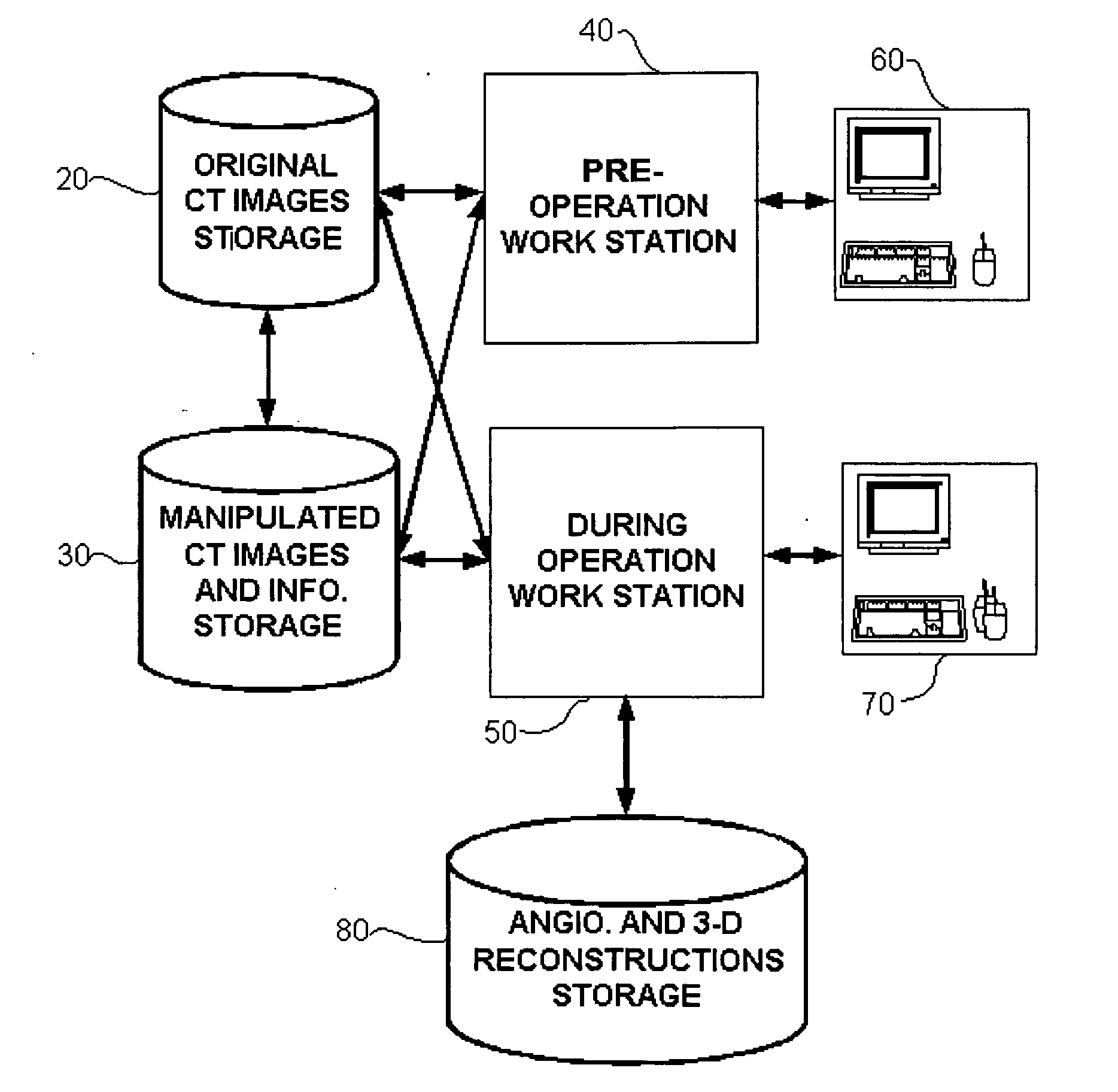
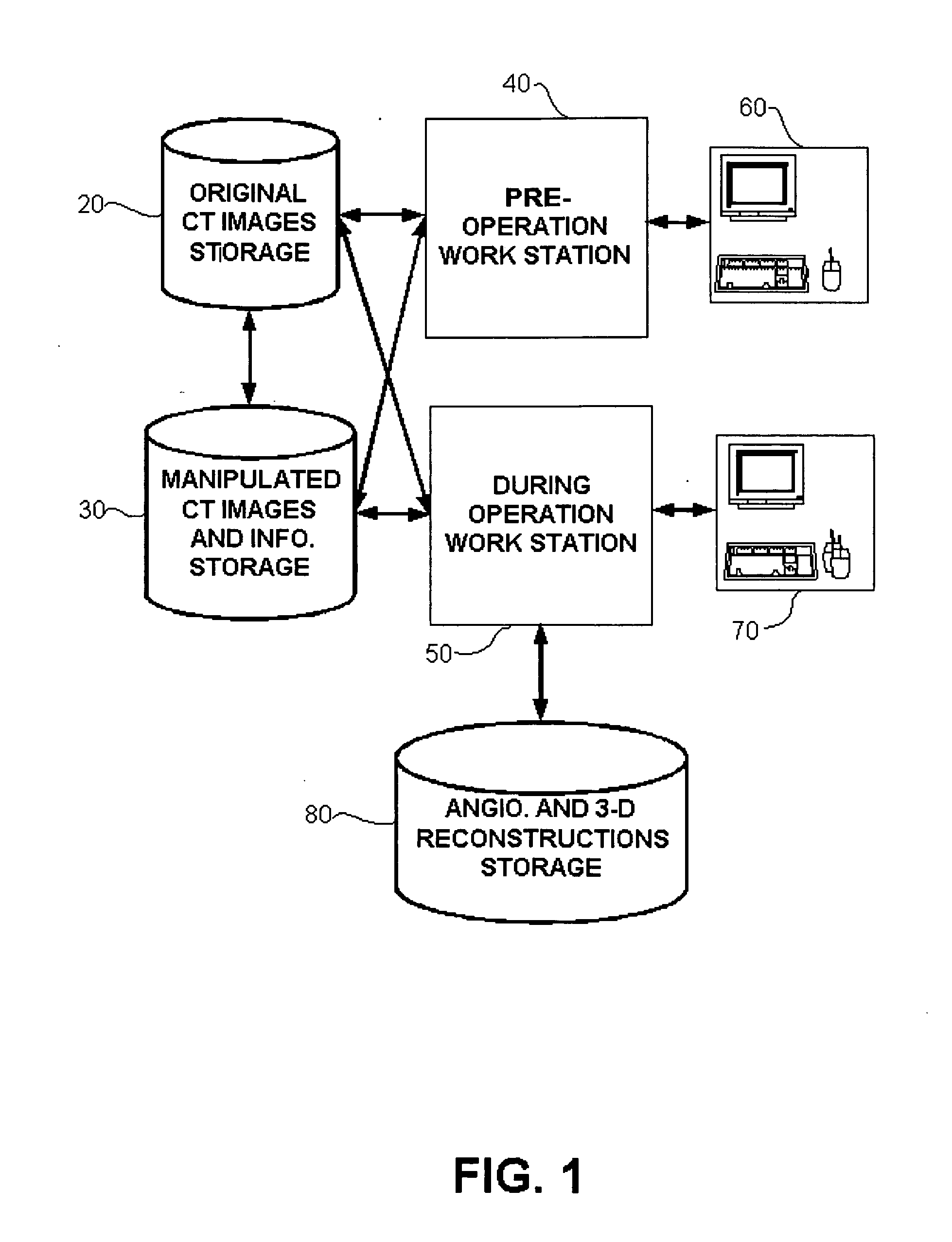
[0018] An apparatus and method for fusing images and information about tubular organs, from CT scans and angiograms, and presentation of the same in 3-dimensions during medical operations is disclosed. The presented information includes different types of sediments deposited inside and outside coronary arteries or other blood vessels, as part of the whole structure of the blood vessels. The apparatus is designed to be used both before and during a medical operation, usually a catheterization, and also to enable the user to mark different areas of interest and pre-defined views prior to the operation. The areas and views will be presented by the system during an operation.

[0019] The preferred embodiment of this invention uses slices taken by a Multi-Slice Computerized Tomography (MSCT) device. The MSCT scanner can simultaneously acquire up to 32, 40, or even 64 slices, thus covering the whole heart area by slices 0.6 mm apart, that were taken during a time frame of 10-20 seconds. Th...


PUM


Abstract

An apparatus and method for fusing images, views and data acquired prior to a medical operation by a medical imaging device with images, views and data acquired by another medical imaging device during the operation. The acquired and fused data includes identification and classification of plaque deposited along blood vessels.

Description

BACKGROUND OF THE INVENTION [0001] 1. Field of the Invention [0002] The present invention relates to medical imaging systems in general, and to an apparatus and method for presenting in-room, real-time updated 3-dimensional arteries model, including plaque. [0003] 2. Discussion of the Related Art [0004] Medical imaging devices are widely used for a number of purposes, both prior to and during medical operations. Pre-operation purposes include evaluation of a patient's status, assessment of required treatment, treatment planning in general and catheterization in particular. During-operation purposes include on-going assessment of the patient's condition, and locating the exact position of invasive tools and devices. [0005] Any currently existing imaging modality has its strengths and weaknesses. Angiograms have high resolution, which enables to depict small vessels (with diameter smaller than 0.8 mm), unnoticeable in other modalities. The high resolution of angiograms and three-dimen...

Claims


Application Information

Patent Timeline
IPC IPC(8): A61B5/05
CPCA61B6/504G06T7/0028G06T7/0038G06T2207/30101G06T7/33G06T7/38
Inventor ZARKH, MICHAELBARLEV, OMERKLAIMAN, MOSHE
Owner PAIEON INC
Why Eureka
Try Eureka