Brachytherapy applicator for delivery and assessment of low-level ionizing radiation therapy and methods of use

a low-level ionizing radiation and applicator technology, applied in the field of brachytherapy applicators for delivery and assessment of low-level ionizing radiation therapy and methods of use, can solve the problems of inability to move the patient for imaging, logistically difficult if not impossible, and inability to accurately diagnose the patien

Active Publication Date: 2006-08-03
XOFT INC
View PDF9 Cites 101 Cited by
  • Summary
  • Abstract
  • Description
  • Claims
  • Application Information

AI Technical Summary

Benefits of technology

[0010] Although the apparatus and methods of this invention are pertinent to radiation therapy utilizing natural radio-isotope sources, the applicator of this invention is preferably for use with systems like those described in copending patent application Ser. No. 10 / 464,140 in which small electronic x-ray sources are utilized in post-operative treatment of proliferative cell disease, and in particular, after breast lumpectomy. Such electronic x-ray sources have the advantage of being controllable by varying input voltage and current, or for that matter, interrupting their radiation output entirely, all of which can be utilized for control of radiation emitted.

Problems solved by technology

These several steps involve considerable time and associated costs and make intraoperative radiation treatment logistically difficult if not impossible.
In the case of breast tumors, moving of the patient for imaging is a problem in itself, because the breast tissue is mobile and the excision cavity may move.
Intraoperative radiation treatment has generally not been possible or practical for several reasons: the need to move the patient to the location of imaging equipment, to obtain the imaging data and transfer that data to a form useable in applicator equipment for performing the irradiation; and the need to obtain data on pathology of the excised tissue or the remaining tissue in the excision cavity, prior to executing a treatment plan.
Obtaining these needed data requires considerable time; in general a patient following tumor excision should be ready for radiation treatment within about M hour, certainly less than 1 hour, and this is not possible with current procedures and equipment.
Current applicators comprise balloons with defined shapes, usually spherical, which can be filled to the appropriate size for the particular cavity, but beyond this size and shape, variation adjustment typically is not possible.
The equipment is not adaptable to an irregularly-shaped excision cavity.
Moreover, the applicator and procedure are not useful for smaller-sized tumors, because of unacceptable surface-to-depth ratio of radiation dosage at near ranges of the radiation source.

Method used


Image

  • Brachytherapy applicator for delivery and assessment of low-level ionizing radiation therapy and methods of use
  • Brachytherapy applicator for delivery and assessment of low-level ionizing radiation therapy and methods of use
  • Brachytherapy applicator for delivery and assessment of low-level ionizing radiation therapy and methods of use

Examples


Embodiment Construction

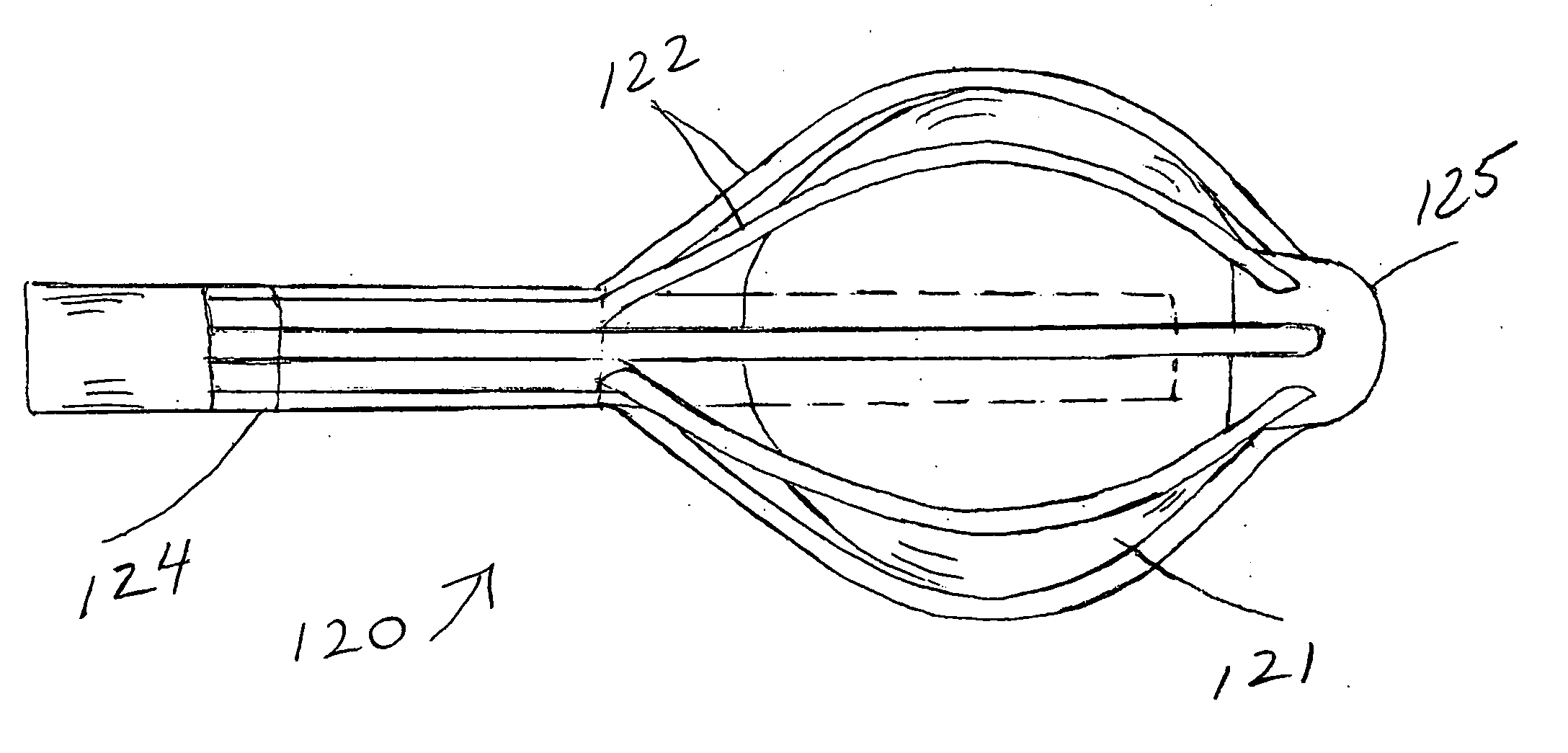
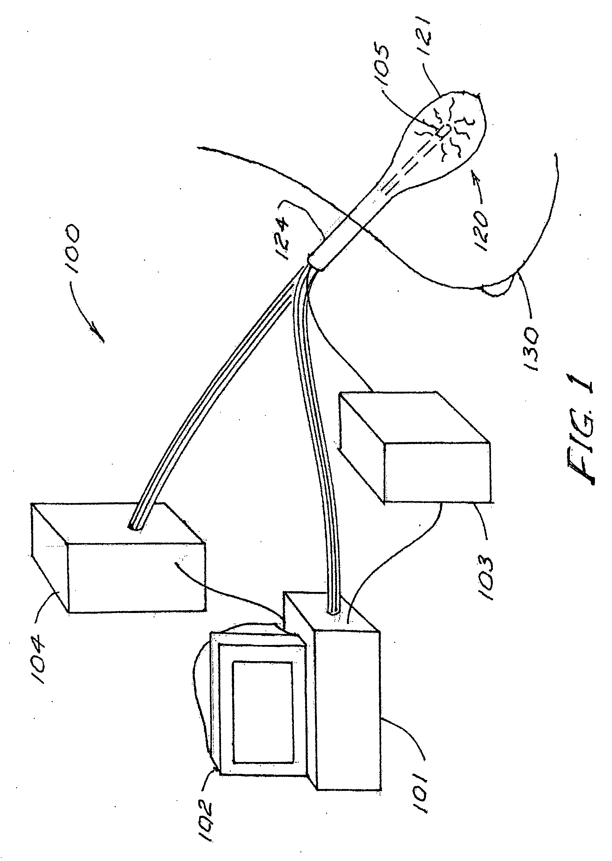
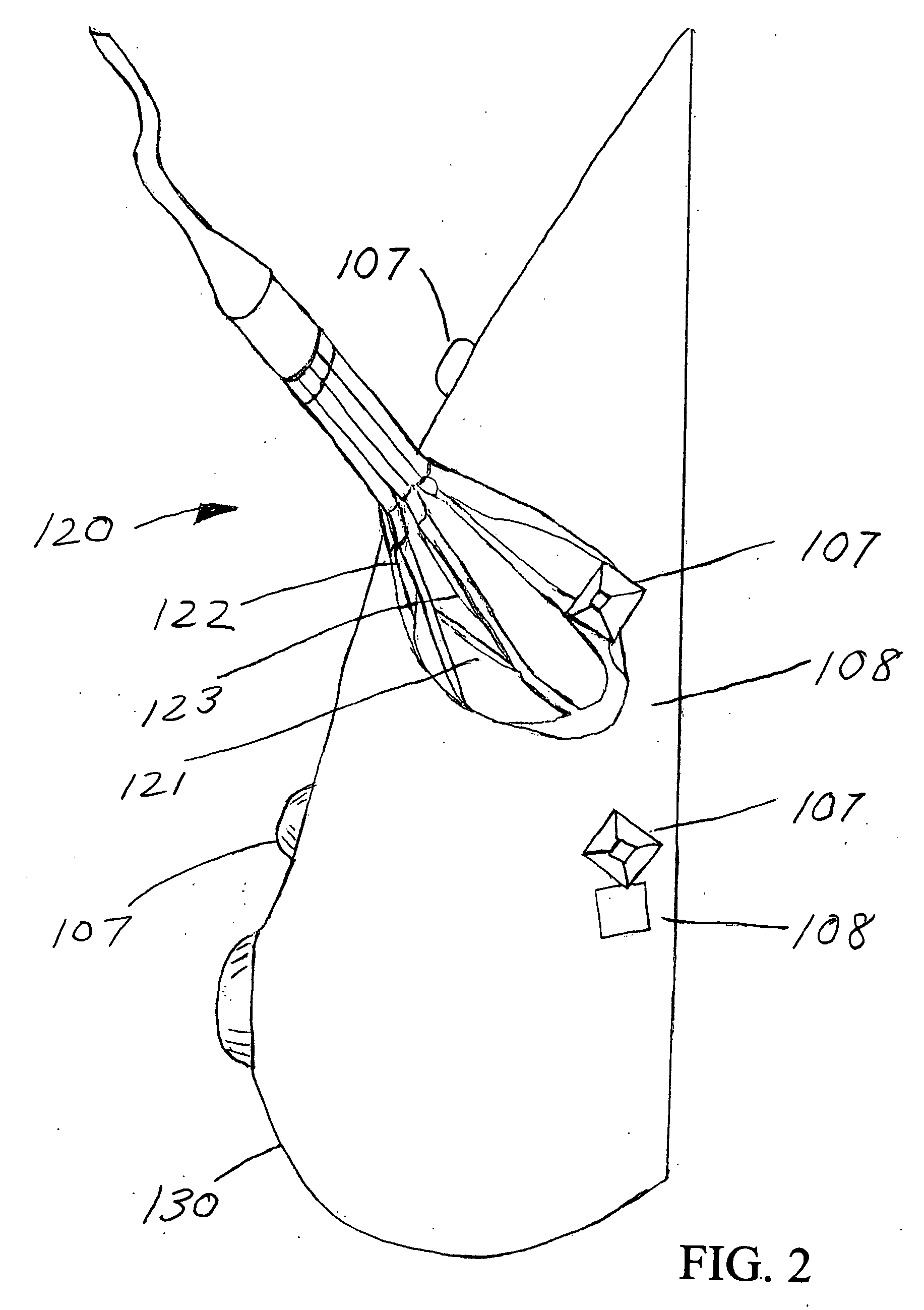
[0023] The system 100 shown in FIG. 1 comprises a processor 101 for making computations based on inputs from other system elements and from the therapist, for indicating system conditions, coordinating and commanding actions of other system components, and for communicating information to the therapist, for example by monitor 102 or alternately, by printed means. Where the radiation source is a miniature x-ray tube (or tubes), a power supply 103 is provided to power the tube (or tubes) in response to commands from the processor. Preferably the system includes a controller 104 to mechanically manipulate the elements of an applicator 120 when the applicator is placed within a tissue volume of the patient for delivery of therapeutic ionizing radiation. The controller acts upon commands from the processor. In the schematic representation of FIG. 1, the applicator 120 is shown positioned in a post-operative cavity within a breast 130. If a controller is not included in the system, manipu...


PUM


Abstract

Applicators for ionizing radiation therapy and their methods of use are provided for expedient preparation of precise treatment plans and for quantitative assessment of therapy delivered to natural or surgically-created, intra-corporeal cavities or lumina. Feedback capability is provided for timely treatment control and for verification of treatment to plan using sensors on the applicators.

Description

[0001] This application is a continuation-in-part of copending application Ser. No. 10 / 464,140, filed Jun. 18, 2003.BACKGROUND OF THE INVENTION [0002] This invention concerns the treatment of breast cancer or otherwise sited cancer, and an applicator capable of feeding back dose data during treatment. The invention also encompasses an efficient procedure for preparation of a radiation treatment plan, and verification and real-time control of treatment to plan following surgical tumor excision. [0003] In treating cancer of the breast, as well as cancer found in other areas of the human body, with the patient under anesthesia, the tumor is surgically excised (with some surrounding tissue) and then typically, the surgical wound is closed and the patient is sent home pending determination of pathology of the excised tumor margins. The need for further excision is evaluated, and if necessary, carried out. A radiation treatment plan is then developed and the patient, in a series of later ...

Claims


Application Information

Patent Timeline
Patent Type & Authority Applications(United States)
IPC IPC(8): A61N5/00A61N5/10
CPCA61N5/1015A61N5/1048A61N2005/1018
Inventor LOVOI, PAUL A.
Owner XOFT INC
Why Eureka
Try Eureka