
System and method for enhanced viewing of rib metastasis

a technology of rib metastasis and enhanced viewing, which is applied in the field of viewing rib metastasis in computed tomography (ct) volume data, can solve the problems of no specific features, difficult identification of rib metastases, and difficult diagnosis process

Active Publication Date: 2007-05-17
SIEMENS MEDICAL SOLUTIONS USA INC
View PDF11 Cites 23 Cited by
  • Summary
  • Abstract
  • Description
  • Claims
  • Application Information

AI Technical Summary

Problems solved by technology

This diagnosis process is very tedious and error prone.
However, there are no specific features which differentiate ribs from one another.
Furthermore, ribs typically cross through the axial planes at an oblique angle making their identification problematic.
Accordingly, accurately identify a particular rib is typically a long and difficult process.

Method used


Image

  • System and method for enhanced viewing of rib metastasis
  • System and method for enhanced viewing of rib metastasis
  • System and method for enhanced viewing of rib metastasis

Examples


Embodiment Construction

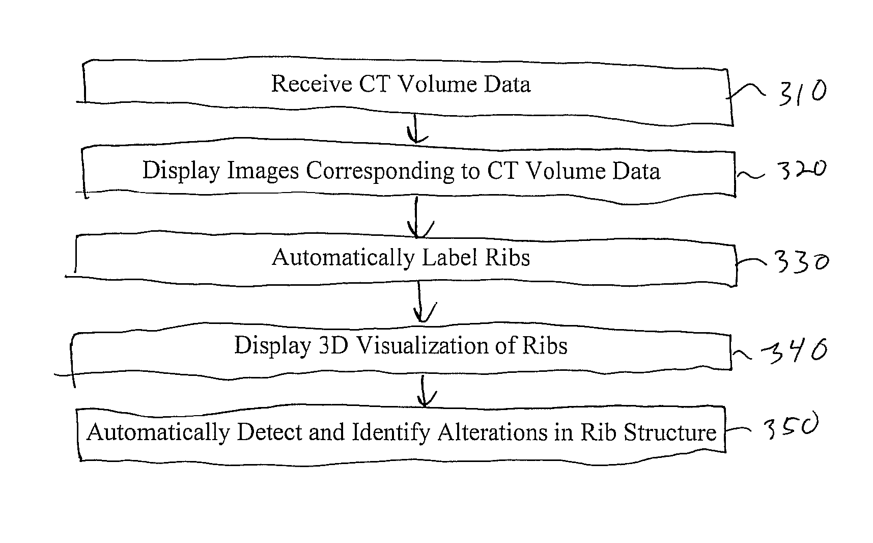
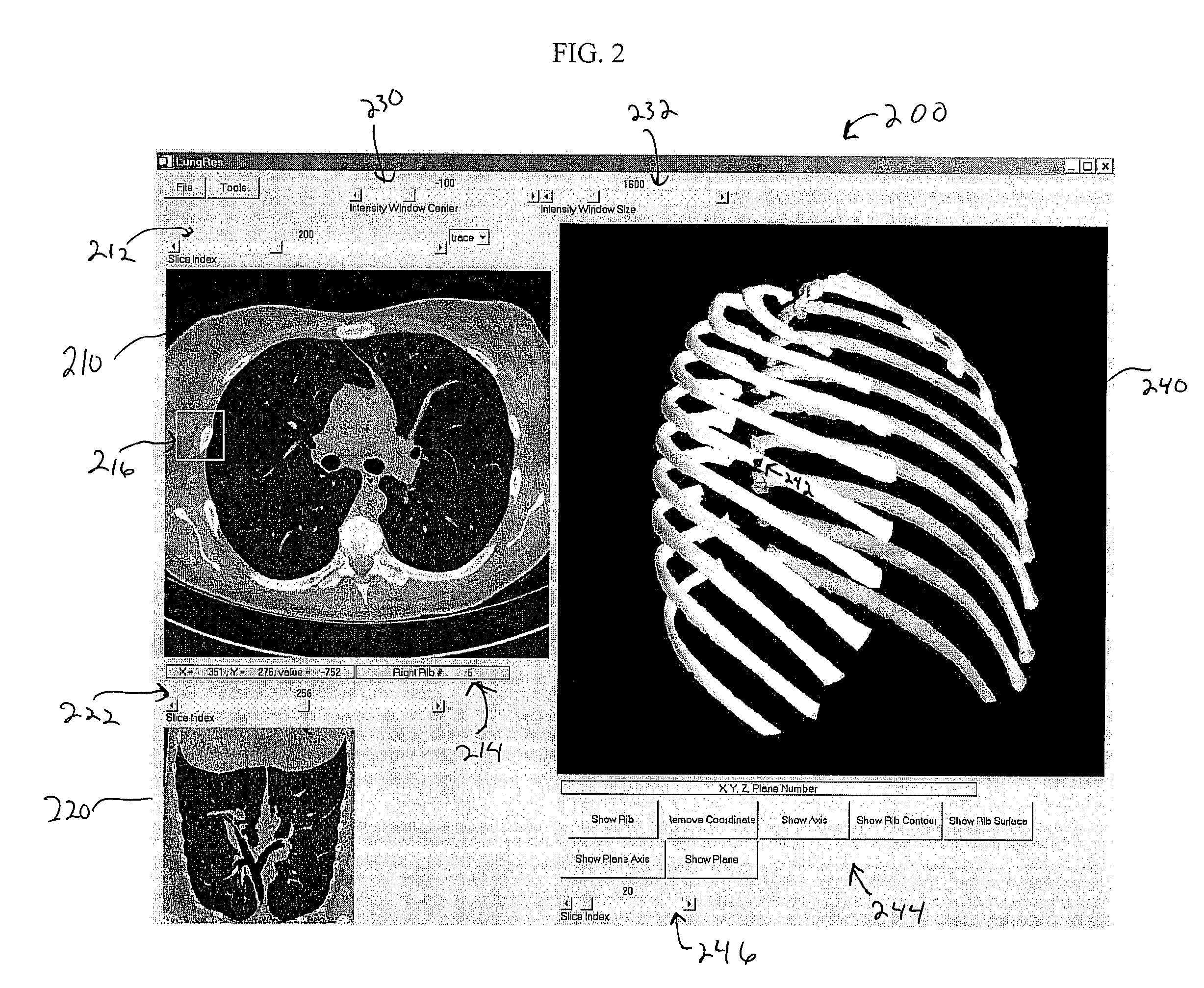
[0018]FIG. 2 illustrates a system interface 200 for the enhanced viewing of rib metastasis according to an embodiment of the present invention. The system interface 200 is displayed by a display portion of a computer system and controlled by a processor of the computer system, which is adapted to execute computer program instructions. FIG. 3 illustrates a method for enhanced viewing of rib metastasis according to an embodiment of the present invention. This method is performed by a rib metastasis visualization system running on a computer system. This method is described while referring to FIGS. 2 and 3.

[0019] At step 310, CT volume data is received. The CT volume data can be CT volume data resulting from a chest CT scan. This CT volume data can be stored in memory of a computer system and loaded to a rib metastasis visualization system running on the computer system (or another computer system). The CT volume data may be stored and loaded in a standard image format. For example, t...


PUM


Abstract

A system and method for enhanced viewing of rib metastasis in CT volume data is disclosed. The system and method receive input CT volume data and display slices of the CT volume data. Ribs are automatically segmented from the CT volume data, ordered and labeled. A 3D visualization of the ribs is generated and displayed. Alterations in the rib structure is automatically detected using shape based analysis of the ribs. The alterations are marked as candidate locations for rib metastasis in the displayed slices and 3D visualization in order to assist in the diagnosis of rib metastasis.

Description

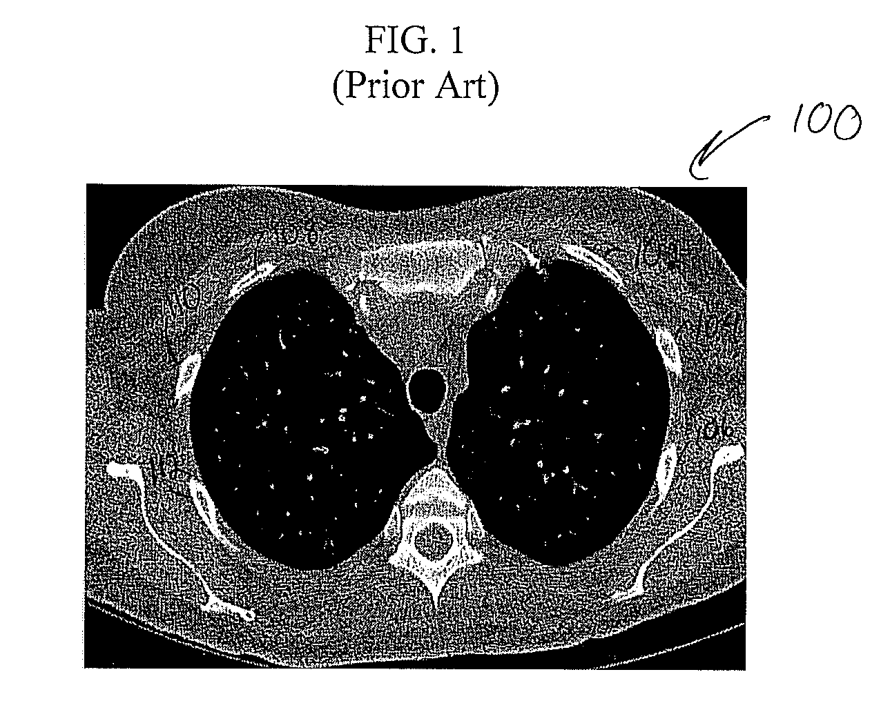
[0001] This application claims the benefit of U.S. Provisional Application No. 60 / 727,573 filed Oct. 17, 2005, the disclosure of which is herein incorporated by reference.BACKGROUND OF THE INVENTION [0002] The present invention relates to viewing ribs in computed tomography (CT) volume data, and more particularly to a system and method for enhanced viewing of rib metastasis using CT volume data. [0003] Computed tomography (CT) is a medical imaging method whereby digital geometry processing is used to generate a three-dimensional image of the internal features of a patient from a large series of two-dimensional X-ray images taken around a single axis of rotation. Such CT imaging results in CT volume data which is a virtual representation of internal anatomical features of a patient. The CT volume data consists of multiple slices, or two-dimensional images, that can be combined to generate a three dimensional image. CT imaging is particularly useful because it can show several types o...

Claims


Application Information

Patent Timeline
IPC IPC(8): G06K9/00
CPCA61B6/03G06T7/0012G06T11/008G06T19/00G06T2207/10081G06T2207/20161G06T2207/30008G06T2207/30096
Inventor SHEN, HONGQING, SHUPING
Owner SIEMENS MEDICAL SOLUTIONS USA INC
Learn More