Mammography Systems and Methods, Including Methods for Improving the Sensitivity and Specificity of the Computer-Assisted Detection (CAD) Process

a technology of compression system and mammography, applied in the field of radiology, can solve the problems of patient discomfort and pain, discomfort of attendants, and procedure for compressing breasts to be uncomfortable and for some, even painful, and achieve the effect of breathing motion and blood flow to the breasts

Inactive Publication Date: 2007-09-27
GALKIN BENJAMIN M
View PDF28 Cites 19 Cited by
  • Summary
  • Abstract
  • Description
  • Claims
  • Application Information

AI Technical Summary

Benefits of technology

[0013] Provided are mammography units, and methods of using the same, for example, in conjunction with imaging of a patient's breast. The mammography units can compress the breast without the need for a traditional compression paddle, or can compress the breast using a traditional compression paddle in conjunction with a novel compression device. Specialized devices for use with the instant mammography units can comprise at least one x-ray transparent inflatable chamber for containing a fluid, for example, a pressurized gas. When fluid is introduced into the chamber, at least one surface of the chamber expands. As the chamber expands, breast motion is limited and the breast and its vasculature are compressed. Fluid may also be released from the chamber, and as the chamber deflates, blood flow to the breast is restored, producing Korotkoff sounds that may be detected by a sound detection device.

Problems solved by technology

This procedure has the effect of forcing the patent's anatomy to contour to the shape of the cassette holder, which often causes patient discomfort and pain.
This requires repositioning the patient for each view with the attendant discomfort due in part to repeat compressions.
It is well known that many women find the procedure for compressing the breast to be uncomfortable and for some, even painful.
Sometimes, adding cushioning materials to compression paddles blocks the light and impedes proper positioning of the breast.
Unfortunately, traditional mammography systems that exert static compression force on a breast that is positioned between a bucky and a compression paddle are incompatible with studies relating to blood flow during mammography screening, as the static compression force at least partially interrupts blood flow within the breast.
A recent study concluded that the use of computer-aided detection (CAD) in mammography is associated with reduced accuracy of interpretation of screening mammograms, and furthermore that the increased rate of biopsy with the use of computer-aided detection is not clearly associated with improved detection of invasive breast cancer.
Improvements to CAD sensitivity and specificity during mammography could reduce incidence of unnecessary biopsies, but such improvements have not yet been developed.

Method used


Image

  • Mammography Systems and Methods, Including Methods for Improving the Sensitivity and Specificity of the Computer-Assisted Detection (CAD) Process
  • Mammography Systems and Methods, Including Methods for Improving the Sensitivity and Specificity of the Computer-Assisted Detection (CAD) Process
  • Mammography Systems and Methods, Including Methods for Improving the Sensitivity and Specificity of the Computer-Assisted Detection (CAD) Process

Examples


Embodiment Construction

[0034] Provided are mammography systems and methods of using the same, for example, in association with the process of imaging of a patient's breast.

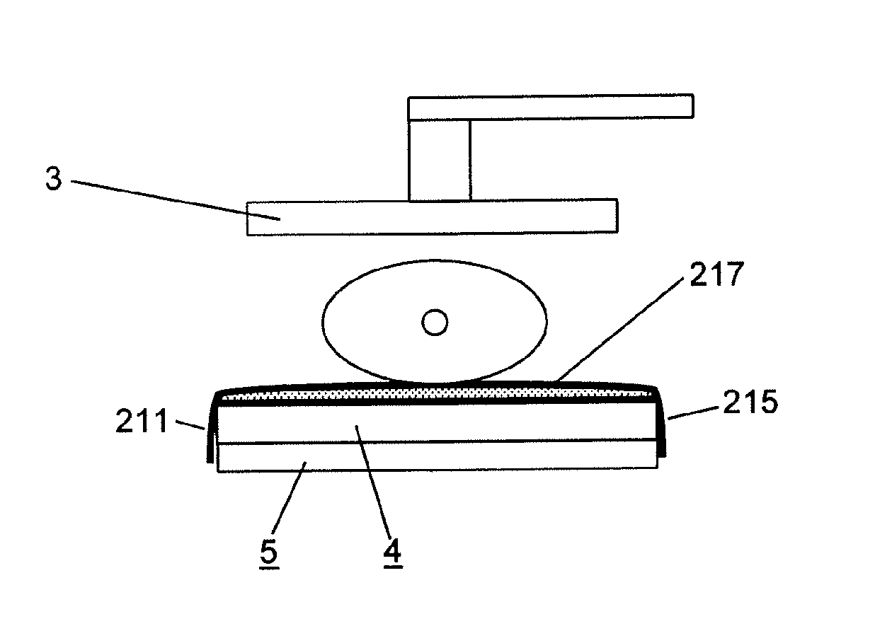
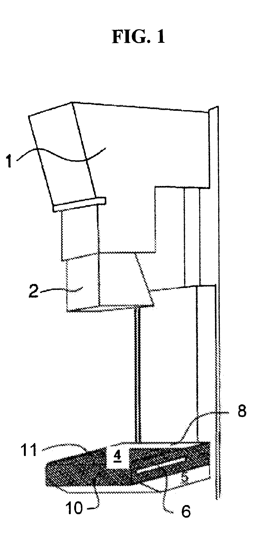
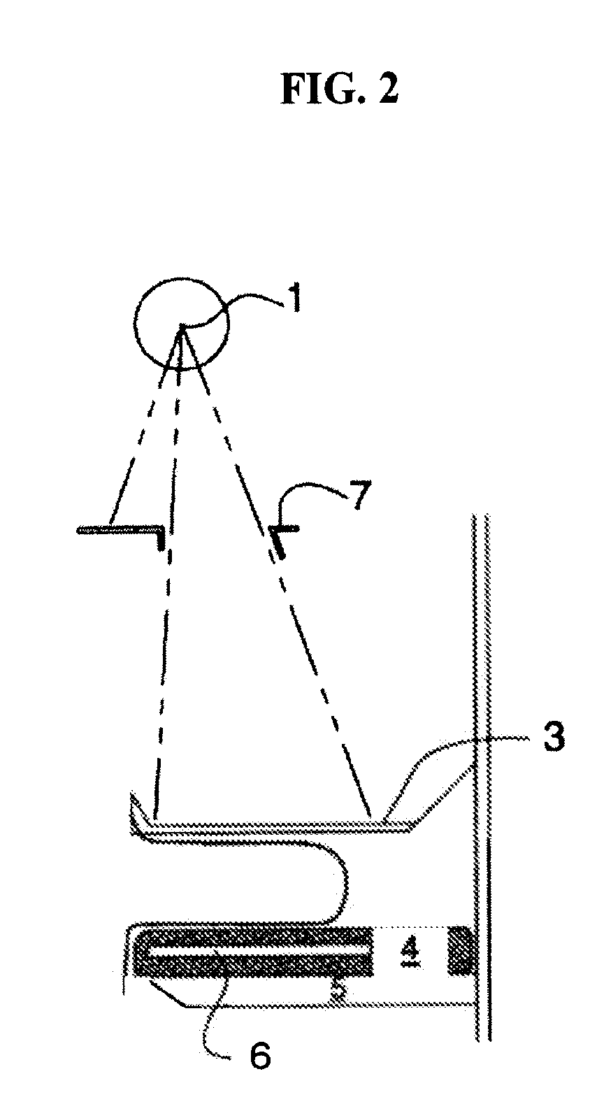
[0035] The instant systems include devices that compress the breast against a bucky without the need for a traditional mammography unit compression paddle. The devices comprise at least one x-ray transparent inflatable chamber for containing a fluid, for example, a pressurized gas. Inflatable chambers can be, for example, medically acceptable balloons. When fluid is introduced into the chamber, at least one surface of the chamber expands. The expansion may be in the direction of the bucky, or may be in the opposite direction, depending on the placement of the device, as described herein.

[0036] For example, in one embodiment, the devices secure the breast to the bucky by wrapping over the top or “tube-side” surface of the breast (so called because it is the surface of the breast that is proximal to the x-ray tube of a mammography unit)...


PUM


Abstract

Provided are systems using compression devices for a mammography unit, and methods of using the same, for example, in conjunction with imaging of a patient's breast. The instant mammography units can comprise at least one x-ray transparent inflatable chamber for containing a fluid. When fluid is introduced into the chamber, at least one surface of the chamber expands, breast motion is limited, and the breast and its vasculature are compressed. Fluid may also be released from the chamber, and as the chamber deflates, blood flow to the breast is restored, producing Korotkoff sounds that may be detected by a sound detection device. The detected sounds may be used to assist a radiologist in identifying regions of interest on a mammogram, and additionally or alternatively may be used to a enhance a computer-assisted detection (CAD) process by contributing an additional data parameter.

Description

CROSS REFERENCE TO RELATED APPLICATIONS [0001] This application is a continuation-in-part of U.S. Ser. No. 11 / 279,280, filed Feb. 11, 2006, and a continuation-in-part of U.S. Ser. No. 11 / 582,243, filed Oct. 17, 2006, which is a continuation of U.S. Ser. No. 11 / 246,419, filed Oct. 7, 2005, which is a continuation-in-part of U.S. Ser. No. 10 / 748,891, filed Dec. 30, 2003, each of which are herein incorporated by reference in their entirety.FIELD OF THE INVENTION [0002] The present inventions relate to the field of radiology and particularly to mammography. More particularly, the present inventions relate to mammography compression systems, and methods of their use. BACKGROUND OF THE INVENTION [0003] Mammography is the process of obtaining x-ray images of the human breast for diagnosis or surgery. It involves positioning a patient's breast in a desired orientation against a cassette holder (also known as a “bucky”) of a mammography unit, compressing the breast with a compression device ...

Claims


Application Information

Patent Timeline
Patent Type & Authority Applications(United States)
IPC IPC(8): A61B6/04
CPCA61B6/0414A61B7/045A61B6/502A61B6/0485
Inventor GALKIN, BENJAMIN
Owner GALKIN BENJAMIN M
Why Eureka
Try Eureka