Volumetric endoscopic coherence microscopy using a coherent fiber bundle

a coherence microscopy and volumetric technology, applied in the field of coherence microscopes, can solve the problems of unsuitable visible light video imaging, unsuitable coherence imaging with coherent fiber bundles, and unsuitable coherence imaging with coherent fiber bundles, and achieve the effect of reducing the spatial coherence of ligh

Active Publication Date: 2008-01-10
THE BOARD OF TRUSTEES OF THE UNIV OF ILLINOIS
View PDF5 Cites 59 Cited by
  • Summary
  • Abstract
  • Description
  • Claims
  • Application Information

AI Technical Summary

Benefits of technology

[0012]b. reducing the spatial coherence of the light prior to coupling the light into the fiber bundle;

Problems solved by technology

Unfortunately, visible light video imaging is unsuitable for this, because of the very shallow penetration of visible light into epithelial tissue.
Unfortunately, however, coherent fiber bundles have proven unsuitable for coherence imaging, mostly because fiber bundle filaments are typically multimode waveguides which scramble the coherence signal, leak light between the filaments, and induce modal dispersion.
Unfortunately, this is not suitable for imaging with a fiber bundle.
This presents problems for conventional beam scanning apparatus because it is designed to scan the beam continuously over the object, usually in a raster-like pattern.
Most of the time, the beam will not illuminate the core of a fiber and no useful signal will be recorded.

Method used


Image

  • Volumetric endoscopic coherence microscopy using a coherent fiber bundle
  • Volumetric endoscopic coherence microscopy using a coherent fiber bundle
  • Volumetric endoscopic coherence microscopy using a coherent fiber bundle

Examples


Embodiment Construction

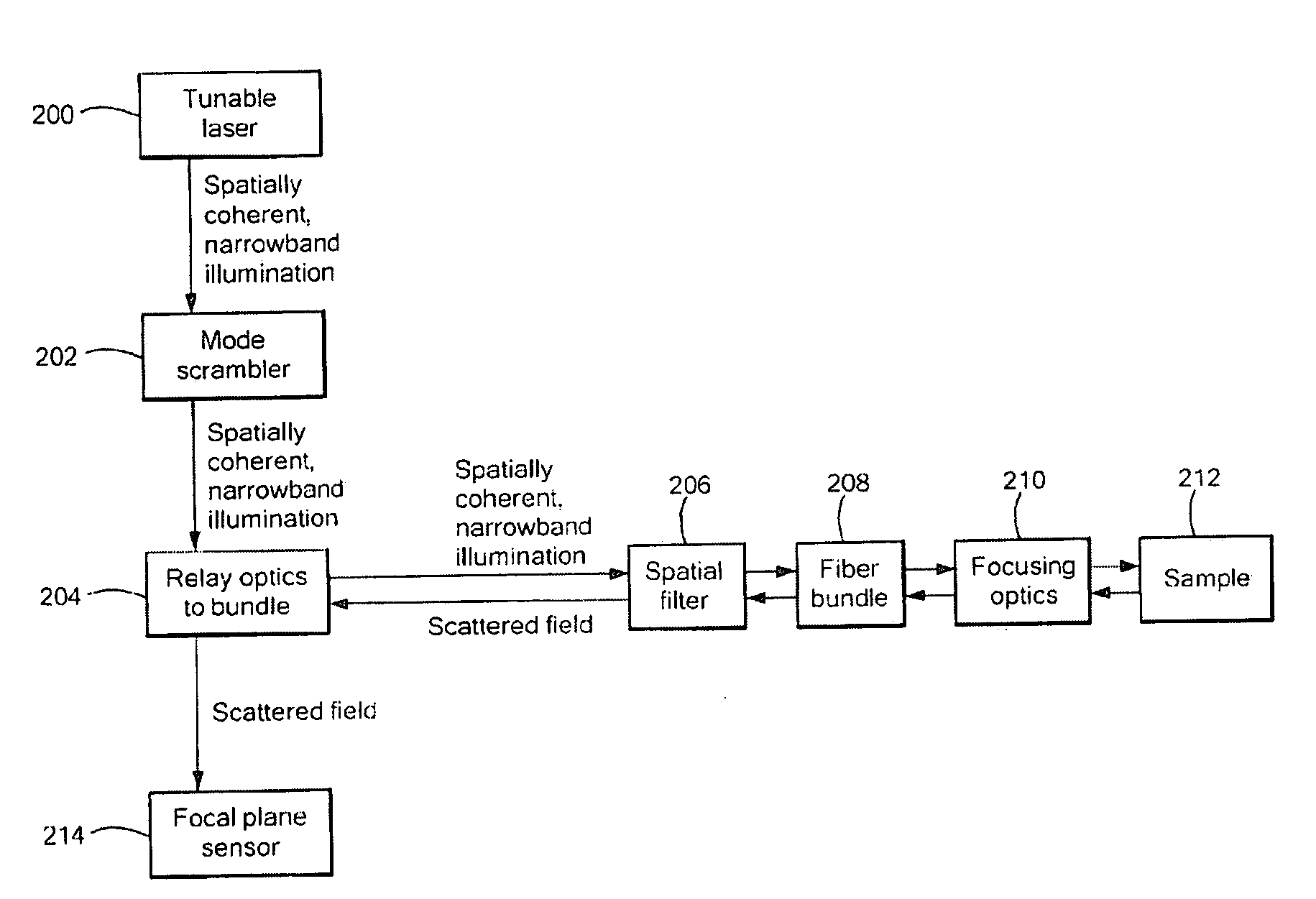
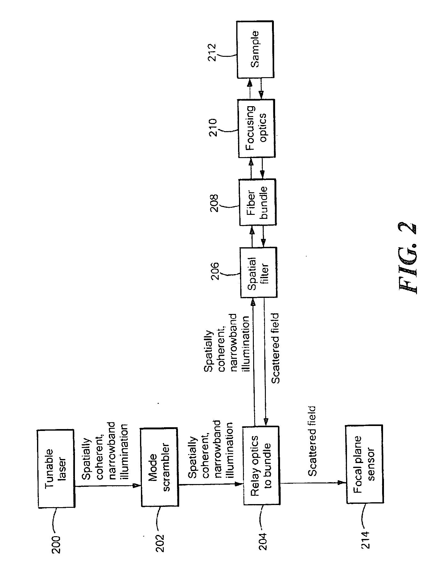
[0037]In accordance with preferred embodiments of the invention, various techniques are provided for overcoming the aforesaid problems with fiber bundles in order to achieve coherence imaging through these bundles. These techniques are simple and inexpensive to implement, and result in an instrument that can acquire 3-D structure of internal tissues at micron-scale resolutions in a fraction of a second. This method is suitable for integration with existing visible light imaging instruments, and even simplifies existing instrumentation, thereby advantageously reducing cost.

[0038]Coherence microscopy, which includes, but is not limited to optical coherence microscopy (OCM) and optical coherence tomography (OCT), uses broadband illumination to peer beneath surfaces that can only be superficially observed with visible light microscopy. It does so, typically, by using near infrared light, although the present invention is not limited with respect to spectral range. Near-infrared light ty...


PUM


Abstract

Methods for employing coherent bundles of optical fibers, whether single- or multi-mode, for optical coherence tomography or optical coherence microscopy. Either a substantially monochromatic source or a broadband source is spatially decohered and / or spatially filtered prior to coupling into the fiber bundle for illumination of a sample. A scatter signal from features disposed beneath the surface of the sample is interfered with a reference signal derived, at either end of the fiber bundle, from the identical source of illumination.

Description

TECHNICAL FIELD AND BACKGROUND ART[0001]The present invention pertains to coherence microscope performed through optical fiber bundles.[0002]Coherent optical fiber bundles that preserve the relative ordering of fibers at their respective ends are commonly used for video imaging of internal structures during internal exploration and surgery. Such means enable a physician or surgeon to see the surface of internal structures using visible light by means of a video camera coupled to the fiber bundle.[0003]A coherent fiber bundle is a bundle of optical fibers that are arranged such that they remain parallel to each other the length of the bundle, in such a way that the nearest neighbor fibers of a particular fiber on one end of the bundle are also the neighbors on the other end. Each of the fibers in the bundle is a waveguide that conducts light from one end of the fiber to the other. The fiber typically consists of a high refractive index core, and a low refractive index cladding, and t...

Claims


Application Information

Patent Timeline
Patent Type & Authority Applications(United States)
IPC IPC(8): G01B9/02
CPCA61B5/0066A61B5/0086A61B5/6852G01N21/4795G02B6/06G02B21/0056G02B21/0064G02B27/58
Inventor MARKS, DANIEL L.BOPPART, STEPHEN A.
Owner THE BOARD OF TRUSTEES OF THE UNIV OF ILLINOIS
Why Eureka
Try Eureka