

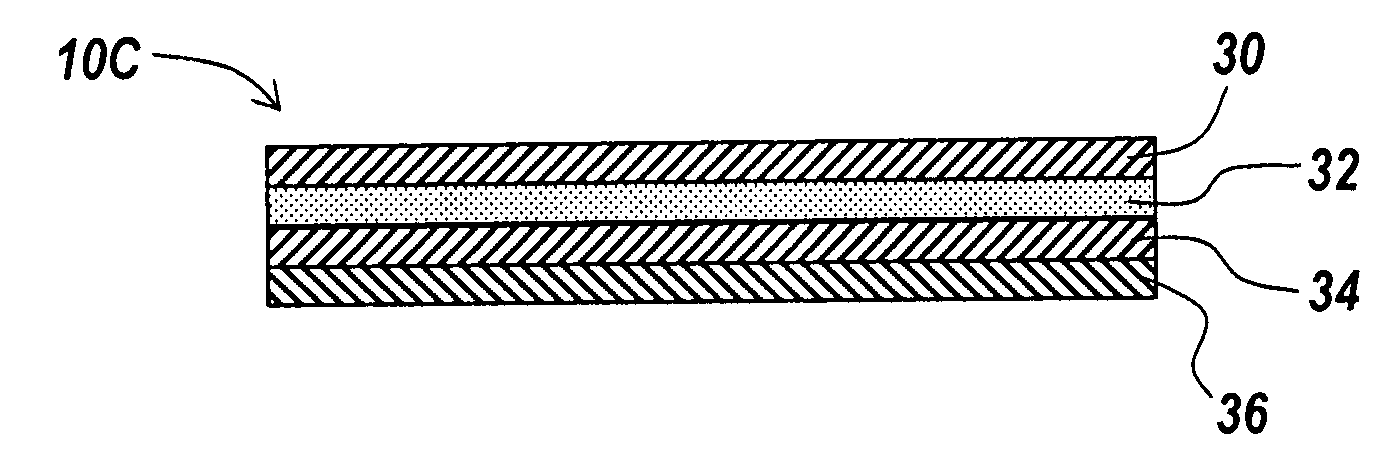
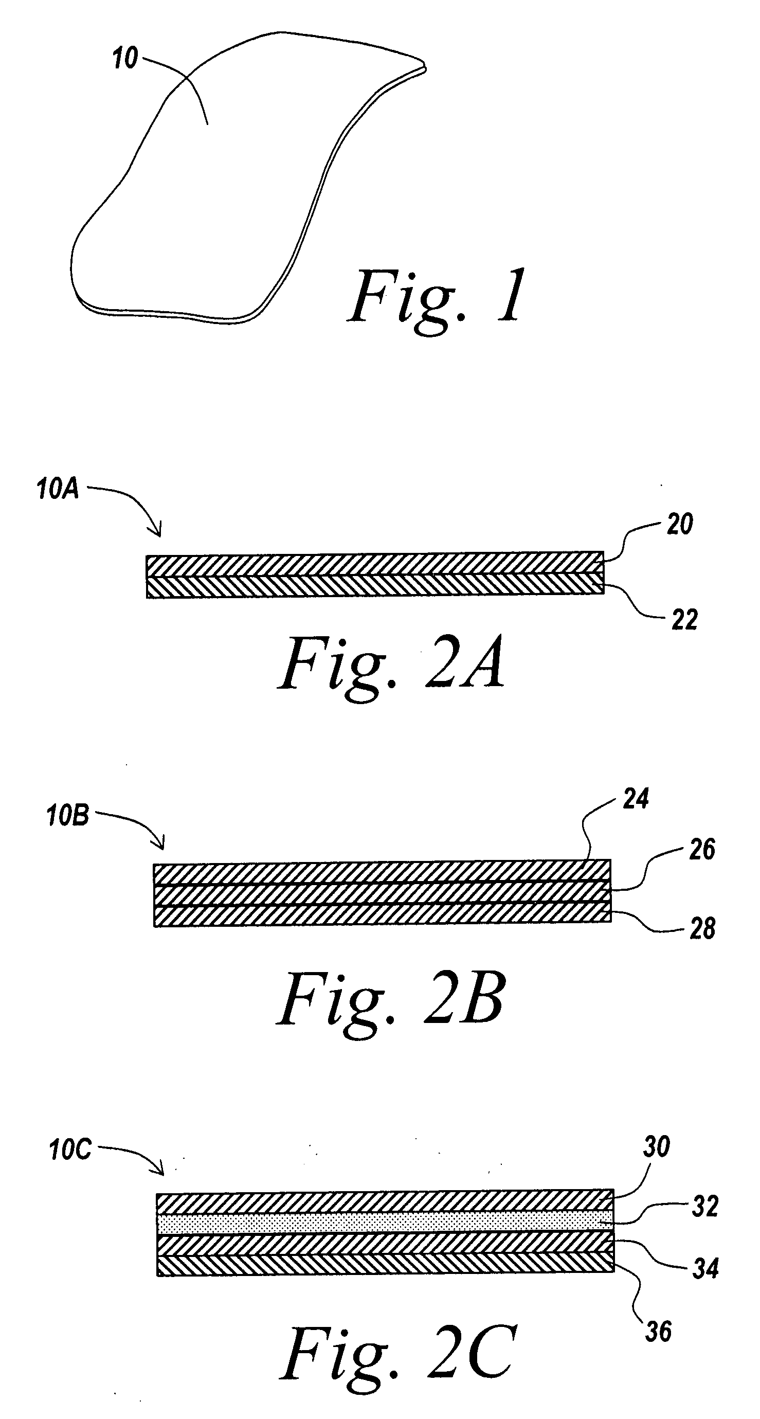
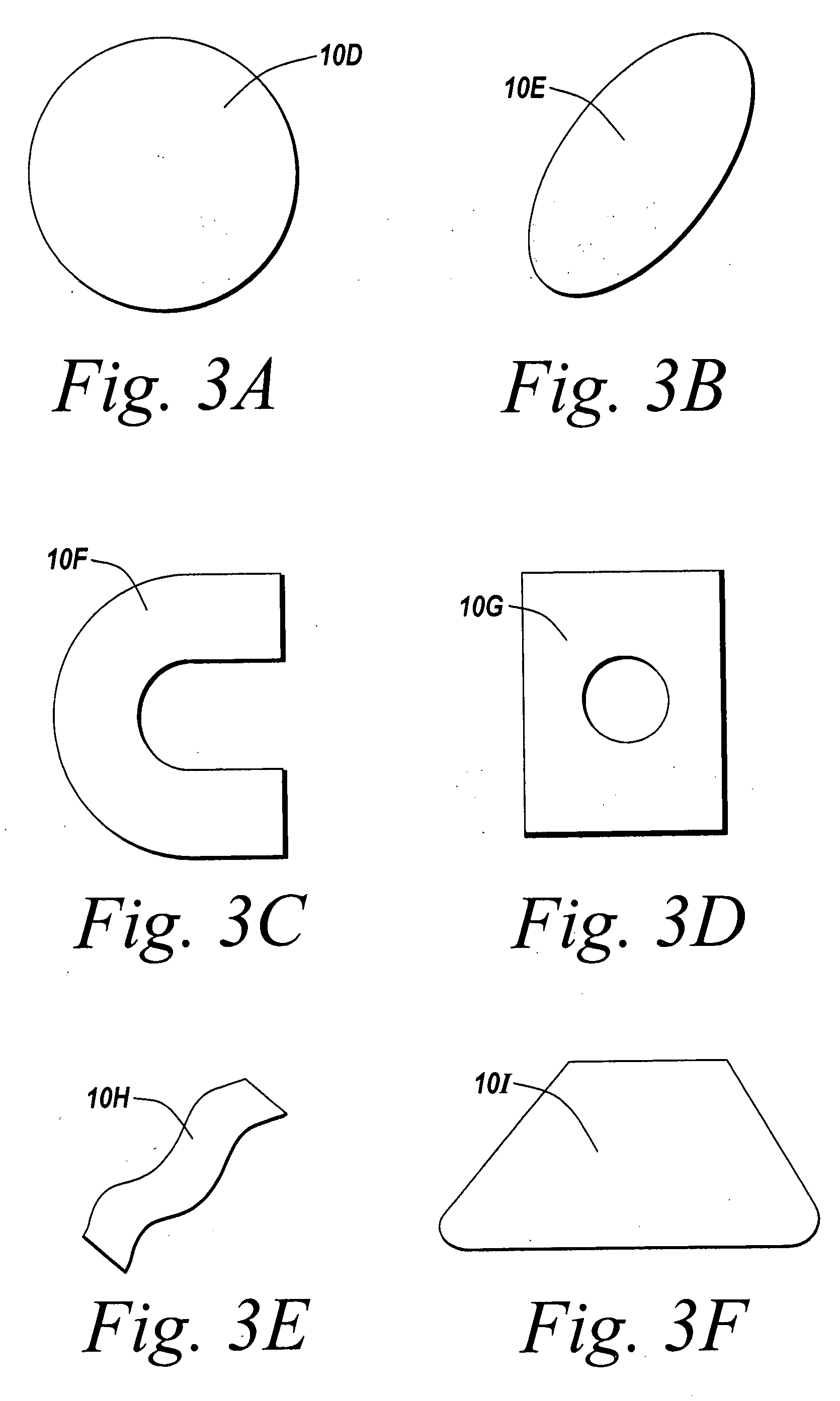
Barrier layer with underlying medical device and one or more reinforcing support structures
a barrier layer and medical device technology, applied in the field of soft tissue devices, can solve the problems of difficult application to the targeted location, difficult handling of adhesion barrier film products, structural weakness, etc., to reduce the degree of adhesion formation, promote tissue growth, and reduce the occurrence of deformation
- Summary
- Abstract
- Description
- Claims
- Application Information
AI Technical Summary
Benefits of technology
Problems solved by technology
Method used
Image

Examples
example # 1
EXAMPLE #1
[0146]An embodiment of the present invention was implemented in a rat model to demonstrate the performance of the barrier layer of the present invention relative to other known surgical mesh devices. The devices were implanted in a rat to repair abdominal wall defects. Healing characteristics, adhesion formation and tenacity, and inflammatory response associated with these materials were compared.
[0147]A polypropylene mesh material (PROLITE™) provided by Atrium Medical Corporation of Hudson, N.H., coated with one embodiment of the barrier layer described herein. The polypropylene mesh with barrier layer was compared with a bare polypropylene control mesh, and DUALMESH® biomaterial provided by W. L. Gore & Associates, Inc.
[0148]Five samples of each mesh type were implanted according to a random schedule. On the day of surgery, the animals were anesthetized with an injection of 50 mg / kg Nembutal IP. The animal was prepped for surgery, and a midline abdominal incision was mad...
example # 2
EXAMPLE #2
[0155]Mechanical testing has been conducted showing an increase in burst strength with the reinforcement element compared to a device without the additional reinforcement element. Testing was conducted using an Instron device to measure burst strength both with and without a 5 mm titanium tack inserted into the device. The material was hydrated in phosphate buffered saline for 1 hour at 37° C. A 1″×3″ strip was laid over a piece of 0.5″ thick rubber backing material, and the 5 mm ProTack™ titanium helical tack was used to fasten the mesh to the underlying rubber substrate. The test strip, with the rubber attached was laid over a cylinder with a 1.875″ opening in the center that was attached to the bottom jaw of the Instron device. A ring of the same diameter was placed on top of the mesh strip and clamped in place to keep the material from moving during the test. A 0.25″ diameter rod was attached to the top Instron jaw, directly above the mesh. The rod was forced through t...
PUM
| Property | Measurement | Unit |
|---|---|---|
| Length | aaaaa | aaaaa |
| Adhesion strength | aaaaa | aaaaa |
| Flexibility | aaaaa | aaaaa |
Abstract
Description
Claims
Application Information