Bionanocomposite for tissue regeneration and soft tissue repair

a biocomposite and soft tissue technology, applied in the field of prosthetic materials, can solve the problems of inability to meet the needs of soft tissue repair biocomposite materials, inability to manufacture biocompatible materials, etc., to achieve the effect of improving overall biocompatibility, promoting cellular attachment, and promoting tissue in-growth

Inactive Publication Date: 2010-04-29
UNIVERSITY OF MISSOURI
View PDF14 Cites 60 Cited by
  • Summary
  • Abstract
  • Description
  • Claims
  • Application Information

AI Technical Summary

Benefits of technology

[0008]In some of the various aspects, the invention is directed to an implant material that provides the necessary strength while promoting cellular attachment, tissue in-growth and integration and improves overall biocompatibility. In one aspect, the inventive implant material (hereinafter called “Bionanocomposite”) is described to include any of a variety of decellularized tissue (selected specifically for a particular implant site) and a variety of nanomaterials, such as polymeric nanofibers, silicon carbide nanowires, gold nanoparticles or combinations thereof, wherein the decellularized tissue and nanomaterials are crosslinked.

Problems solved by technology

The availability of biocompatible materials for soft tissue repair applications such as hernia repair, meniscus tissue replacement, and vascular grafts, is a critical issue for the medical society due to the large number of patients requiring these types of repairs.
This bulge may be asymptomatic, unsightly, and may cause pain when contracting the abdominal musculature.
The most concerning scenario is entrapment of the viscera within the defect, known as incarceration, which can be quite painful, cause bowel obstruction, or lead to strangulation of the bowel and potential intestinal death, resulting in a surgical emergency.
In the past decade, this theory has been challenged.
Mesh shrinkage may uncover the original defect and lead to a recurrence of the hernia.
When mesh is placed in the abdominal wall, the robust fibrotic response may cause chronic pain associated with either nerve entrapment in the scar plate or mesh contraction.
Abdominal wall mobility may become limited.
In general, implanted biomaterials utilized for soft tissue repair suffer from poor tissue integration, which permits sliding and rubbing of the material on the cells and tissues.
This lack of control at the biomaterial-tissue interface and the body's natural response to a foreign body results in repeated cellular injury and a chronic inflammatory response.
This may lead to decreased function, chronic pain, and eventual implant removal.
New soft tissue repair materials have utilized collagen scaffolds, but purified collagen is mechanically weak and chemically crosslinked collagen has inadequate biocompatibility.

Method used


Image

  • Bionanocomposite for tissue regeneration and soft tissue repair
  • Bionanocomposite for tissue regeneration and soft tissue repair
  • Bionanocomposite for tissue regeneration and soft tissue repair

Examples


example 1

Decellularized Porcine Diaphragm Tendon Crosslinked with AuNP or SiCNW

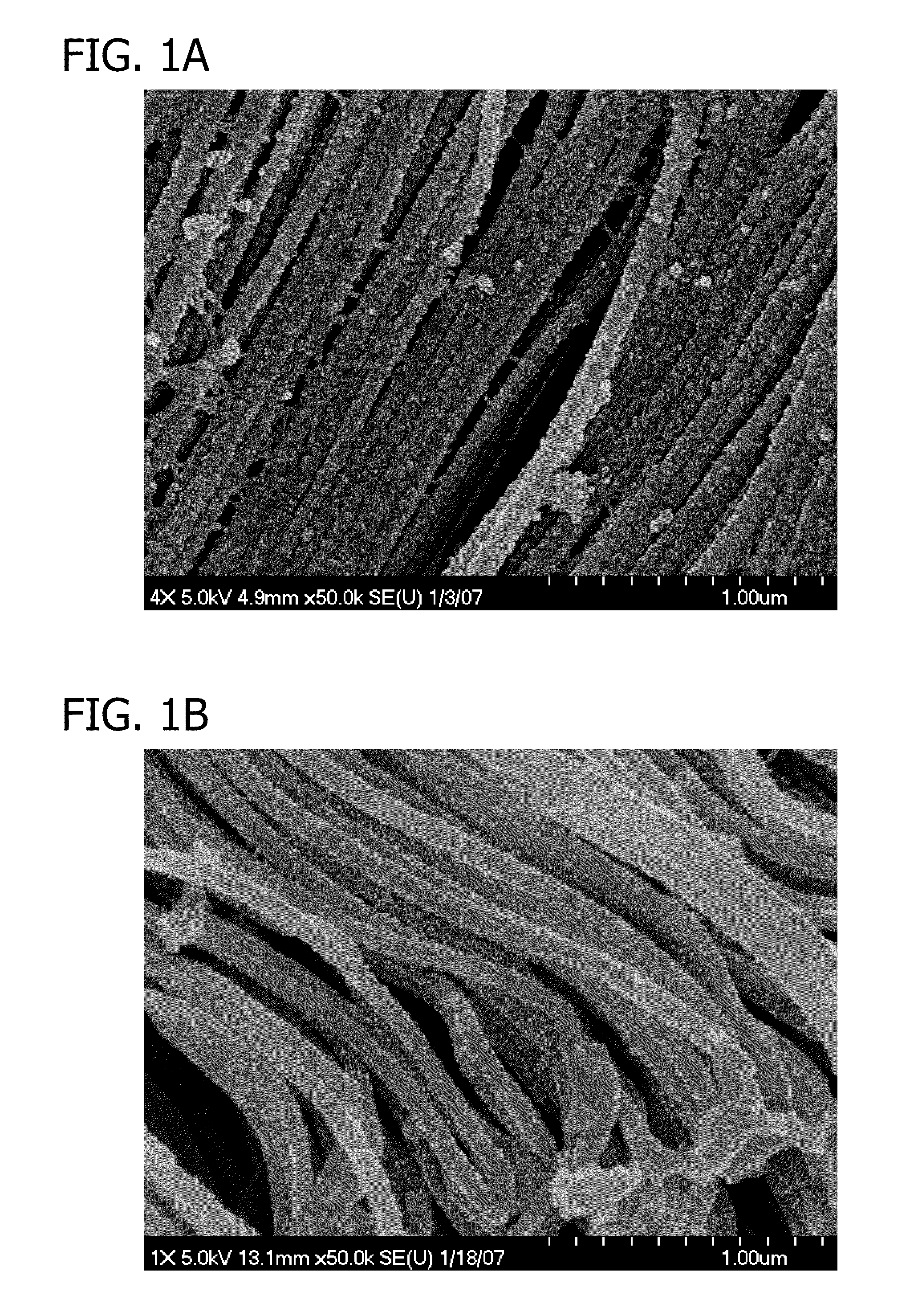
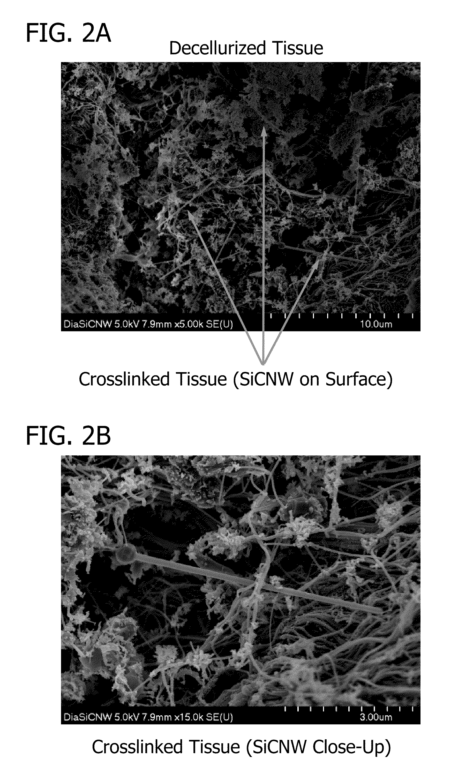
[0090]Decellularization of central tendon portion of porcine diaphragm. When selecting and storing natural tissue, the surrounding muscle of the porcine diaphragm is first removed so that only the collagen rich central tendon portion remains. The resulting natural tissue is placed immediately into Tris buffer solution (pH 8.0) containing 5 mM ethylenediaminetetraacetic acid (EDTA), 0.4 mM phenylmethanesulfonyl fluoride (PMSF), and 0.2% (w / v) sodium azide and stored at 4° C. until ready to use.

[0091]In the decellularization step, a 7 cm×7 cm piece of natural tissue is placed into Tris buffer solution (pH 8.0) containing 5 mM ethylenediaminetetraacetic acid (EDTA), 0.4 mM phenylmethanesulfonyl fluoride (PMSF), 0.2% (w / v) sodium azide, and 1% (v / v) tri-n-butyl phosphate (TnBP). The reaction is placed on a shaker table at room temperature for 24 hours at 225 rpm.

[0092]In the rinsing step, the resulting tissue is rinse...

example 2

In Vivo Implant Study in Rats

[0112]Experimental design. Forty-five male, Sprague-Dawley rats were divided into the following five treatment groups. Fifteen rats were sacrificed at each of the three time points (seven, twenty-one, and ninety-seven days). The abdominal walls of the rats and any remaining scaffold materials were recovered at these times and subjected to histological analysis to determine whether differences existed between the inflammatory response to the scaffolds, fibroblast infiltration, and neovascularization.

[0113]“Control” rats treatment group (one per time point): Rats in this treatment group did not undergo surgery, so tissues recovered from these animals served as examples of normal, healthy abdominal wall. “Sham” rats treatment group (two per time point): Rats in this treatment group underwent the surgical procedure but no scaffold materials were implanted. “Decellularized” scaffolds treatment group (four per time point): Rats in this treatment group received...

example 3

AuNP-Crosslinked Bionanocomposite, Surgisis, and Permacol Study

[0156]Experimental design. The following four biologic tissue scaffold materials were implanted into the abdominal walls of fifteen female, Landrace pigs. The abdominal wall of each pig was divided into four regions separated from each other by at least one inch on each side. A 16 cm2 piece of each of the four types of scaffolds was placed into these quadrants, and the placement location of each type of scaffold was randomly determined for each pig. Five pigs were sacrificed at each of the three time points (one, three, and six months). Full thickness sections of the abdominal walls and any remaining scaffold materials were recovered at these times and subjected to histological analysis.

[0157]“Non-crosslinked” (Surgisis) scaffolds: This scaffold material was comprised of several layers of non-crosslinked porcine small intestine submucosa. (Cook Biotech Incorporated, West Lafayette, Ind.) “Slightly crosslinked” (AuNP-cros...


PUM


Abstract

The present invention provides a bionanocomposite including a pre-selected decellularized tissue crosslinked with a pre-selected nanomaterial. Also provided is a process for fabricating the bionanocomposite. Additionally, applications for using the bionanocomposite as soft tissue repair materials or scaffolds for tissue engineering are described.

Description

FIELD OF INVENTION[0001]The present invention relates to prosthetic materials, methods of fabrication, and applications thereof. More specifically, the present invention relates to a series of biocompatible materials that can be used in soft tissue repair in a living body.BACKGROUND OF INVENTION[0002]The availability of biocompatible materials for soft tissue repair applications such as hernia repair, meniscus tissue replacement, and vascular grafts, is a critical issue for the medical society due to the large number of patients requiring these types of repairs. For example, millions of inguinal hernia repairs are performed each year worldwide with 750,000 inguinal and 150,000 ventral repairs performed in the United States alone.[0003]Hernias are by definition a breakdown of the tough connective tissue that encases the abdominal musculature, known as fascia. As a result, there is a bulging of the intra-abdominal viscera through the abdominal wall defect, with a wide variation of res...

Claims


Application Information

Patent Timeline
Patent Type & Authority Applications(United States)
IPC IPC(8): A61F2/06A61F2/08A61K9/00A61K35/12
CPCA61L27/3633A61L2400/12A61L31/005A61L27/38
Inventor GRANT, SHEILA ANNDEEKEN, COREY RENEERAMSHAW, BRUCE JOHNBACHMAN, SHARON LIEBERAMASWAMY, ARCHANAFEARING, NICOLE MARIE
Owner UNIVERSITY OF MISSOURI
Why Eureka
Try Eureka