
Method and apparatus for performing an open wedge, high tibial osteotomy

a high tibial osteotomy and open-wedge technology, applied in the field of surgical methods and equipment, can solve the problems of difficult to create the wedge-like opening in the bone with the necessary precision, difficult to stabilize the upper and lower portions of the tibia relative to one another, and procedural challenges for surgeons

Inactive Publication Date: 2008-08-28
ARTHREX
View PDF87 Cites 20 Cited by
  • Summary
  • Abstract
  • Description
  • Claims
  • Application Information

AI Technical Summary

Benefits of technology

The present invention provides a novel method and apparatus for performing an open wedge, high tibial osteotomy. This technique involves cutting the bone along a cutting plane, creating a wedge-like opening in the bone, and inserting a wedge-shaped implant into the opening. The wedge-shaped implant has keys that are inserted into corresponding keyholes in the tibia, allowing for stability of the tibia with the desired orientation. The invention allows for reorienting the lower portion of the tibia relative to the femur, adjusting the manner in which load is transferred from the femur to the tibia. The technical effects of the invention include improved outcomes, reduced complications, and simplified procedures for performing the osteotomy.

Problems solved by technology

While both closed wedge osteotomies and open wedge osteotomies provide substantial benefits to the patient, they are procedurally challenging for the surgeon.
Among other things, with respect to open wedge osteotomies, it can be difficult to create the wedge-like opening in the bone with the necessary precision and with a minimum of trauma to the surrounding tissue (e.g., the neurological and vascular structures at the back of the knee).
Furthermore, with open wedge osteotomies, it can be difficult to stabilize the upper and lower portions of the tibia relative to one another and to maintain them in this position while healing occurs.

Method used


Image

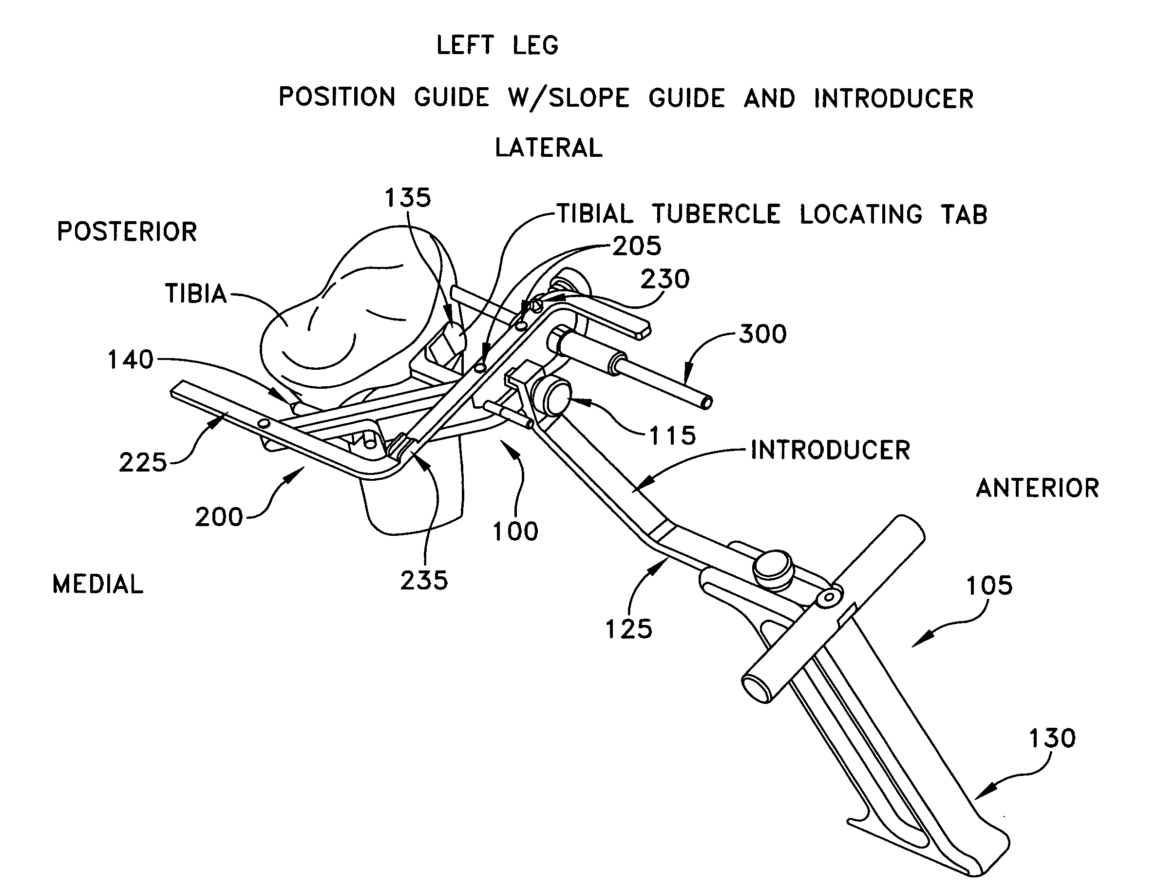
  • Method and apparatus for performing an open wedge, high tibial osteotomy
  • Method and apparatus for performing an open wedge, high tibial osteotomy
  • Method and apparatus for performing an open wedge, high tibial osteotomy

Examples


Embodiment Construction

Overview of an Open Wedge, High Tibial Osteotomy

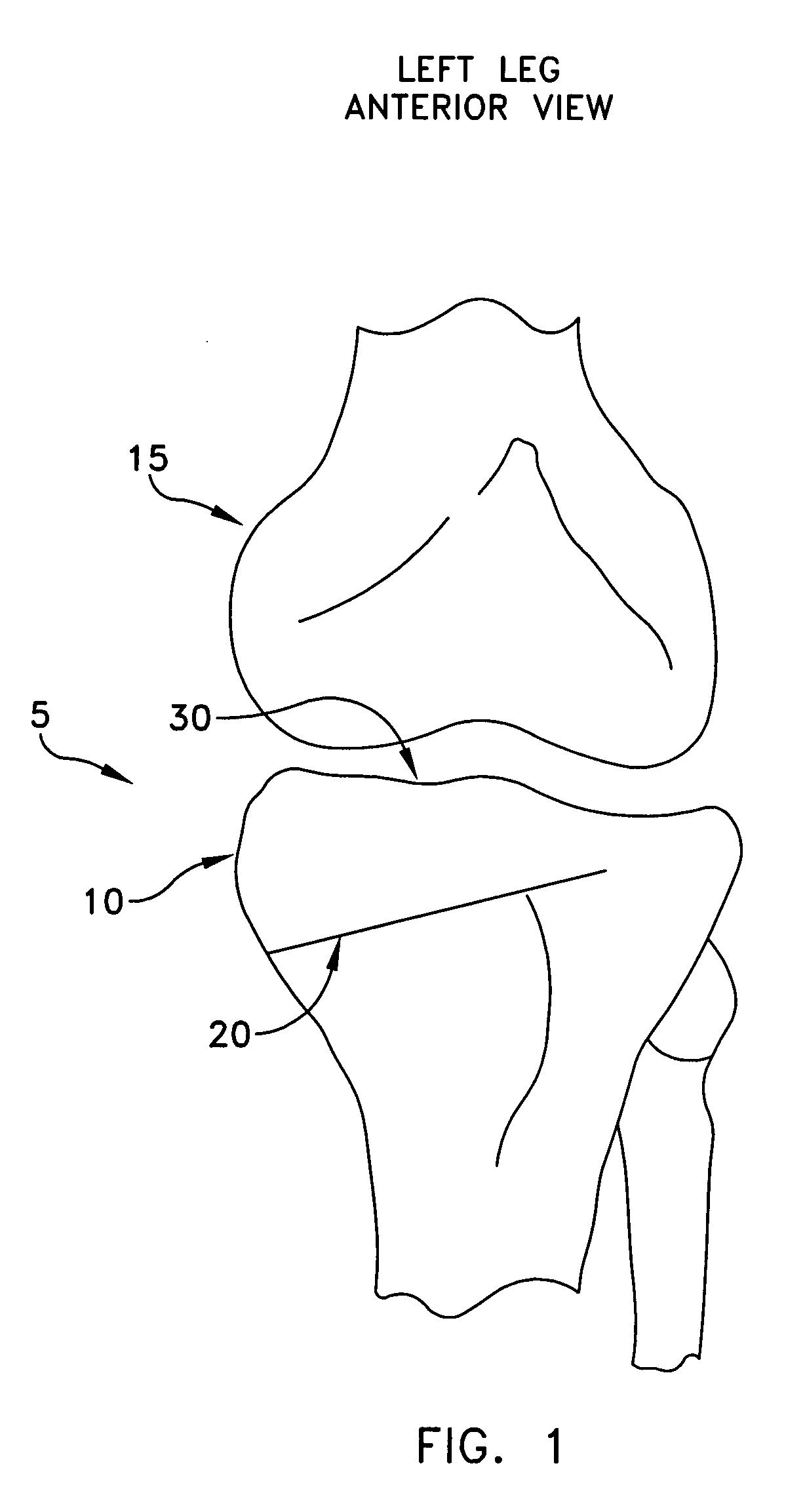
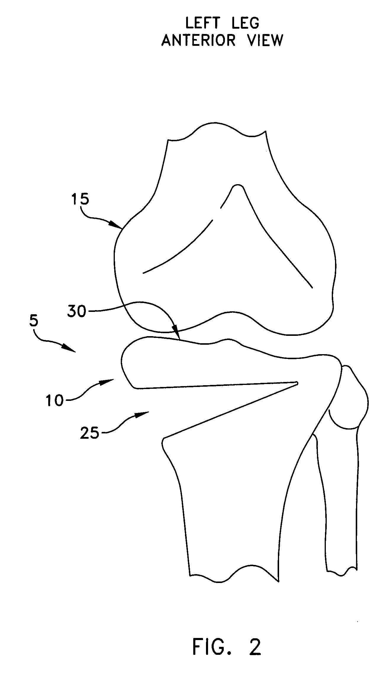
[0069]Looking first at FIGS. 1-3, there is shown a knee joint 5 upon which an open wedge osteotomy is to be performed. Knee joint 5 generally comprises a tibia 10 and a femur 15. In accordance with the present invention, the open wedge osteotomy is effected by first making a cut 20 (FIG. 1) into the upper tibia, and then manipulating the lower portion of the tibia so as to open a wedge-like opening25 (FIG. 2) in the bone, with the wedge-like opening 25 being configured so as to adjust the manner in which load is transferred from the femur to the tibia. In this respect, it should be appreciated that a variety of methods are well known in the art for determining the degree of correction necessary to correctly re-align the weight-bearing axis of the knee. Furthermore, cut 20 and wedge-like opening 25 may be formed in a variety of ways well known in the art.

[0070]Among other things, the present invention provides a new and improved method ...


PUM


Abstract

A method for performing an open wedge, high tibial osteotomy, the method comprising:identifying a cutting plane through the tibia and a boundary line for terminating a cut made along the cutting plane, wherein the boundary line is located within the tibia, parallel to the anterior-posterior slope of the tibia and parallel to the sagittal plane of the patient;positioning a hollow cylinder adjacent to an exterior surface of the tibia and co-axial with the boundary line;positioning a fluoroscope so that its field of view is parallel to the anterior-posterior slope of the tibia, parallel to the sagittal plane of the patient, and co-axial with the hollow cylinder;imaging with the fluoroscope and observing the profile of the hollow cylinder so as to confirm that the hollow cylinder is aligned co-axial with the boundary line;advancing an apex pin through the hollow cylinder and into the tibia along the boundary line so as to provide a positive stop at the boundary line for limiting cutting along the cutting plane;cutting the tibia along the cutting plane, with the cut terminating at the boundary line;moving the tibia on either side of the cut apart so as to form a wedge-like opening in the tibia; andstabilizing the tibia.A method for performing an open wedge, high tibial osteotomy, the method comprising:identifying a cutting plane through the tibia and a boundary line for terminating a cut made along the cutting plane, wherein the boundary line is located within the tibia, parallel to the anterior-posterior slope of the tibia and parallel to the sagittal plane of the patient;positioning a hollow apex pin adjacent to an exterior surface of the tibia and co-axial with the boundary line;positioning a fluoroscope so that its field of view is parallel to the anterior-posterior slope of the tibia, parallel to the sagittal plane of the patient, and co-axial with the hollow apex pin;imaging with the fluoroscope and observing the profile of the hollow apex pin so as to confirm that the hollow apex pin is aligned co-axial with the boundary line;advancing the hollow apex pin into the tibia along the boundary line so as to provide a positive stop at the boundary line for limiting cutting along the cutting plane;cutting the tibia along the cutting plane, with the cut terminating at the boundary line;moving the tibia on either side of the cut apart so as to form a wedge-like opening in the tibia; andstabilizing the tibia.

Description

REFERENCE TO PENDING PRIOR PATENT APPLICATIONS[0001]This patent application claims benefit of pending prior U.S. Provisional Patent Application Ser. No. 60 / 861,869, filed Nov. 30, 2006 by Kelly Ammann et al. for METHOD AND APPARATUS FOR PERFORMING AN OPEN WEDGE, HIGH TIBIAL OSTEOTOMY (Attorney's Docket No. NOVAK-20 PROV).[0002]The above-identified patent application is hereby incorporated herein by reference.FIELD OF THE INVENTION[0003]This invention relates to surgical methods and apparatus in general, and more particularly to surgical methods and apparatus for performing open wedge, high tibial osteotomies of the knee.BACKGROUND OF THE INVENTION[0004]Osteotomies of the knee are an important technique for treating knee osteoarthritis. In essence, knee osteotomies adjust the geometry of the knee joint so as to transfer weight bearing load from arthritic portions of the joint to relatively unaffected portions of the joint.[0005]Knee osteotomies are also an important technique for add...

Claims


Application Information

Patent Timeline
Patent Type & Authority Applications(United States)
IPC IPC(8): A61F5/00
CPCA61B17/15A61B17/152A61B17/1615A61B17/1675A61B17/1764A61B2017/00004A61B2017/1602A61B2019/304A61B17/1732A61B2090/034
Inventor AMMANN, KELLYNOVAK, VINCENT P.SCHNEIDER, ROBERT
Owner ARTHREX
Learn More