
Image diagnosis support system

Active Publication Date: 2008-10-16
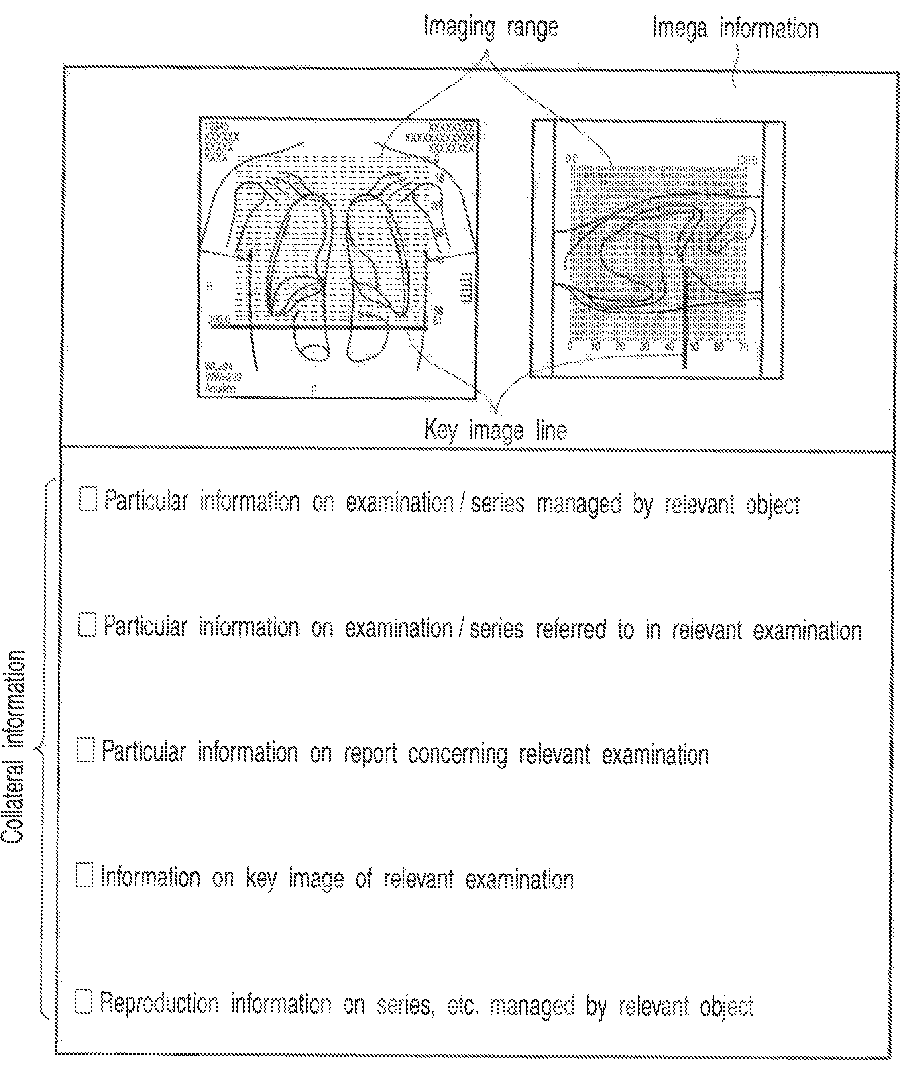
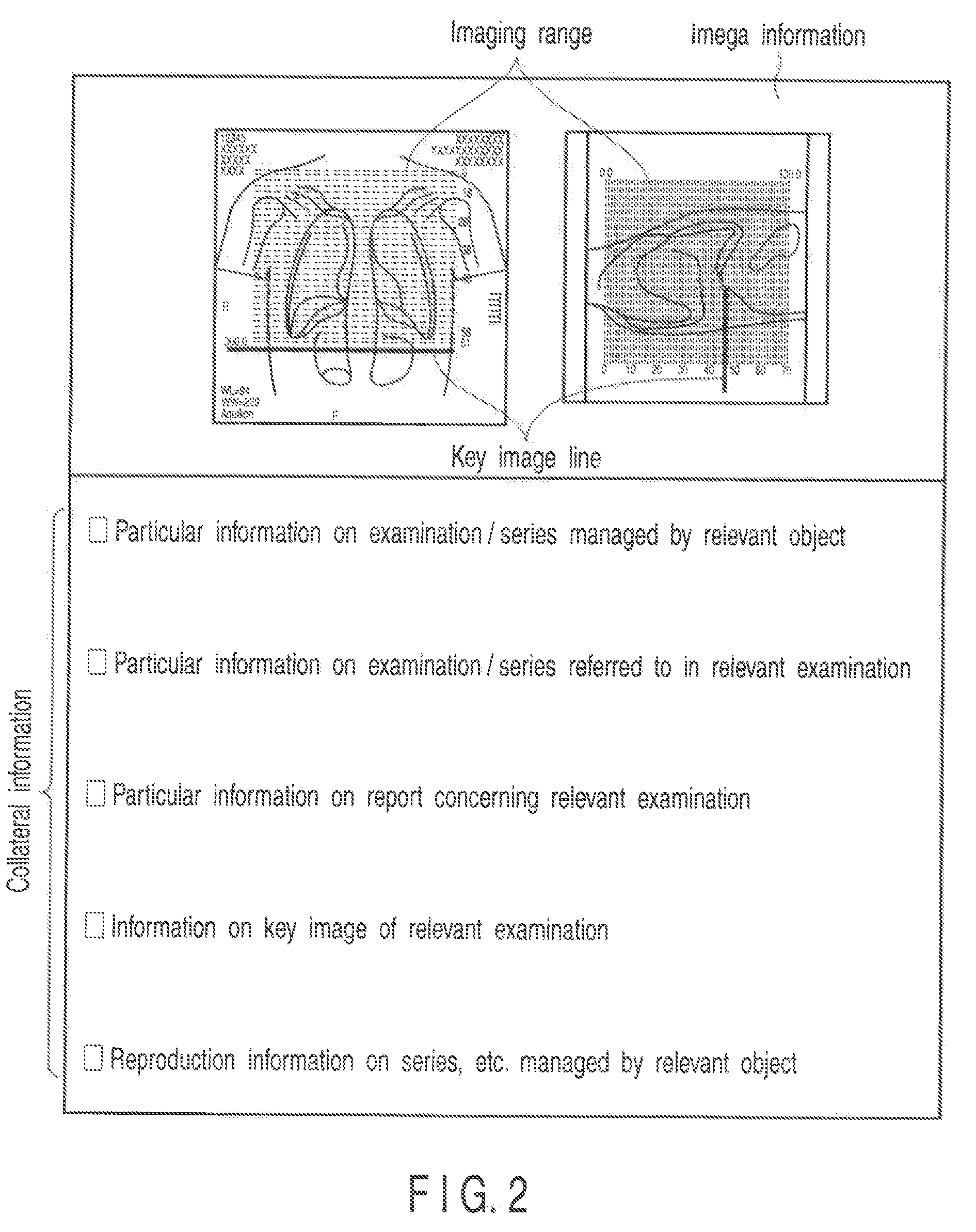
TOSHIBA MEDICAL SYST CORP
View PDF11 Cites 22 Cited by
  • Summary
  • Abstract
  • Description
  • Claims
  • Application Information

AI Technical Summary

Benefits of technology

[0010]The present invention has been made in view of such circumstances, and is directed to provide an image diagnosis support system capable of always conforming a key image serving as the basis of a diagnosis to, for example, a key image specified by information included in an object.

Problems solved by technology

However, for example, the following problems may occur if the information for identifying the key image or the key image itself is included in the object.

Method used


Image

  • Image diagnosis support system
  • Image diagnosis support system
  • Image diagnosis support system

Examples


second embodiment

[0091]Next, a second embodiment of the present invention is described. The present embodiment controls and manages the admission of the editing of information on a key image of an object, the type of the edit processing, and the contents targeted for edit processing in accordance with a combination of user information and device information (i.e., a scene where the key image is used) so that an image actually treated as the key image during the generation of a report may conform to a key image specified by information on the key image of the object.

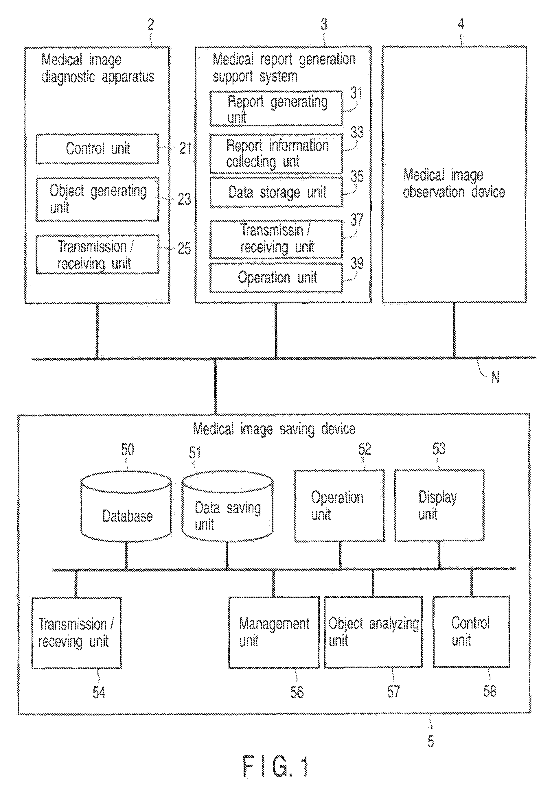
[0092]FIG. 5 is a diagram showing the configuration of an in-hospital network system 1 which realizes an image diagnosis support system according to the second embodiment. By comparison with FIG. 1, the configuration of a medical image saving device 5 is different.

[0093]Furthermore, a database 50 stores an edit processing table and an edit target table used in the image diagnosis support function.

[0094]Here, the edit processing table defi...

third embodiment

[0109]Next, a third embodiment is described. In the image diagnosis support system according to the second embodiment, after an instruction to edit a key image has been issued on the side of the medical report generation support system 3, the medical image saving device 5 determines and controls / manages the admission of the edit processing, the type of the edit processing and the contents targeted for edit processing. In contrast, in an image diagnosis support system according to the present embodiment, before an instruction to edit a key image is issued on the side of a medical report generation support system 3, a medical image saving device 5 determines the admission of the edit processing, etc. in accordance with a combination of user information and device information, and the result of the determination is provided to the side of the medical report generation support system 3.

[0110]That is, the medical image saving device 5 receives the user information and the device informat...


PUM


Abstract

The admission of the editing of information on a key image of an object, the type of edit processing, and the contents targeted for edit processing are controlled in accordance with a combination of user information and device information (i.e., a scene where the key image is used).

Description

CROSS-REFERENCE TO RELATED APPLICATIONS[0001]This application is based upon and claims the benefit of priority from prior Japanese Patent Application No. 2007-105331, filed Apr. 12, 2007, the entire contents of which are incorporated herein by reference.BACKGROUND OF THE INVENTION[0002]1. Field of the Invention[0003]The present invention relates to a system for managing, in an examination carried out using a medical image diagnostic apparatus, an object which has, as its contents, information set in the apparatus in imaging (imaging information), a positioning image used in the imaging, information on an image (key image) serving as a basis in a past diagnosis, information on the past examinations referred to in the imaging, etc.[0004]2. Description of the Related Art[0005]Recently, the special field of medical practice is segmentized. For example, an image diagnosis is separated into various tasks, such as the acquisition of a diagnostic image of a patient, the interpretation of th...

Claims


Application Information

Patent Timeline
Patent Type & Authority Applications(United States)
IPC IPC(8): G06K9/00G06K9/54G06K9/03A61B5/00A61B6/03G06T1/00G16H10/60G16H15/00G16H30/20G16H40/67
CPCG06F19/321G06F19/3487G06Q10/10G16H15/00G16H40/67G16H30/20
Inventor MATSUE, KENJIKAZUNO, MUNEYASUNIWA, KENICHIIWASA, AKIRA
Owner TOSHIBA MEDICAL SYST CORP
Learn More