Methods of Identifying Agents Having Antiangiogenic Activity
a technology of antiangiogenic activity and agent, which is applied in the direction of biochemistry apparatus and processes, peptide/protein ingredients, genetic material ingredients, etc., to achieve the effects of inhibiting angiogenesis, inhibiting chemotaxis, and inhibiting angiogenesis
- Summary
- Abstract
- Description
- Claims
- Application Information
AI Technical Summary
Benefits of technology
Problems solved by technology
Method used
Image

Examples
example 1
Experimental Methods
[0073]The following materials and methods were used in the experiments described in Examples 2-12.
[0074]Materials—Protective antigen (PA) was purchased from List Biological Laboratories (Campbell, Calif.). Alexa Fluor 594 phalloidin was purchased from Molecular Probes (Eugene, Oreg.). 8CPT-2Me-cAMP and PKA activator N6-Benzoyladenosine-3′,5′cyclic monophosphate (6-Bnz-cAMP) were from Biolog Life Science Institute (Bremen, Germany). Rabbit polyclonal antibody against Phospho-CREB (Serl33) was from Cell Signaling Technology (Beverly, Mass.). Rabbit polyclonal antibody against Rap1 and a mouse monoclonal antibody against α-tubulin were from Santa Cruz Biotechnology (San Diego, Calif.). Mouse anti-human CD31 was from DAKO (Carpinteria, Calif.). Forskolin, IBMX and recombinant human endostatin were from Sigma-Aldrich (Saint Louis, Mo.). Di-TSPa was from Abbott Laboratories (Abbott Park, Ill.). Recombinant human vascular endothelial growth factor (rhuVEGF) was from Alp...
example 2
Anthrax Edema Toxin Activates cAMP and Induces Cytoskeletal Changes in Vascular Endothelial Cells
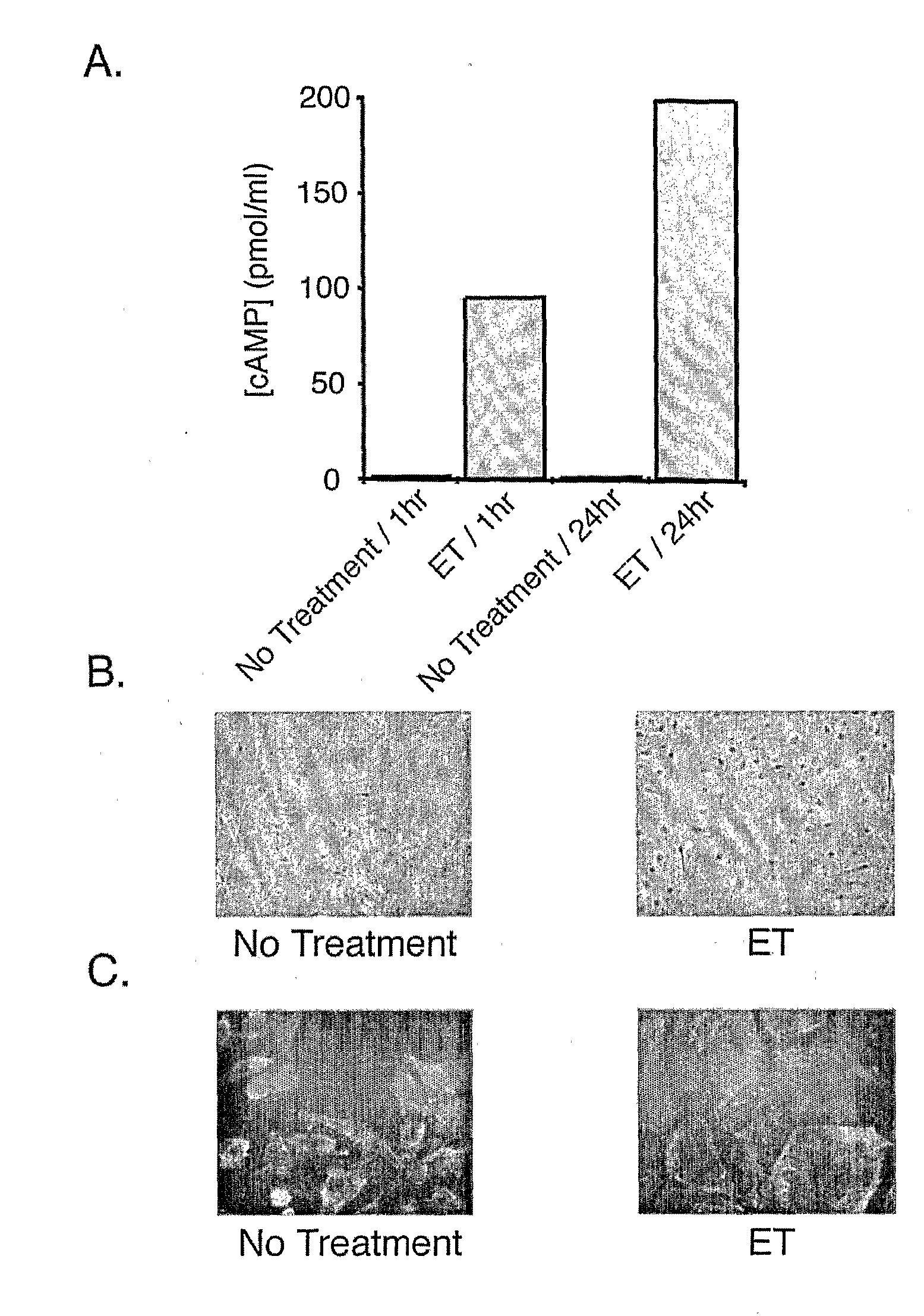
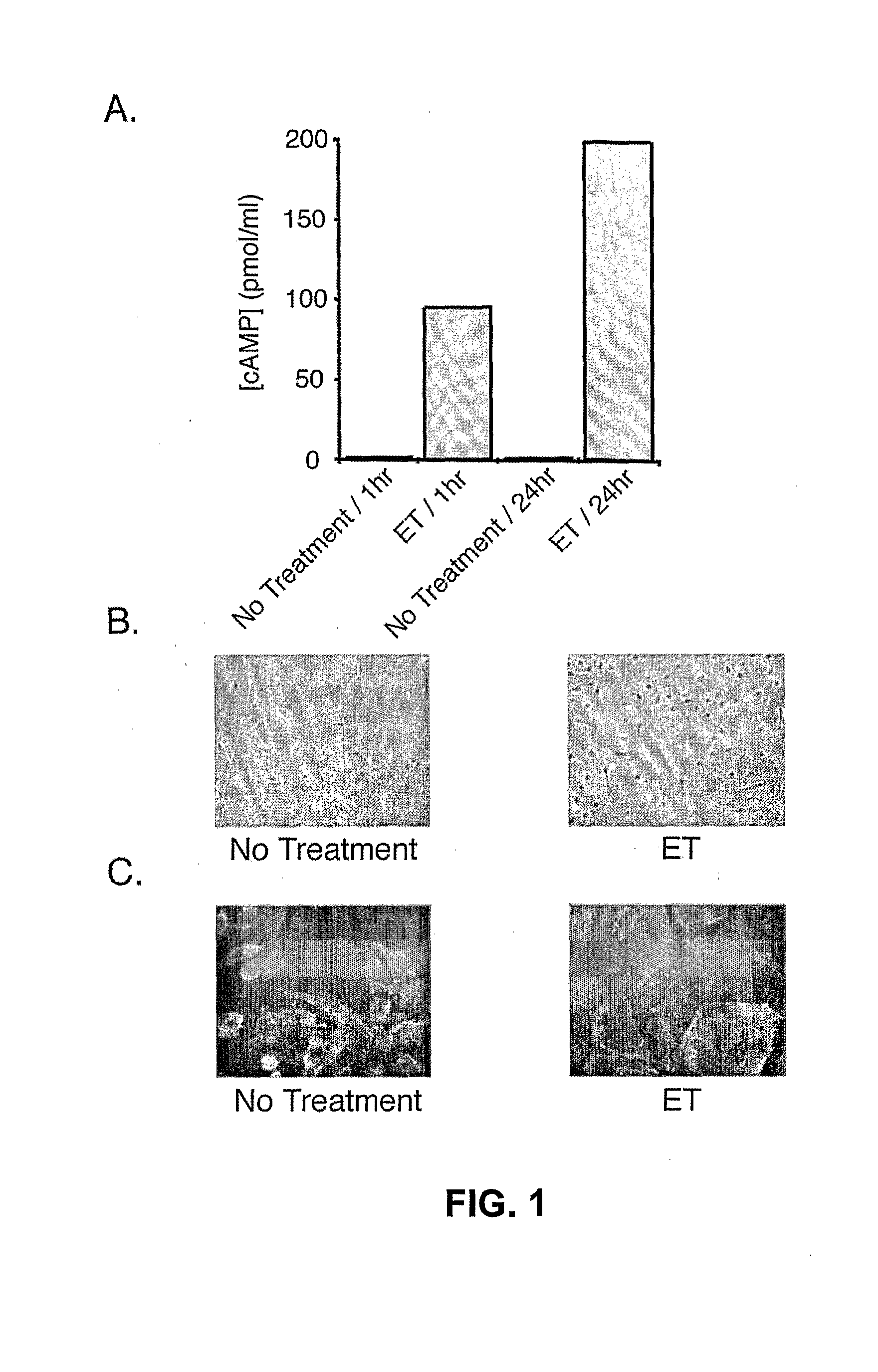
[0089]As shown in FIG. 1A, treatment of human microvascular endothelial cells (HMVECs) with ET resulted in the rapid intracellular accumulation of cAMP that more than doubled by one day following treatment. As shown in FIG. 1B, examination of HMVEC cell morphology revealed a dramatic change in shape from the normal oval morphology to a flattened morphology. Similar results were observed upon treatment of human umbilical vein endothelial cells (HUVECs) with ET (data not shown). As shown in FIG. 1C, this change in morphology and the underlying actin cytoskeleton was more clearly illustrated by staining actin in ET-treated versus untreated HMVECs with Alexa-labeled phalloidin. Taken together, these results indicate that EF can induce significant morphological and cytoskeletal changes in vascular endothelial cells.
example 3
Anthrax Edema Toxin Inhibits Chemotaxis and Tube Formation but not Cell Proliferation in Vascular Endothelial Cells
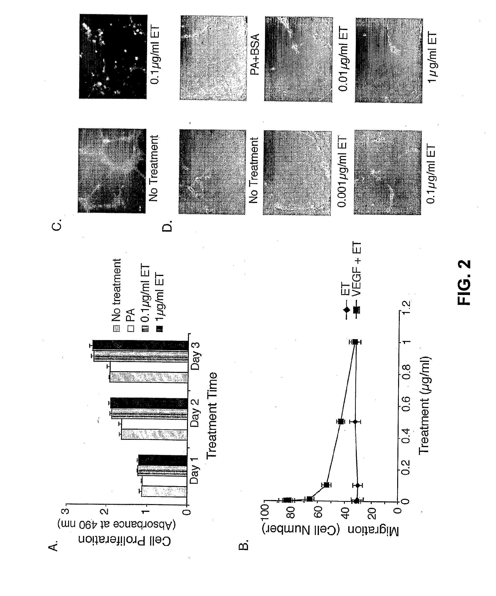
[0090]A cell proliferation assay was used to analyze the possibility of cell growth inhibition by cAMP. As shown in FIG. 2A, not only was there no suppression of cell growth, but ET actually appeared to enhance cellular proliferation at a low but statistically significant level. These results suggest that the changes in cell shape are unlinked to growth defects.
[0091]As an in vitro measure of chemotactic migration, HMVECs were placed in a Boyden chamber and the number of cells that migrate toward a VEGF stimulus were quantitated. As shown in FIG. 2B, when cells were pretreated with ET, the number of cells that underwent VEGF-induced migration decreased in a dose-dependent manner with an ID50 of 0.07 μg / ml. All modes of migration were not inhibited by ET, as the fraction of cells migrating in the absence of VEGF was the same independent of ET treatment. Thus, ET inhibiti...
PUM
| Property | Measurement | Unit |
|---|---|---|
| concentration | aaaaa | aaaaa |
| total volume | aaaaa | aaaaa |
| volume | aaaaa | aaaaa |
Abstract
Description
Claims
Application Information
 - R&D Engineer
- R&D Manager
- IP Professional
- Industry Leading Data Capabilities
- Powerful AI technology
- Patent DNA Extraction
Browse by: Latest US Patents, China's latest patents, Technical Efficacy Thesaurus, Application Domain, Technology Topic.
© 2024 PatSnap. All rights reserved.Legal|Privacy policy|Modern Slavery Act Transparency Statement|Sitemap