Radiosurgical Ablation of the Myocardium

a radiosurgical and myocardium technology, applied in the field of heart treatment, can solve the problems of low cardiac output, severe symptoms, and inconvenient treatment, and achieve the effect of reducing the risk of heart disease, and improving the quality of li

Active Publication Date: 2009-03-26
VARIAN MEDICAL SYSTEMS
View PDF15 Cites 10 Cited by
  • Summary
  • Abstract
  • Description
  • Claims
  • Application Information

AI Technical Summary

Benefits of technology

[0012]In some embodiments, creating the target shape includes registration by combining two or more images selected from the group consisting of a Computer Tomography (CT) scan, a Magnetic Resonance Image (MRI) scan, a sonogram, an X-ray, an angiogram and an electrophysiologic map. In some embodiments, creating the target shape includes using one or more fiducials.
[0013]In another aspect, the invention provides a system for treatment of an anatomical site related to

Problems solved by technology

The rate is not constant, however, and changes during and following exercise, with fear or anxiety, or for other reasons.
Arrhythmias interfere with the heart's ability to pump blood, and can result in severe symptoms, including death.
Patients with chronic atrial fibrillation may suffer from symptomatic tachycardia or low cardiac output, have a risk of thromboembolic complications, and are at risk for death.
However, medical research has uncovered some devastating complications, including stroke, congestive heart failure, and cardiomyopathy.
During

Method used


Image

  • Radiosurgical Ablation of the Myocardium
  • Radiosurgical Ablation of the Myocardium
  • Radiosurgical Ablation of the Myocardium

Examples


example 1

Cardiac Motion at Potential Sites for Non-invasive Ablation of Arrhythmias

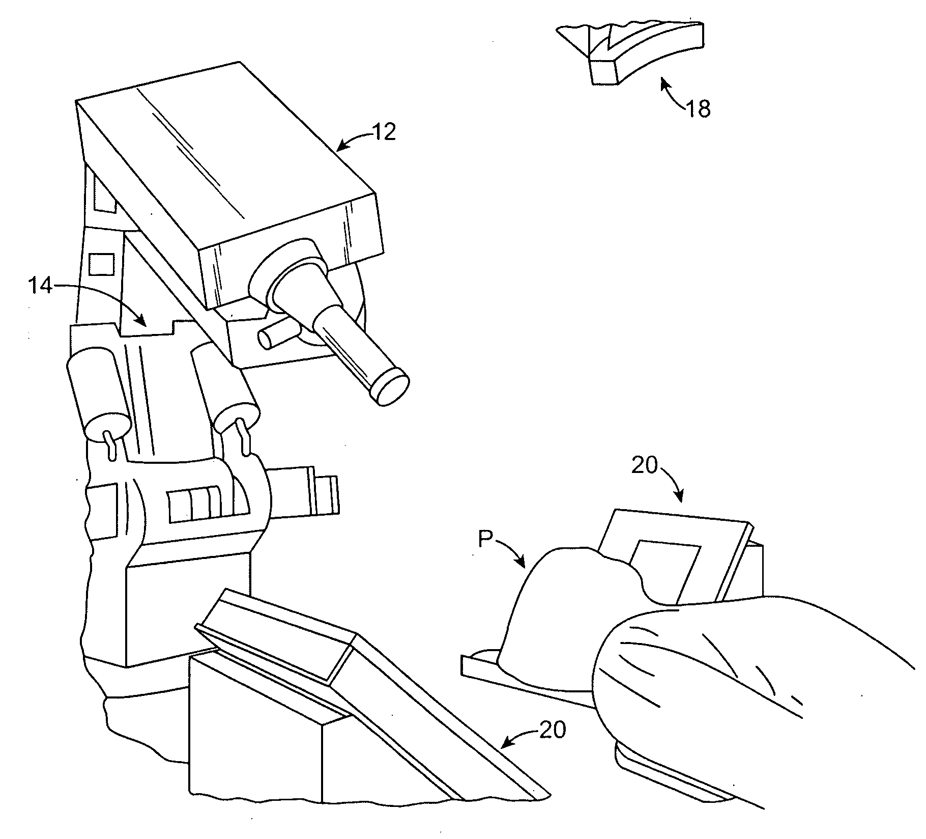
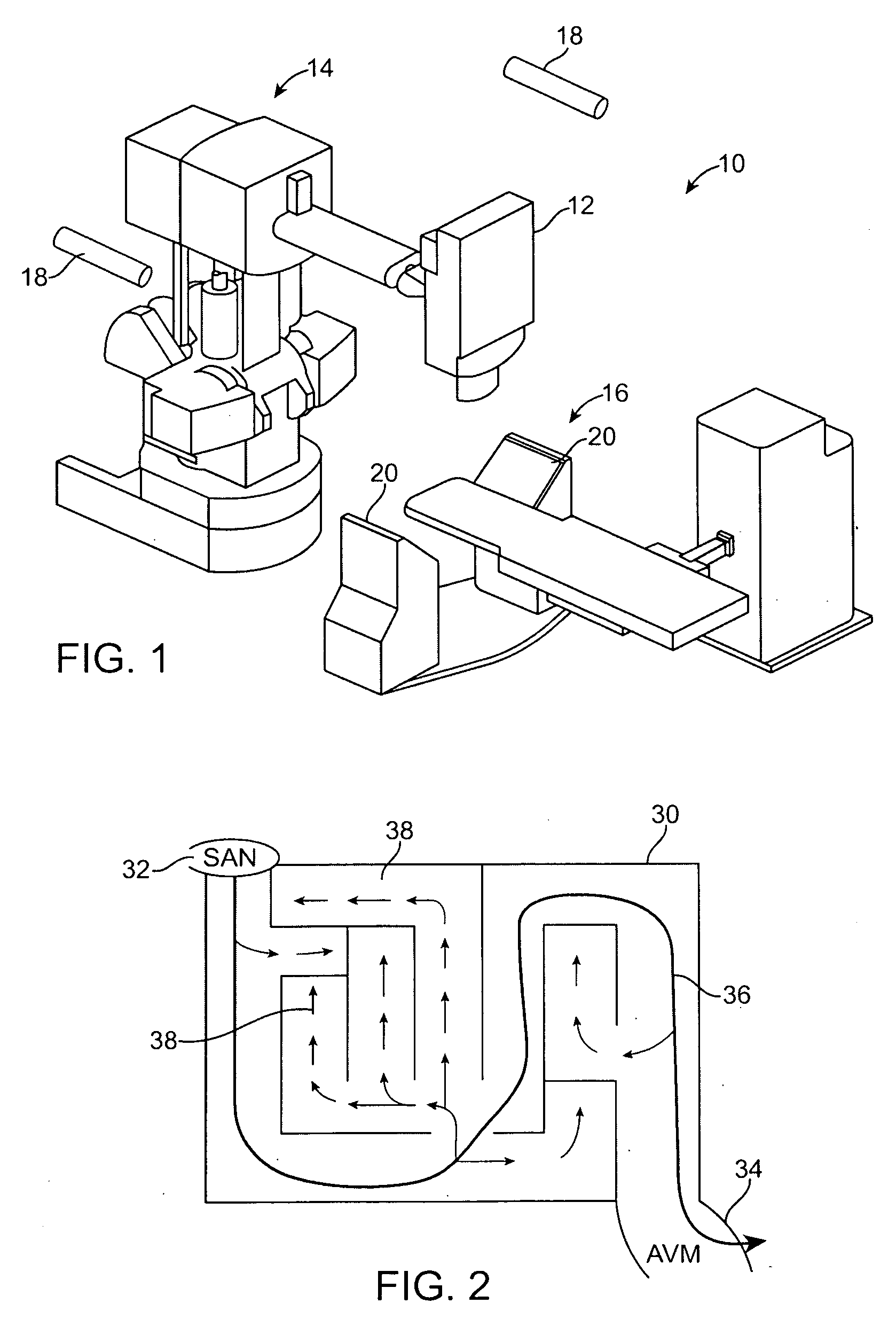
[0059]Introduction: A non-invasive method for ablating cardiac tissue using externally focused radiation was done on an animal. This method compensates for motion associated with respiration by using a robotic arm to control the path of a high energy x-ray beam from a linear accelerator. Respiration is monitored by a vest with LEDs worn on the chest and other motion is detected by x-ray imaging in 2 planes. This method has been used successfully to treat lung tumors. However, it is not known to what extent targeting cardiac tissues for the treatment of arrhythmias may require compensation for cardiac contractile motion also.

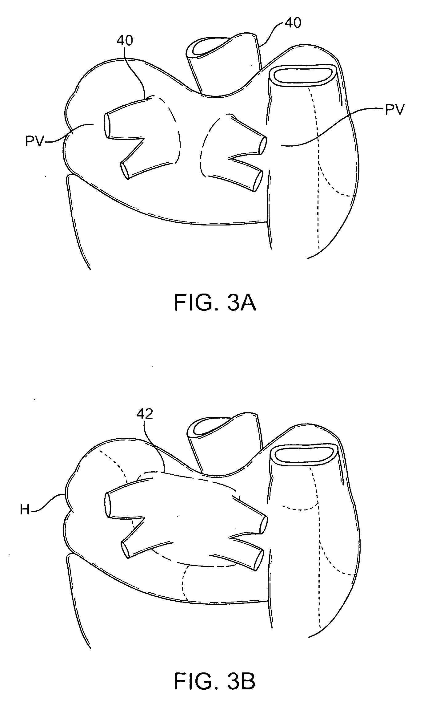
[0060]Purpose: To determine the relative magnitudes of cardiac and respiratory movement at potential arrhythmia targets such as the left superior pulmonary vein (PV), AV junction (AVJ) and the cavo-tricuspid isthmus (CTI).

[0061]Method: Hanford-Sinclair mini-swine (40-70 kg) (n=5) were studi...

example 2

Non-invasive ablation of the Left Superior Pulmonary Vein-Left Atrial junction using stereotactic focused radiation

[0065]Introduction: High energy x-ray irradiation from a linear accelerator mounted on a robot arm (Cyberknife) has been used to produce ablation of cardiac tissue in swine. This device is currently used in patients for diverse indications requiring careful targeting, from tumors to trigeminal neuralgia.

[0066]Conclusions: Selective targeting of cardiac arrhythmogenic sites can be done using stereotactically focused external radiation. The left superior pulmonary vein can be ablated without compensation for cardiac contractility.

[0067]Purpose: To determine if this method of energy delivery can be used to target potential arrhythmogenic sites such as the Left superior pulmonary vein-left atrial junction.

[0068]Method: Hanford-Sinclair mini-swine (40-70 kg) (n=9) were studied under anesthesia. The animals underwent radiation (x-rays, 40-80 Gray) using a computer controlled ...


PUM


Abstract

The invention provides a non-invasive system and method for treatment of the heart. In a first aspect, a method for treatment of an anatomical site related to arrhythmogenesis of a heart of a patient comprises creating a target shape encompassing the anatomical site, directing particle beam radiation or x-ray radiation from outside the patient toward the target shape wherein one or more doses of radiation ablates the target shape and disregarding at least one orientation of cardiac motion while creating the target shape or directing the particle beam or both.

Description

CROSS-REFERENCES TO RELATED APPLICATIONS[0001]This application claims the benefit under 35 USC 119(e) of U.S. Provisional Application No. 60 / 975,373, filed Sep. 26, 2007, entitled “Radiosurgical Ablation of the Myocardium”, the full disclosure of which is incorporated herein by reference. This application is related to U.S. Pat. No. 6,889,695 entitled “Method for Non-Invasive Heart Treatment”, the contents of which are incorporated herein by reference.BACKGROUND OF THE INVENTION[0002]The present invention is generally related to treatment of the heart, and in particular, non-invasive treatment of the heart using radiosurgical ablation.[0003]A typical adult human heart beats at a rate of about 70 beats per minute. The rate is not constant, however, and changes during and following exercise, with fear or anxiety, or for other reasons. A heart can also pump arrhythmically for many reasons, including damage to the heart's electrical conduction system. Arrhythmias interfere with the hear...

Claims


Application Information

Patent Timeline
IPC IPC(8): A61N5/10A61B5/055
CPCA61N5/1049A61N5/1067A61N5/1039A61N2005/1061A61N5/1037A61N2005/1059
Inventor SUMANAWEERA, THILAKAMAGUIRE, PATRICKYOUNG, RODERICK A.
Owner VARIAN MEDICAL SYSTEMS
Why Eureka
Try Eureka