Apparatus and method for achieving micropuncture

a micro-puncture and endovascular technology, applied in the direction of guide wires, catheters, guide needles, etc., can solve the problems of lining the blood vessel, thrombosis and tissue or organ damage, unnecessarily complex guidewire placement systems, and potentially dangerous to patients, so as to minimize tissue trauma and reduce the trauma at the puncture site.

Inactive Publication Date: 2009-07-23
PRESSURE PROD MEDICAL SUPPLIES INC
View PDF19 Cites 25 Cited by
  • Summary
  • Abstract
  • Description
  • Claims
  • Application Information

AI Technical Summary

Benefits of technology

[0007]The illustrated embodiment of the invention is an apparatus and method of using an assembly comprising a longitudinally openable or separable, small diameter, hollow micropuncture device or microneedle for puncture into a vascular or body cavity while minimizing tissue trauma at a puncture site, and a stepped guidewire. The term, “micropuncture device”, shall be used throughout this specification and claims to refer to a needle, microneedle, and / or an assembly of elements which include a microneedle, a needle or an element that functions at least in part as a microneedle or needle having a maximum gauge size of 20, or can accommodate a 0.025 inch maximum sized diameter guidewire. The terms, “non-micropuncture vascular device”, “larger diameter vascular instrument”, “larger medical device”, “large instrument” are defined as a vascular or medical instrument which is too stiff or too large in diameter to be effectively or reliably guided into the vascular system or other body cavity through a micropuncture device or by means of guidewires which are usable in micropuncture devices. The term “microguidewire” refers to a guidewire or a portion of a guidewire that is 0.025 inch diameter or smaller whereas the term “larger guidewire” refers to a guidewire or a portion of a guidewire that is larger than 0.025 inch diameter. The micropuncture device has an inner diameter; and the stepped guidewire has a distal portion for telescopic disposition through the inner diameter of the micropuncture device or needle, and a proximal portion with a diameter larger than the inner diameter of the micropuncture device or needle. The stepped guidewire is provided for guiding a larger diameter vascular instrument. By use of the illustrated embodiment tissue trauma at the puncture site is substantially reduced.

Problems solved by technology

Each time a guidewire is introduced it carries the risk of damaging the lining of the blood vessel which may result in thrombosis and tissue or organ damage.
The current systems for micropuncture guidewire placement are thus unnecessarily complex and potentially dangerous to the patient.

Method used


Image

  • Apparatus and method for achieving micropuncture
  • Apparatus and method for achieving micropuncture
  • Apparatus and method for achieving micropuncture

Examples


Embodiment Construction

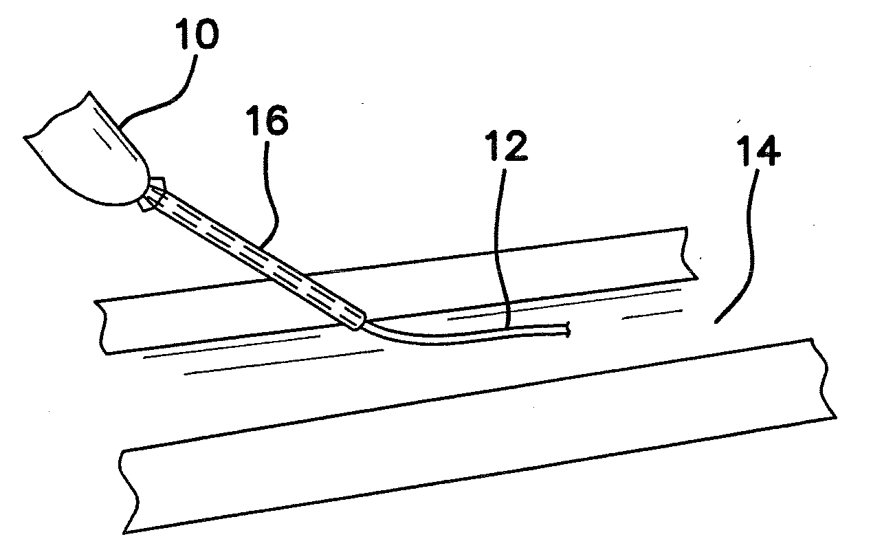
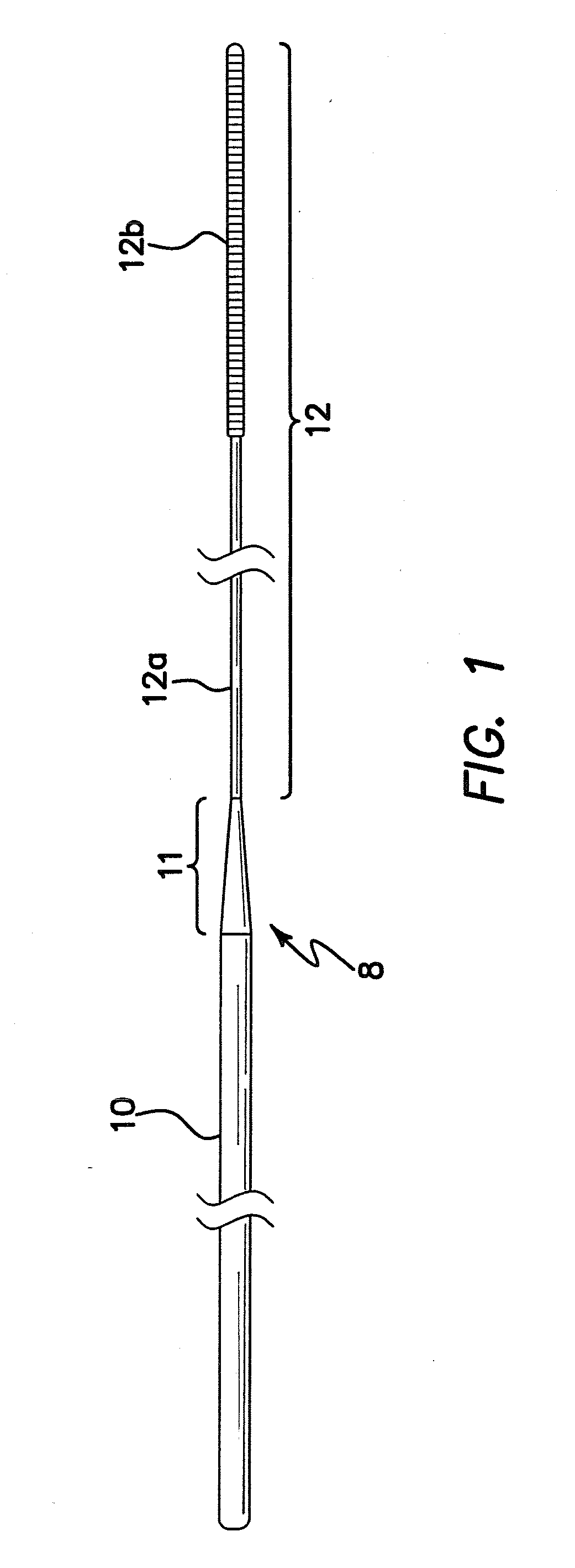
[0043]The invention is directed to a system and method for using a microneedle and stepped guidewire to micropuncture a body cavity or endovascular cavity and dispose a microguidewire therein for subsequent disposition of a larger guidewire and instrument such as an introducer or catheter while reducing tissue trauma at the puncture site and to the vascular system.

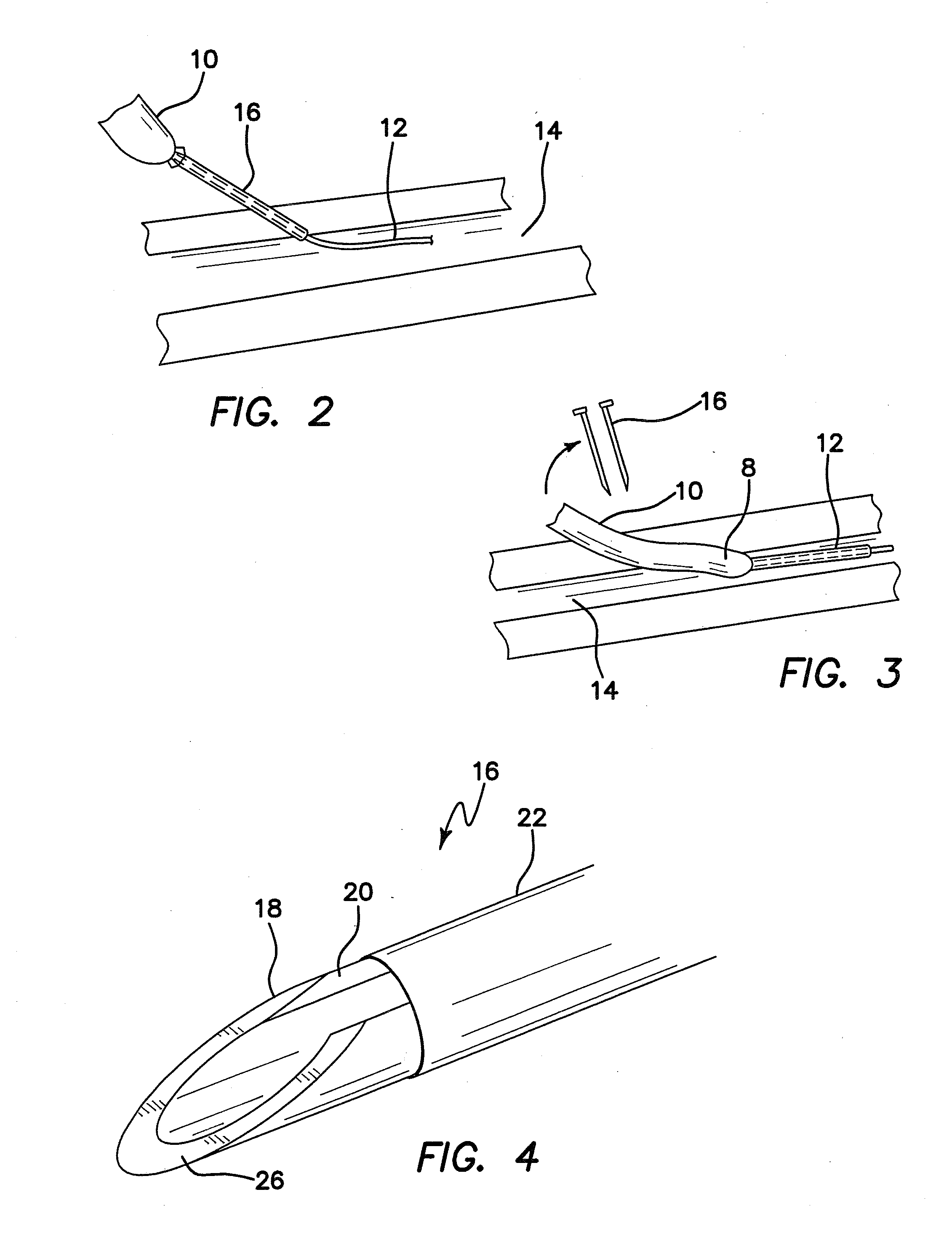
[0044]The illustrated embodiment comprises a splittable microneedle 16 as shown in FIG. 2 and a stepped or preferably a steeply tapered guidewire, generally denoted by reference numeral 8, as shown in FIGS. 1-3. As shown in FIG. 1 guidewire 8 has a distal portion 12 comprised of two subportions, namely proximal subportion 12a with a diameter of approximately 0.014 inch and an enlarged distal tip subportion 12b approximately 5 cm in length and approximately 0.018 inch in diameter. In another embodiment portions 12a and 12b may have the same diameters, e.g. 0.18 inch. The two subportions 12a and 12b of distal portion 12 coll...


PUM


Abstract

A method comprises the steps of inserting a microneedle or micropuncture device into a body cavity to achieve a micropuncture into the cavity while reducing tissue trauma at a puncture site. A stepped or tapered guidewire having a reduced diameter distal portion is telescopically disposed through the inner diameter of the microneedle or Micropuncture device. A proximal portion of the guidewire has a diameter larger than the inner diameter of the microneedle or Micropuncture device and extends to a proximal end of the guidewire. The proximal portion is adapted for guiding a larger diameter instrument into and through the vascular system. The microneedle or micropuncture device is withdrawn from the micropuncture while leaving the distal portion of the guidewire extending through the micropuncture. The microneedle or micropuncture device is removed from the guidewire without removing the microneedle or Micropuncture device over the proximal end of the guidewire. As a result, tissue trauma at the puncture site is substantially reduced.

Description

BACKGROUND OF THE INVENTION [0001]1. Field of the Invention[0002]The invention relates to the field of micropuncture endovascular devices and methods, and in particular to micropuncture endovascular systems and guidewires for vascular introduction and methods of using the same.[0003]2. Description of the Prior Art[0004]The prior art methodology used to make a micropuncture is as follows: (1) First the vasculature is punctured with a microneedle. (2)Then placement of a microguidewire through the needle is achieved. (3)Then a coaxial catheter pair is advanced over the microguidewire into the blood vessel. (4) Then the microguidewire is removed and then (5) The inner sleeve of the coaxial catheter pair is removed and discarded. (6) Then a large guidewire is introduced into the vasculature through the remaining outer sleeve (which has an inside diameter large enough to accommodate a large guidewire) of the coaxial catheter pair, (7) Then the retained outer sleeve must be removed. Then t...

Claims


Application Information

Patent Timeline
Patent Type & Authority Applications(United States)
IPC IPC(8): A61M25/09
CPCA61M25/06A61M25/0606A61M2025/09133A61M25/09041A61M2025/09083A61M25/09
Inventor KURTH, PAUL A.ARMOUR, ANDREW W.
Owner PRESSURE PROD MEDICAL SUPPLIES INC
Why Eureka
Try Eureka