
Method of Assessing Blood Volume Using Photoelectric Plethysmography

a technology of plethysmography and blood volume, applied in the field of methods, can solve the problems of unacceptably frequent complications, remarkably little work done to document or quantify this phenomenon, and lack of a continuous measurement method of the phenomenon

Inactive Publication Date: 2010-01-21
YALE UNIV
View PDF28 Cites 38 Cited by
  • Summary
  • Abstract
  • Description
  • Claims
  • Application Information

AI Technical Summary

Problems solved by technology

There is growing evidence that invasive monitors of volume status, such as the pulmonary artery catheter, may be a source of unacceptably frequent complications.
To date though, there has been remarkably little work done to document or quantify this phenomenon.
1) Lack of a method of continuous measurement of the phenomenon.
2) Reliance on positive pressure and mechanical ventilation; and the requirement of ventilator maneuvers such as periods of apnea.
3) Lack of recognition of the venous contribution to the plethysmographic signal.
4) The lack of algorithms resistant to artifacts.
Where flow is compromised (e.g. by hypovolemia or vasoconstriction), the pulse oximeter may be unable to function.
While the plethysmograph has been examined previously as a potential anesthesia monitoring device, remarkably little research has been done on this ubiquitous signal.

Method used


Image

  • Method of Assessing Blood Volume Using Photoelectric Plethysmography
  • Method of Assessing Blood Volume Using Photoelectric Plethysmography
  • Method of Assessing Blood Volume Using Photoelectric Plethysmography

Examples


Embodiment Construction

[0047]The detailed embodiments of the present invention are disclosed herein. It should be understood, however, that the disclosed embodiments are merely exemplary of the invention, which may be embodied in various forms. Therefore, the details disclosed herein are not to be interpreted as limiting, but merely as the basis for the claims and as a basis for teaching one skilled in the art how to make and / or use the invention.

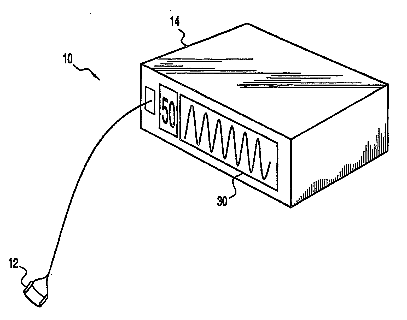
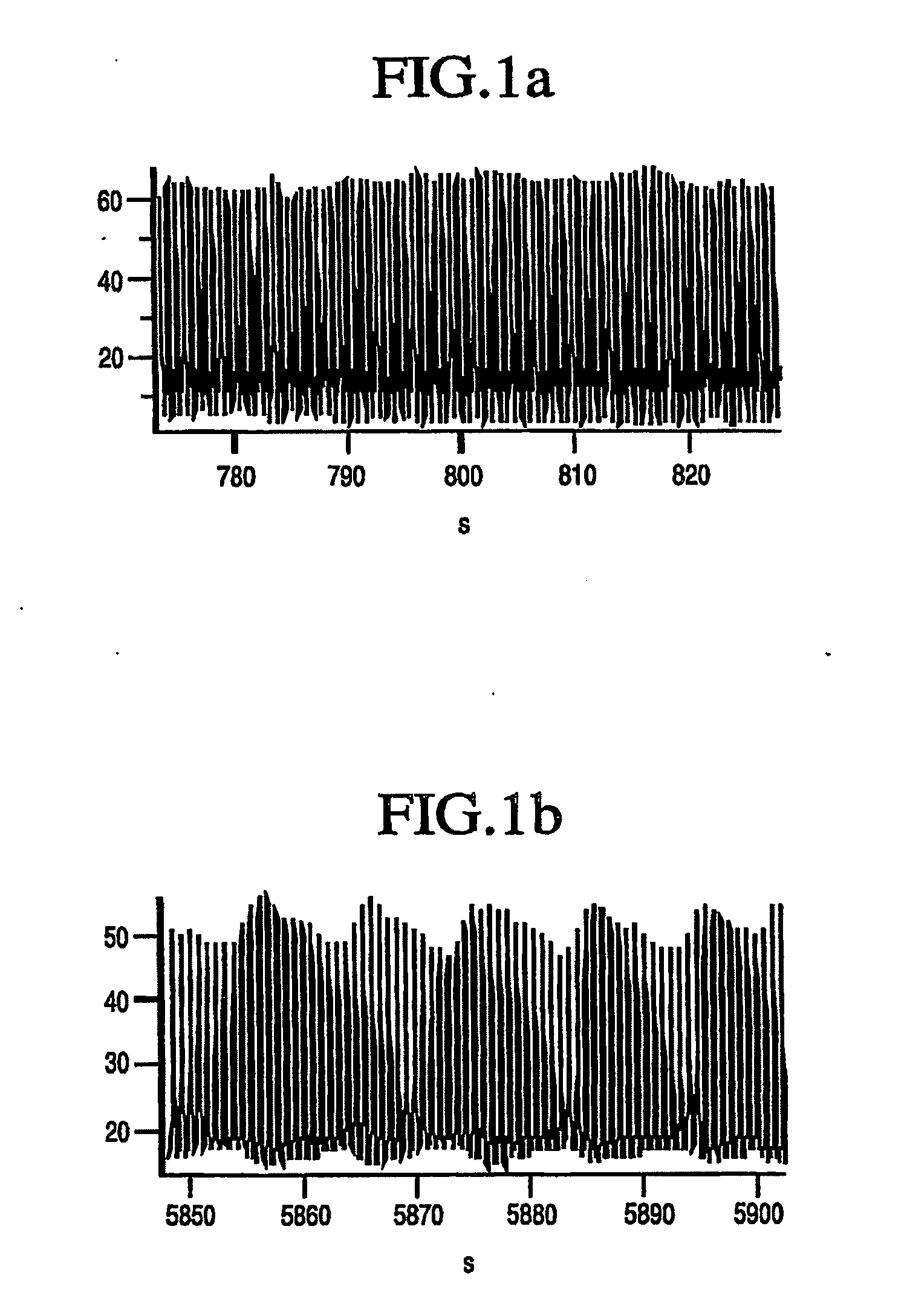
[0048]With reference to FIGS. 1 to 5, and in accordance with a first embodiment of the present invention, a method for assessing blood volume through the analysis of a cardiovascular waveform, in particular, a pulse oximeter waveform, is disclosed. As the following disclosure will clearly demonstrate, the present invention monitors relative blood volume or effective blood volume. As such, the present method is preferably applied in analyzing blood volume responsiveness (that is, whether a patient needs to be given blood or other fluid).

[0049]In accordance with a ...


PUM


Abstract

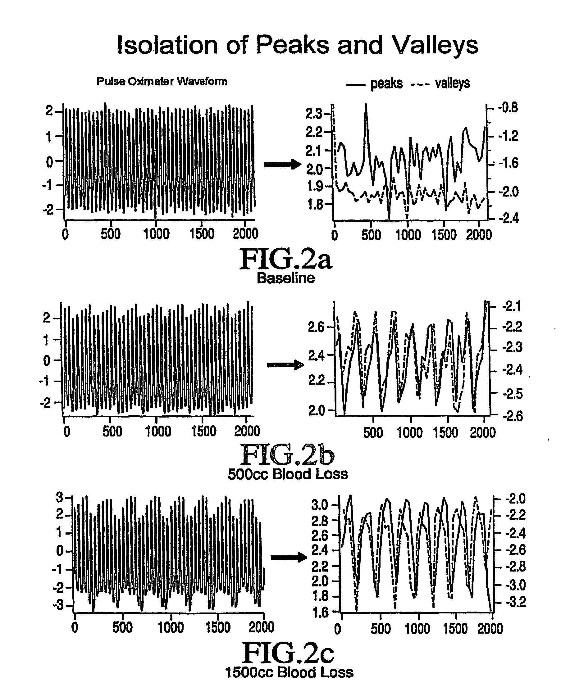
A method and system for assessing blood volume within a subject includes generating a cardiovascular waveform representing physiological characteristics of a subject and determining blood volume of the subject by analyzing the cardiovascular waveform. The step of analyzing includes generating a first trace of the per heart-beat maximums of the cardiovascular waveform, which is representative of the systolic pressure upon the cardiovascular signal, generating a second trace of the per heart-beat minimums of the cardiovascular waveform, which is representative of the diastolic pressure upon the cardiovascular signal, and comparing the respective first trace and the second trace to generate an estimate of relative blood volume within the subject. In accordance with an alternate method of analyzing harmonic analysis is applied to the cardiovascular waveform, extracting a frequency signal created by ventilation and applying the extracted frequency signal in determining blood volume of the subject.

Description

BACKGROUND OF THE INVENTION[0001]1. Field of the Invention[0002]The invention relates to methods for studying and utilizing flow waveforms in the peripheral vasculature. In particular, the invention relates to a method for assessing blood volume by analyzing photoelectric plethysmographic waveforms of the pulse oximeter.[0003]2. Description of the Prior Art[0004]There is growing evidence that invasive monitors of volume status, such as the pulmonary artery catheter, may be a source of unacceptably frequent complications. Dalen J & Bone R, Is it time to pull the pulmonary artery catheter?, JAMA 1996; 276:916-14; Connors A, Speroff T & Dawson N, The effectiveness of right heart catheterization in the initial care of critically ill patients, JAMA 1996; 276:889-97. The potential loss of this important monitor from routine peri-operative care necessitates the search for another means of monitoring a patient's blood volume status.[0005]It has been known for quite some time that ventilatio...

Claims


Application Information

Patent Timeline
Patent Type & Authority Applications(United States)
IPC IPC(8): A61B5/02A61B5/0402A61B5/00A61B5/024
CPCA61B5/0059A61B5/02416A61B5/1455G06F19/3406G06F19/366A61B5/026A61B5/14551A61B5/7282A61B5/7405A61B5/742A61B5/746A61B5/0205G16H40/63G16H10/40Y02A90/10
Inventor SHELLEY, KIRK H.SILVERMAN, DAVID G.SHELLEY, ADAM J.STOUT, ROBERT G.
Owner YALE UNIV
Learn More