Method and system with encapsulated imaging and therapy devices, coupled with an extracorporeal imaging device

a therapy device and imaging technology, applied in the field of encapsulated imaging and therapy devices, coupled with an extracorporeal imaging device, can solve the problems of insufficient diagnosis of the endoscope capsule diagnostic tool, just like the diagnostic tool of the conventional mechanical sliding endoscope, and the task cannot be resolved through optical image acquisition, so as to achieve image-supported diagnosis and therapy. , the effect of effective diagnosis

Inactive Publication Date: 2010-02-04
SIEMENS AG
View PDF18 Cites 3 Cited by
  • Summary
  • Abstract
  • Description
  • Claims
  • Application Information

AI Technical Summary

Benefits of technology

[0012]An object of the invention is to overcome the aforementioned disadvantages and to provide a system that enables an effective, image-supported diagnosis and therapy. The object is achieved by a medical system for image-supported diagnosis or therapy of the body of a patient, possessing

Problems solved by technology

However, there are cases in which the diagnostic tools of an endoscopy capsule, just like the diagnostic tools of conventional, mechanical sliding endoscopy, are not sufficient for a complete diagnosis.
Such a task cannot be resolved through the optical image acquisition method of endoscopy.
The greatest danger in the removal of polyps is the possible presence of larger vessels tangential to or contained in that polyp that could lead to more severe bleeding upon a careless removal of the polyp.
Although in classical endoscopy an appropriate instrument can be introduced into the operating canal in order to stem the bleeding, this is not possible in the case of capsule endoscopy.
In addition to the interruption of the treatment and the costs therefore incurred for lost time that is caused, an exact assessment using the external image acquisition system is often not possible due to the lack of coordinates of the assessment position.
This thus leads to a radiation exposure of more or less large adjacent areas.
Conversely, irregularities or pathologies cannot necessarily be detected with radiological image acquisition systems.

Method used


Image

  • Method and system with encapsulated imaging and therapy devices, coupled with an extracorporeal imaging device
  • Method and system with encapsulated imaging and therapy devices, coupled with an extracorporeal imaging device
  • Method and system with encapsulated imaging and therapy devices, coupled with an extracorporeal imaging device

Examples


Embodiment Construction

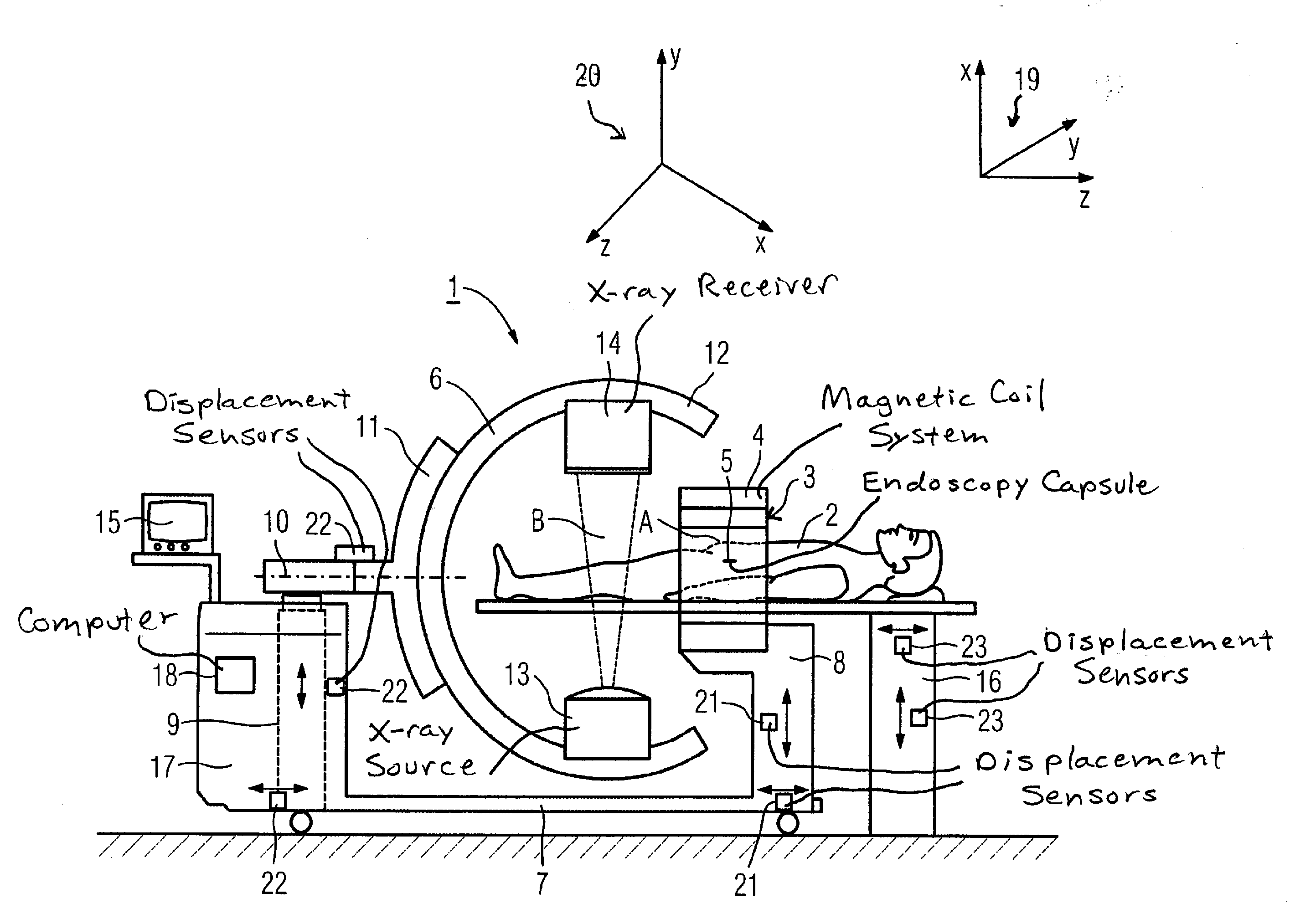
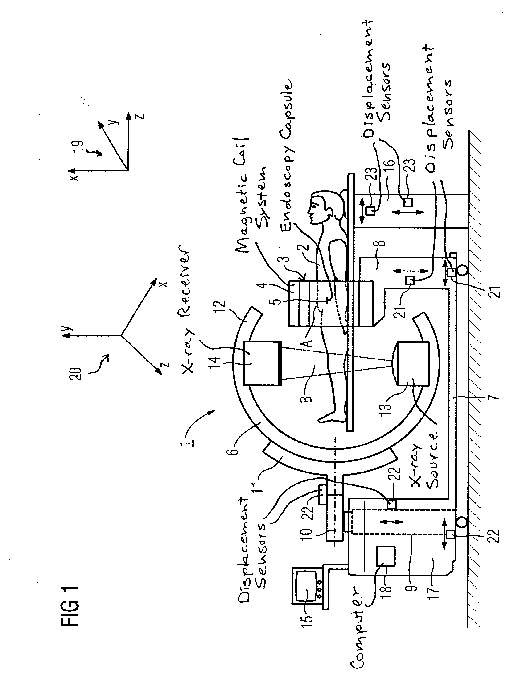
[0031]FIG. 1 shows the medical system 1 according to the invention as a mechanically coupled system in which an endoscopy system 3 and an image acquisition apparatus 6 (shown by way of example in the form of a C-arm) are mechanically coupled with one another via a base 7. The endoscopy system 3 essentially is composed of a magnetic coil system 4 and a cylindrical endoscopy capsule 5 that can move freely in a working volume A via the magnetic coil system 4. The working volume A is the space within the magnetic coil system 4 in which the gradient fields generated by the magnetic coil system 4 act on the endoscopy capsule 5. The position and, if applicable, the alignment of the endoscopy capsule 5 in the longitudinal axis are determined via a position detection system (not designated in detail) which is integrated into the magnetic coil system 4. The position detection is mapped in the coordinate system 20 of the endoscopy system 3. The endoscopy capsule 5 is equipped with an image acq...


PUM


Abstract

A medical system has an endoscopy system and an extracorporeal imaging system and a patient positioning device. The endoscopy system includes an intracorporeally movable capsule that is navigable within the body of a patient by a magnetic coil system within a tube-like working volume formed by the magnetic coil system. An encapsulated imaging unit in the endoscopy capsule obtains image data associated with a medical finding. The spatial coordinates of the medical finding identified by the encapsulated imaging unit are relayed to the extracorporeal image acquisition system to allow an extracorporeal image to be obtained based on those spatial coordinates.

Description

BACKGROUND OF THE INVENTION[0001]1. Field of the Invention[0002]The present invention concerns a device for implementation of minimally invasive diagnoses, and when necessary, additional therapeutic procedures inside the body of a patient.[0003]2. Description of the Prior Art[0004]In medicine it is frequently necessary to conduct a medical procedure inside (normally in the body of) a (normally living) person or animal, which medical procedure can be a diagnosis or a treatment, for example. The target of such a medical procedure is often a hollow organ in the patient; in particular the gastrointestinal tract is subjected to such an examination. Such medical measures have been conducted for a long time with the aid of endoscopes which are (invasively or non-invasively) inserted into a patient from the outside, either via bodily orifices of the patient or through small incisions, and are mechanically controlled and positioned.[0005]For catheter-free or tube-less endoscopy, endoscopy ca...

Claims


Application Information

Patent Timeline
Patent Type & Authority Applications(United States)
IPC IPC(8): A61B1/04G06K9/00
CPCA61B1/00158A61B1/041A61B6/4441A61B5/073A61B5/062
Inventor GRAUMANN, RAINERKUTH, RAINER
Owner SIEMENS AG
Why Eureka
Try Eureka