
Methods of organ regeneration

a technology of organ regeneration and organs, applied in the field of organ regeneration, can solve the problems of inability to prevent end-stage diseases or permanently reverse insulitis, inability to clinically apply primarily, and inability to achieve the effect of permanently restoring insulitis, enhancing post-receptor tnf-alpha action, and enhancing tnf-alpha bioavailability or stability

Inactive Publication Date: 2010-06-17
THE GENERAL HOSPITAL CORP
View PDF4 Cites 11 Cited by
  • Summary
  • Abstract
  • Description
  • Claims
  • Application Information

AI Technical Summary

Benefits of technology

[0009]In another aspect, the invention features a method for treating, stabilizing, or preventing an autoimmune disease (e.g., an established autoimmune disease) and / or increasing or maintaining the number of functional cells of a predetermined type in a mammal. This method involves (a) administering to the mammal a composition that selectively kills a predetermined subpopulation of blood cells, in an amount sufficient to selectively kill at least 10%, preferably at least 75%, of the subpopulation of blood cells in the mammal, and (b) prior to, after, or concurrently with step (a), administering to the mammal cells that have the potential to differentiate into the predetermined type or that are of the predetermined cell type. In one embodiment, the method includes determining, prior to step (a), whether the blood of the mammal contains a subpopulation of blood cells with higher than normal sensitivity to the composition to be administered; if this is the case, the decision to employ the composition is reinforced.
[0010]In another aspect, the invention features a method for treating, stabilizing, or preventing an autoimmune disease (e.g., an established autoimmune disease) and / or increasing or maintaining the number of functional cells of a predetermined type in a mammal. This method involves (a) administering to the mammal a first composition that selectively kills a pre-determined subpopulation of stimulated blood cells, in an amount sufficient to selectively kill at least 10%, preferably at least 75%, of the subpopulation of stimulated blood cells in the mammal, (b) prior to, after, or concurrently with step (a), administering to the mammal a second composition that selectively kills a predetermined subpopulation of unstimulated blood cells, in an amount sufficient to selectively kill at least 10%, preferably at least 75%, of the subpopulation of unstimulated blood cells in the mammal, and (c) prior to, after, or concurrently with steps (a) or (b), administering to the mammal cells that have the potential to differentiate into the predetermined type or that are of the predetermined cell type. In one embodiment, prior to step (a), the method includes determining whether the blood of the mammal contains a subpopulation of stimulated blood cells with increased sensitivity to the first composition; if this is the case, the decision to employ the first composition is reinforced. In another embodiment, prior to step (b), the method includes determining whether the blood of the mammal contains a subpopulation of unstimulated blood cells with increased sensitivity to the second composition; if this is the case, the decision to employ the second composition is reinforced.
[0047]With respect to the therapeutic methods of the invention, it is not intended that the administration of one or more compounds (e.g., purified or unpurified donor cells, MHC class I and peptide, and an inducer of lymphopenia) to a patient be limited to a particular mode of administration, dosage, or frequency of dosing; the present invention contemplates all modes of administration, including intramuscular, intravenous, intraperitoneal, intravesicular, intraarticular, intralesional, subcutaneous, or any other route sufficient to provide a dose adequate to increase the number of desired cells. The compound(s) may be administered to the patient in a single dose or multiple doses. When multiple doses are administered, the doses may be separated from one another by, for example, one day, one week, one month, or one year. For example, a compound that induces lymphopenia may be administered once a week for, e.g., 2, 3, 4, 5, 6, 7, 8, 10, 15, 20, or more weeks. It is to be understood that, for any particular subject, specific dosage regimes should be adjusted over time according to the individual need and the professional judgment of the person administering or supervising the administration of the compositions. For example, the dosage of donor cells can be increased if more cells of a particular cell type are needed, e.g., if the glucose levels of a diabetic patient have not returned to normal or if an ongoing process decreases the number or activity of cells of the predetermined cell type. The dosage of a compound that induces lymphopenia can also be increased if autoimmune cells remain in the patient, for example, if a blood sample from the patient contains autoantibodies or contains cells with increased sensitivity to TNF-alpha, indicating that autoimmune cells are still present in the patient. Conversely, the dosage of donor cells or lymphopenia-inducing compounds can be decreased if a desired number of cells are present in the patient or if autoimmune cells are no longer present. If desired, conventional treatments may be used in combination with the therapies of the present invention. For example, diet and exercise can be used to facilitate the control of glucose levels in diabetic patients.
[0049]By “treating, stabilizing, or preventing a disease, disorder, or condition” is meant preventing or delaying an initial or subsequent occurrence of a disease, disorder, or condition; increasing the disease-free survival time between the disappearance of a condition and its reoccurrence; stabilizing or reducing an adverse symptom associated with a condition; reducing the severity of a disease symptom; slowing the rate of the progression of a disease; or inhibiting or stabilizing the progression of a condition. Desirably, at least 10, 20, 30, 40, 60, 80, 90, or 95% of the treated subjects have a complete remission in which all evidence of the disease disappears. In another preferred embodiment, the length of time a patient remains free of disease symptoms after being diagnosed with a condition and treated with a therapy of the invention is at least 10, 20, 40, 60, 80, 100, 200, or even 500% greater than (i) the average amount of time an untreated patient remains free of disease symptoms or (ii) the average amount of time a patient treated with another therapy remains free of disease symptoms. Desirably, the number of disease-causing white blood cells decreases by at least 10, 20, 30, 40, 60, 80, 95, or 100%.
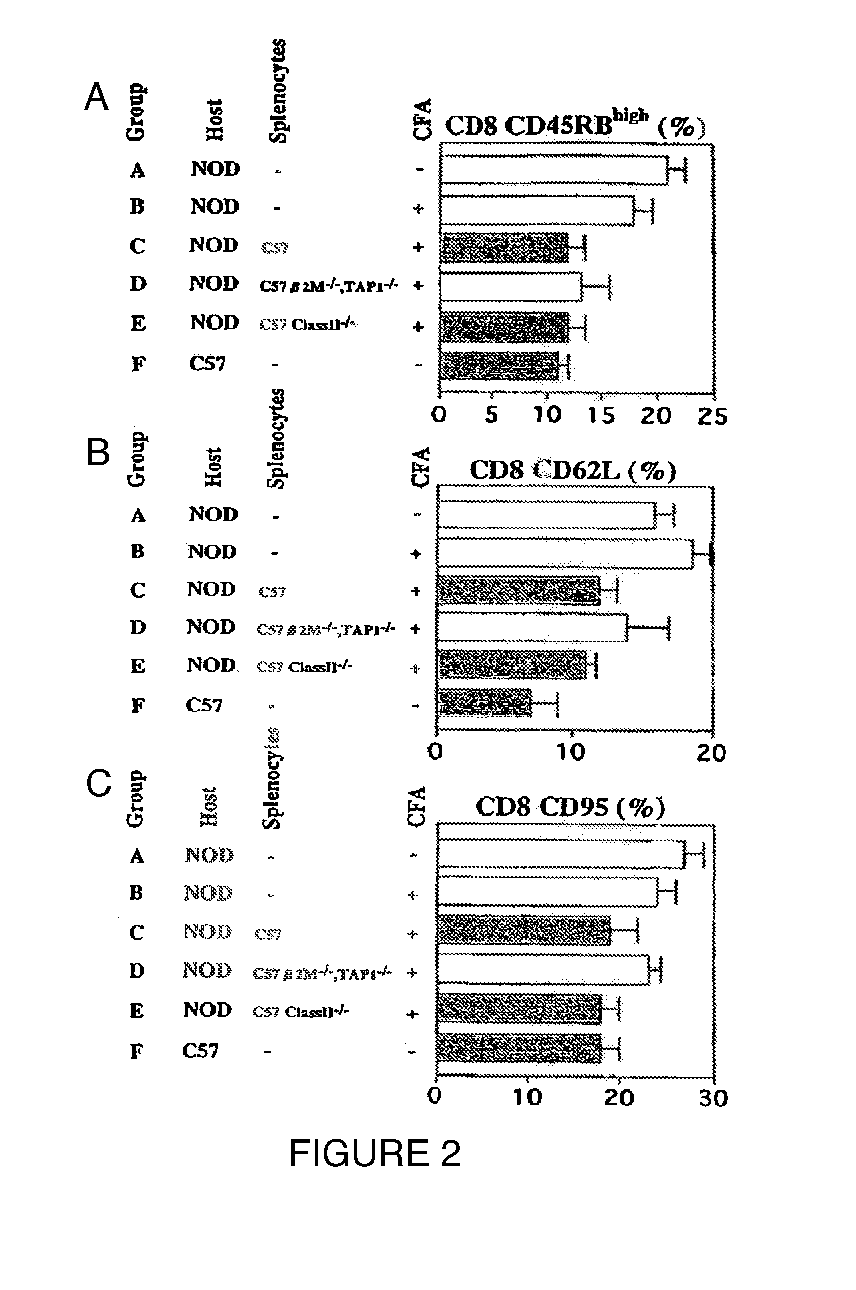
[0058]A “TNF-alpha inducing agent,” is desirably a compound that results in the expression of endogenous TNF-alpha, enhances secretion of TNF-alpha, or enhances bioavailability or stability of TNF-alpha. However, TNF-alpha agonists, agents that stimulate TNF-alpha signaling or enhance post-receptor TNF-alpha action, or agents that act on pathways that cause accelerated cell death of autoimmune cells, are also included in this definition. Stimulation of TNF-alpha induction (e.g., by administration of CFA) is desirably carried out prior to, after, or during administration (via implantation or injection) of cells in vivo.

Problems solved by technology

Such damage may result from an existing autoimmune disease, or may be the result of a non-autoimmune insult.
While an autoimmune disease may be successfully treated, the individual may nonetheless have significant tissue damage as a result of the prior autoimmune attack.
This approach is not clinically applicable primarily because syngeneic islets are not available.
Furthermore, existing cell replacement strategies have not prevented end-stage diseases or permanently reversed insulitis.
Furthermore, diabetic host treatments such as body irradiation and bone marrow transplantation are unacceptably toxic, rendering the short-term alternative of insulin therapy more attractive.
Recently, islet transplantation has achieved limited success in clinical trials, such as that observed for allogenic transplants combined with multi-drug immunosuppression therapy, with type 1 diabetic patients having a sustained return to normoglycemia over a 6 month period.
However, these moderately successful islet transplants show failures after about one year, speculated to be due in part to the drug therapy itself inducing insulin resistance.

Method used


Image

  • Methods of organ regeneration
  • Methods of organ regeneration
  • Methods of organ regeneration

Examples


example 1

CFA Treatment and Islet Transplantation in NOD Mice

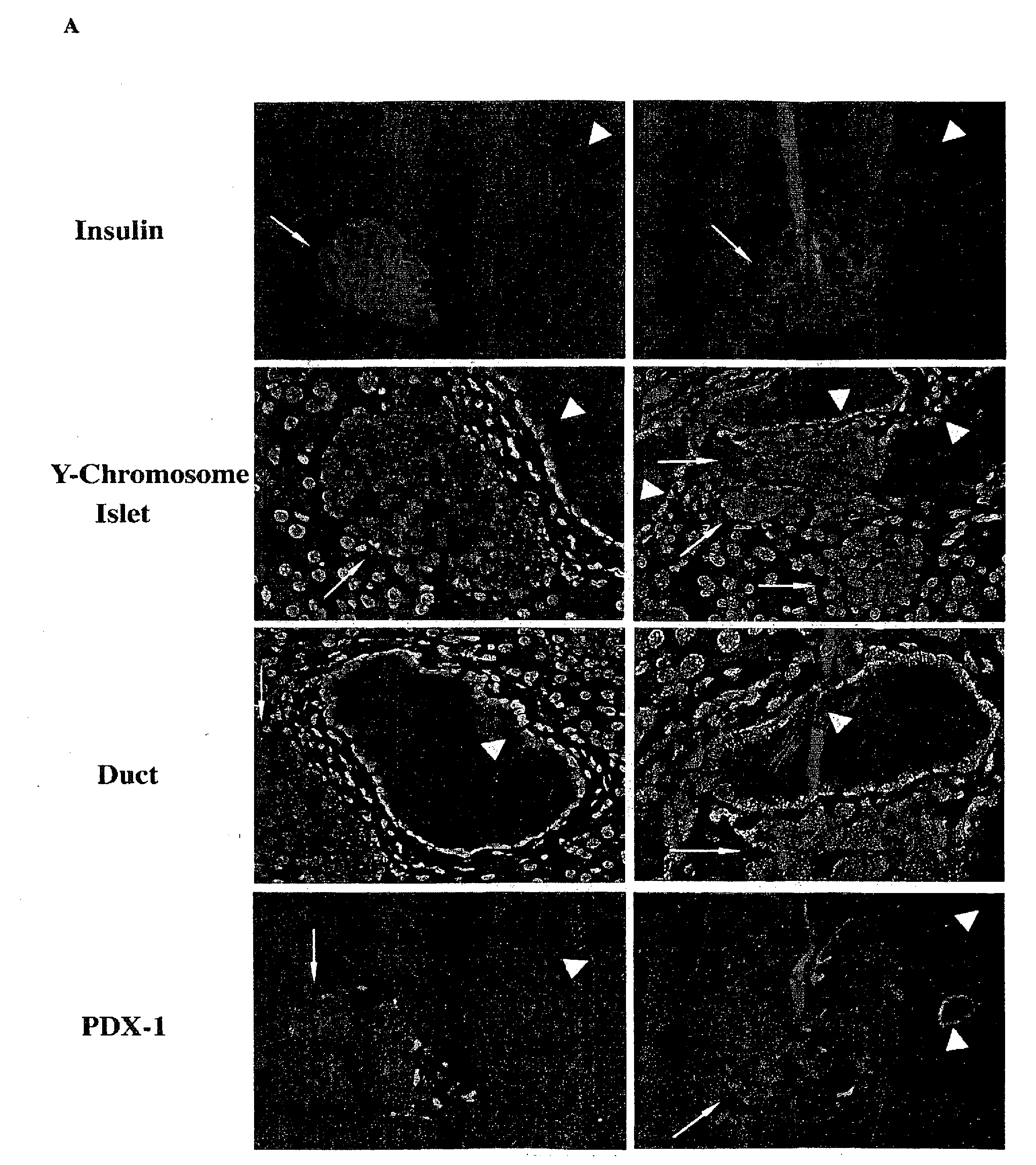
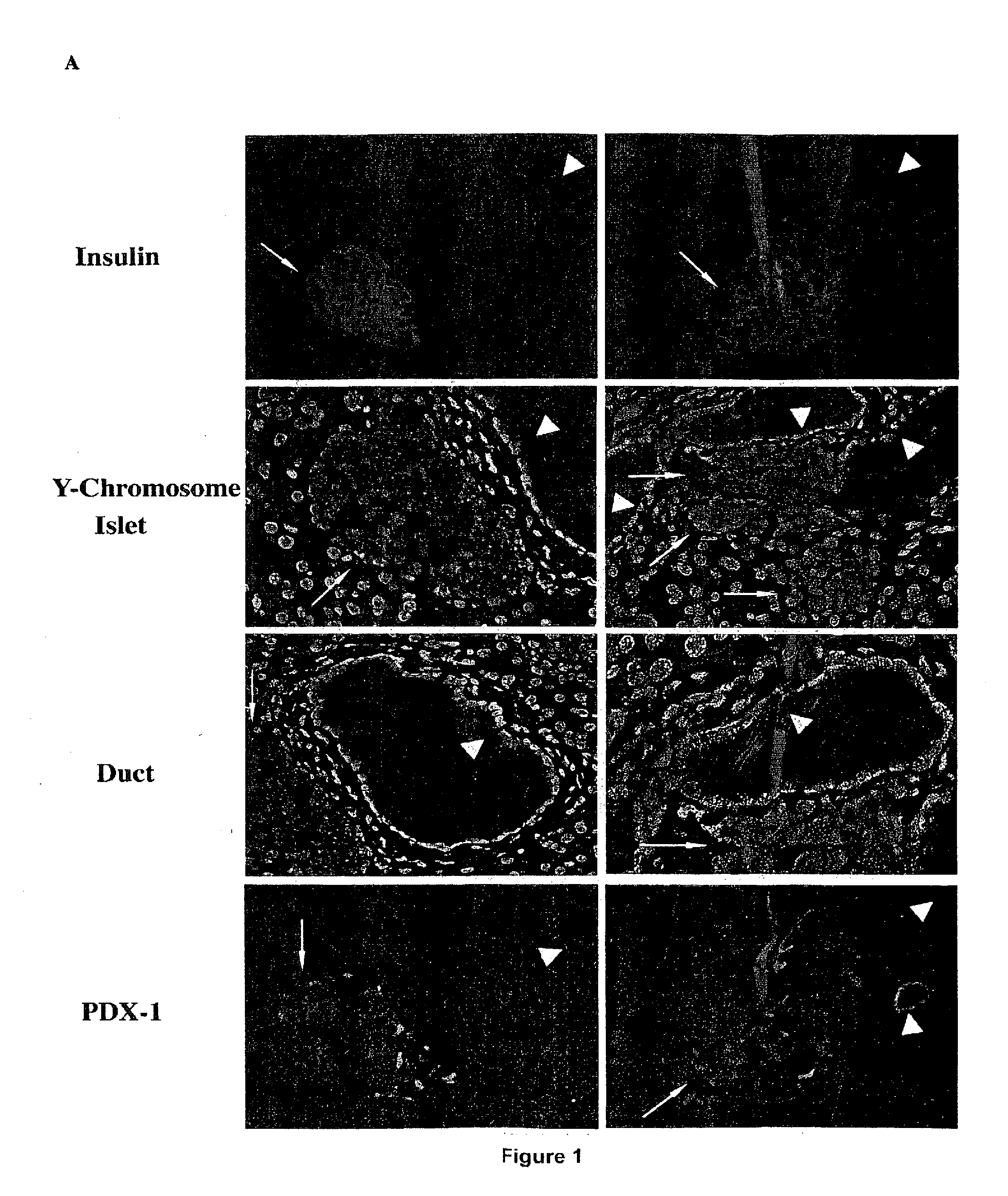
[0087]Hosts for the transplantation experiments were severely diabetic female NOD mice, usually greater than 20 weeks of age, which exhibited blood glucose concentrations of greater than 400 mg / dl for at least seven days and had been treated by daily administration of insulin for that length of time. Islet transplants were placed unilaterally in the kidney capsule to facilitate non-lethal removal and histological examination. Islets from 6-8 week-old pre-diabetic donor females or from normal C57 donor mice were rapidly rejected by diabetic NOD hosts in all cases, usually by day 9. Although C57 donor islets with transient ablation of class I survive indefinitely in non-immunosuppressed diabetic and non-autoimmune diabetic hosts, the survival time for β2M− / − C57 islets in diabetic NOD females is only about two to three times that of normal C57 islets. This observation confirms that the ablation of the donor cells is related to the cur...

example 2

Optimizing a Curative Therapy in the Diabetic NOD Mouse

[0099]In order to examine the differential effects of CFA, BCG, TNF-α, and splenocyte administration, late stage NOD mice (>15 weeks of age) were randomly assigned to one of four treatment groups, i.e., one injection of CFA, one injection of BCG (4 mg / kg), one injection of 10 ug TNF-α, or one injection of F1 splenocytes (1×10⁶ cells, IV) obtained from normal donors. The treated NOD mice were serially sacrificed on day 2, day 7, and day 14 (Table 2). An examination of pancreatic histology evaluated the effects of the interventions on invasive insulitis (Table 3).

[0100]Table 2 shows that on day 2 both a single injection of CFA and a single injection of low dose TNF-α (10 μg) had eliminated completely all subpopulations of cells with in vitro TNF-α sensitivity. At day 7 and day 14, this population of TNF-α sensitive cells was again evident. Simultaneous pancreatic histology of these cohorts confirmed a dramatic reduction in insulit...

example 3

Functional Impact of Donor Cell Radiation on Restoration of Normoglycemia in Severely Diabetic NOD Mice

[0105]Severely hyperglycemic NOD mice were originally treated with CFA to induce TNF-alpha and simultaneously exposed to functional complexes of MHC class I molecules and allogeneic peptides presented on either viable splenocytes or on islets. Normoglycemia for a 40-day treatment period, a critical parameter of this approach, was induced either by the implantation of temporary alginate encapsulated islets or subrenally placed syngeneic islet transplants. Both methods of glucose control can be surgically removed to test for restored endogenous pancreatic function. All original experiments were designed to treat established NOD female mice with severe hyperglycemia and utilized a 40-day time period of tight, artificial glucose control. Utilization of this temporary glucose control permitted sufficient endogenous pancreatic re-growth of the endogenous pancreas to sustain normoglycemia...


PUM


Abstract

The invention features methods for increasing or maintaining the number of functional cells of a predetermined type, for example, insulin producing cells of the pancreas, blood cells, spleen cells, brain cells, heart cells, vascular tissue cells, cells of the bile duct, or skin cells, in a mammal (e.g., a human patient) that has injured or damaged cells of the predetermined type.

Description

CROSS REFERENCE TO RELATED APPLICATIONS[0001]This application claims priority from U.S. Provisional Application No. 60 / 392,687, filed Jun. 27, 2002.BACKGROUND OF THE INVENTION[0002]The invention relates to repairing and regenerating damaged tissue in a human. Such damage may result from an existing autoimmune disease, or may be the result of a non-autoimmune insult. We have previously shown that eliminating autoimmune cells and re-educating the immune system are important components of an effective treatment of an autoimmune disease (described in U.S. patent application Ser. Nos. 09 / 521,064, 09 / 768,769, and Ryu et al., Journal of Clinical Investigations, “Reversal of Established Autoimmune Diabetes by Restoration of Endogenous Beta Cell Function,” 108:31-33, 2001), which are hereby incorporated by reference). While an autoimmune disease may be successfully treated, the individual may nonetheless have significant tissue damage as a result of the prior autoimmune attack.[0003]Many tis...

Claims


Application Information

Patent Timeline
Patent Type & Authority Applications(United States)
IPC IPC(8): A61K45/00A61P37/06A61K31/711A61K35/12A61K35/14A61K35/26A61K35/28A61K35/32A61K35/34A61K35/36A61K35/39A61K35/44A61K38/00A61K38/17A61K38/18A61K38/19A61K38/21A61K48/00A61P1/00A61P1/04A61P1/16A61P3/00A61P3/10A61P5/14A61P7/06A61P9/14A61P11/00A61P13/12A61P17/06A61P17/14A61P19/02A61P21/04A61P25/00A61P27/02A61P29/00A61P35/00A61P37/00C12N5/08
CPCA61K35/26A61K39/04A61K35/39A61K35/44A61K38/1866A61K38/191A61K35/28A61K35/30A61K38/1774A61K2300/00A61P1/00A61P1/04A61P1/16A61P11/00A61P13/10A61P13/12A61P15/00A61P15/08A61P17/00A61P17/06A61P17/14A61P19/00A61P19/02A61P19/04A61P21/00A61P21/04A61P25/00A61P27/02A61P29/00A61P3/00A61P35/00A61P37/00A61P37/06A61P5/14A61P5/16A61P7/06A61P9/00A61P9/14A61P3/10Y02A50/30
Inventor FAUSTMAN, DENISE
Owner THE GENERAL HOSPITAL CORP
Learn More