
Method of displaying diagnostic image, image processing device, and medical image system for performing the same

Active Publication Date: 2012-06-28
SAMSUNG ELECTRONICS CO LTD
View PDF2 Cites 15 Cited by
  • Summary
  • Abstract
  • Description
  • Claims
  • Application Information

AI Technical Summary

Benefits of technology

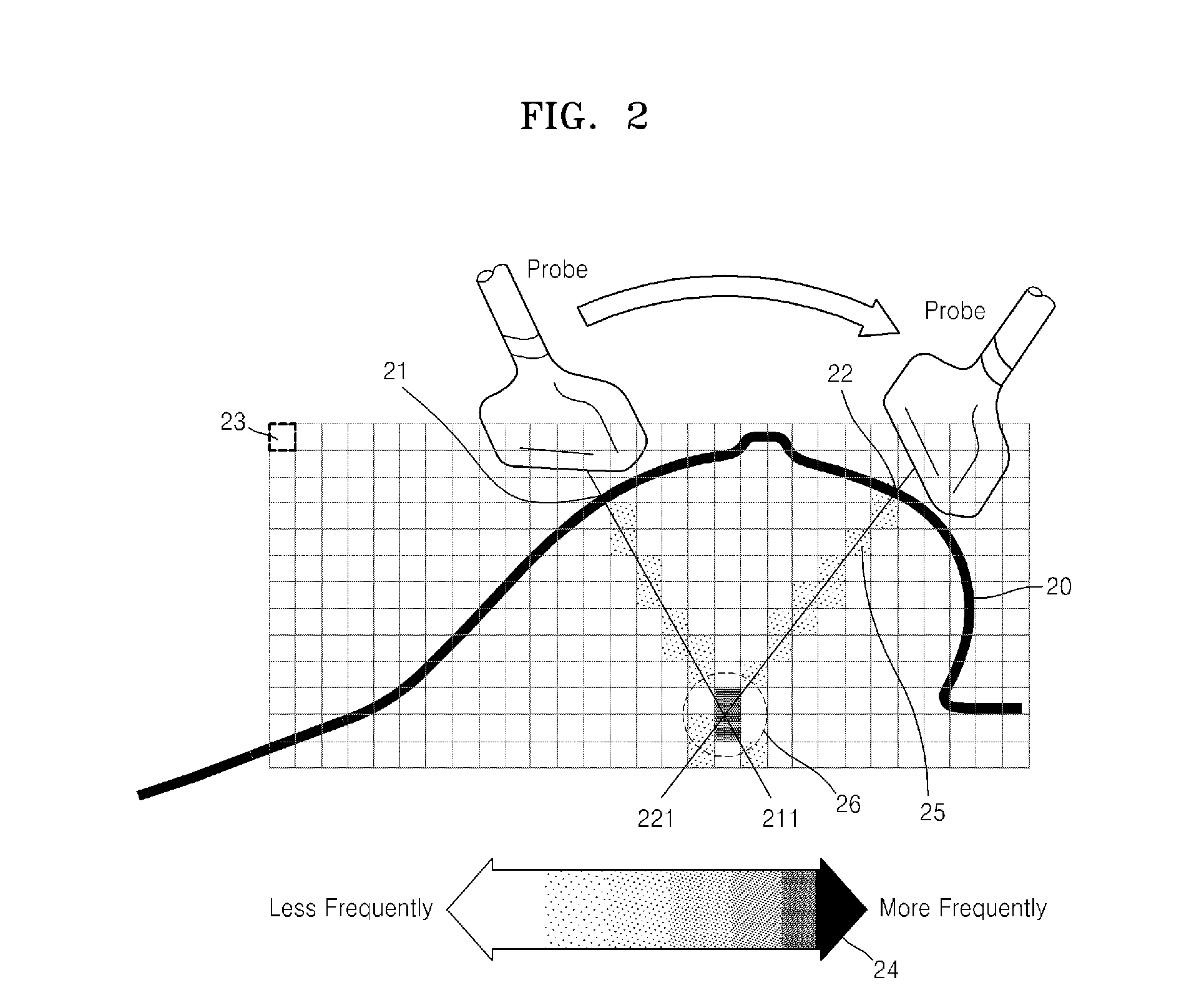
[0014]The method may further include providing information indicating that the examination regions of the diagnostic image contains an examination region having a collision frequency that is equal to or less than a threshold value to the user.

Problems solved by technology

In this case, during manipulation of the probe, the medical professional may inadvertently overlook some examination regions of the subject, or may not perfectly examine some examination regions, which may result in a misdiagnosis.

Method used


Image

  • Method of displaying diagnostic image, image processing device, and medical image system for performing the same
  • Method of displaying diagnostic image, image processing device, and medical image system for performing the same
  • Method of displaying diagnostic image, image processing device, and medical image system for performing the same

Examples


Embodiment Construction

[0038]The following detailed description is provided to assist the reader in gaining a comprehensive understanding of the methods, apparatuses, and / or systems described herein. Accordingly, various changes, modifications, and equivalents of the systems, apparatuses and / or methods described herein will be suggested to those of ordinary skill in the art. Also, descriptions of well-known functions and constructions may be omitted for increased clarity and conciseness.

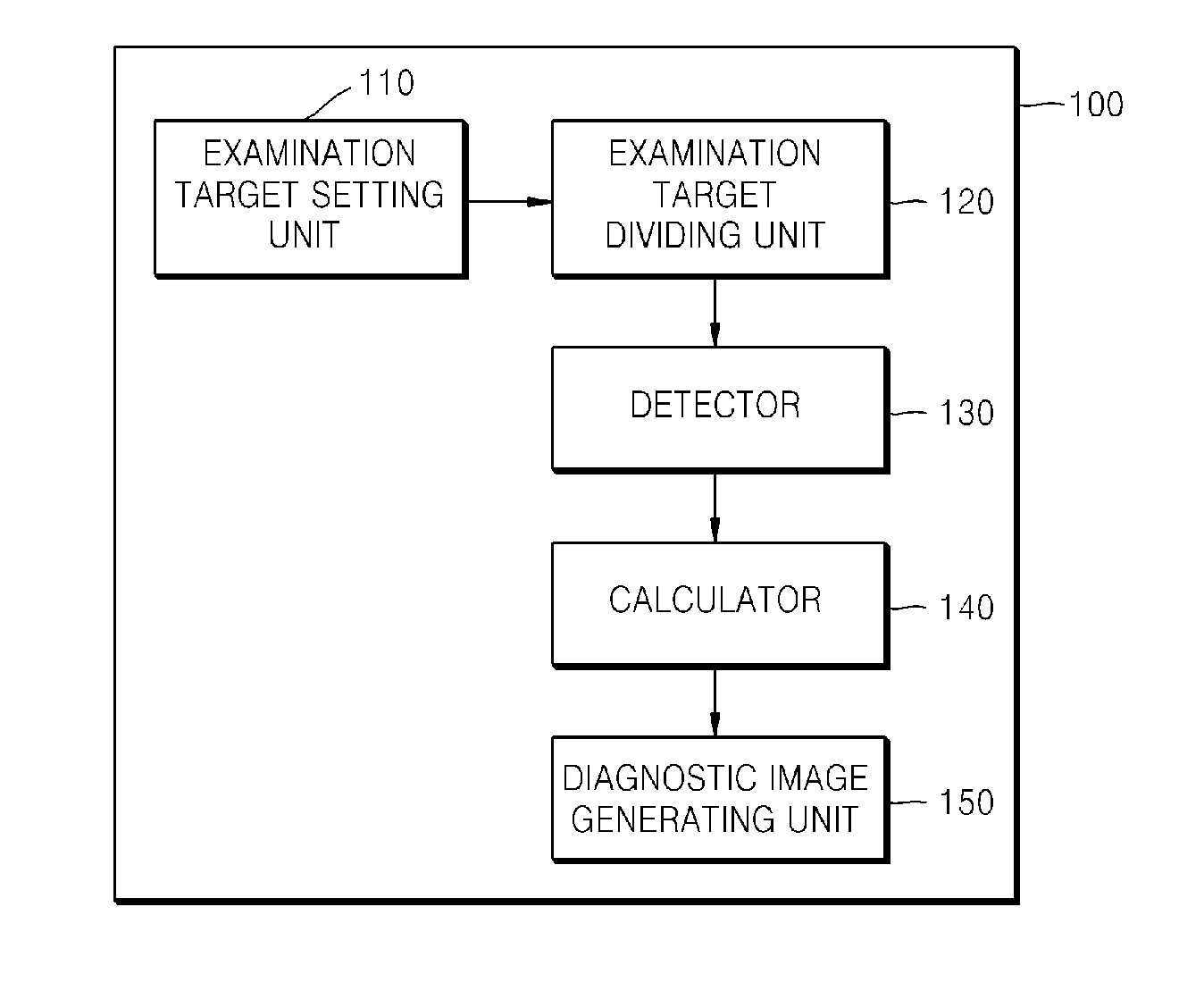
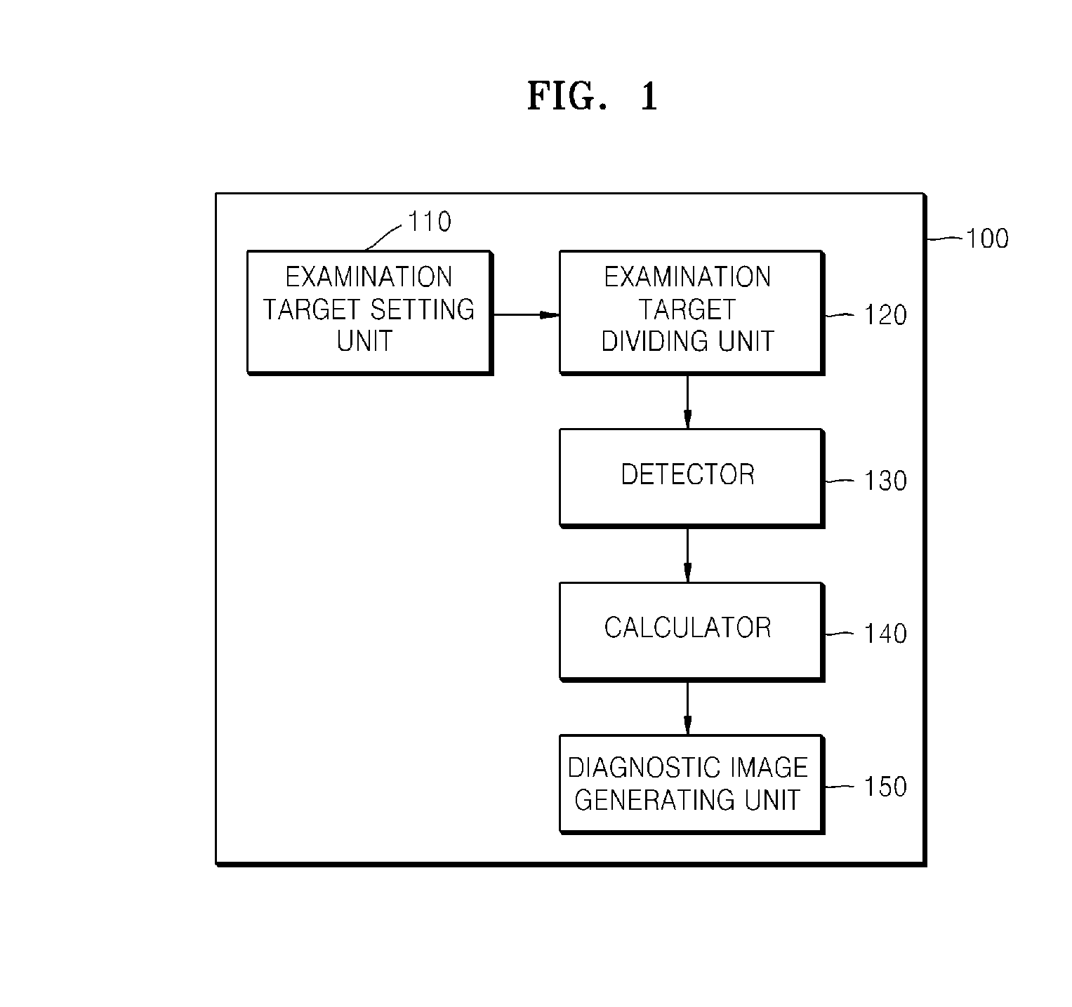
[0039]FIG. 1 is a diagram illustrating an example of an image processing device 100. Referring to FIG. 1, the image processing device 100 includes an examination target setting unit 110, an examination target dividing unit 120, a detector 130, a calculator 140, and a diagnostic image generating unit 150.

[0040]Elements related to the example are illustrated in FIG. 1. It is understood to those skilled in the art that other general elements may be further included.

[0041]The examination target setting unit 110, the examinatio...


PUM


Abstract

A method of displaying a diagnostic image is provided. The method includes setting an examination target of a subject, dividing the examination target into a plurality of examination regions, detecting whether a signal region indicated by a signal transmitted from a probe collides with each of the plurality of examination regions, calculating a collision frequency of each of the plurality of examination regions, based on a result of the detecting, and displaying a diagnostic image that indicates a sectional view of the subject and includes information indicating a degree of the collision frequency of each of the plurality of examination regions included in the sectional view, based on the calculation result.

Description

CROSS-REFERENCE TO RELATED APPLICATIONS[0001]This application claims the benefit of Korean Patent Application No. 10-2010-0134908, filed on Dec. 24, 2010, in the Korean Intellectual Property Office, the entire disclosure of which is incorporated herein by reference for all purposes.BACKGROUND[0002]1. Field[0003]The following description relates to a method of displaying a diagnostic image, an image processing device, and a medical image system for performing the same.[0004]2. Description of the Related Art[0005]A medical professional, such as a doctor, examines a subject by using a probe, and determines whether there is a lesion based on an image obtained from the probe. In this case, during manipulation of the probe, the medical professional may inadvertently overlook some examination regions of the subject, or may not perfectly examine some examination regions, which may result in a misdiagnosis.SUMMARY[0006]According to an aspect, a method of displaying a diagnostic image is prov...

Claims


Application Information

Patent Timeline
Patent Type & Authority Applications(United States)
IPC IPC(8): A61B8/14
CPCA61B8/0825A61B8/463A61B8/429A61B8/4254
Inventor PARK, MOON-HO
Owner SAMSUNG ELECTRONICS CO LTD
Learn More