Medical imaging apparatus and medical image diagnosis apparatus
a diagnostic apparatus and imaging technology, applied in the field of medical imaging apparatus and medical image diagnosis apparatus, can solve the problems of difficult to find the desired examination from among the examination records related to many patients, take time to find the desired shot, and not be able to sufficiently represent the characteristics of the examination in a single thumbnail, so as to achieve the effect of easy finding
- Summary
- Abstract
- Description
- Claims
- Application Information
AI Technical Summary
Benefits of technology
Problems solved by technology
Method used
Image

Examples
first embodiment
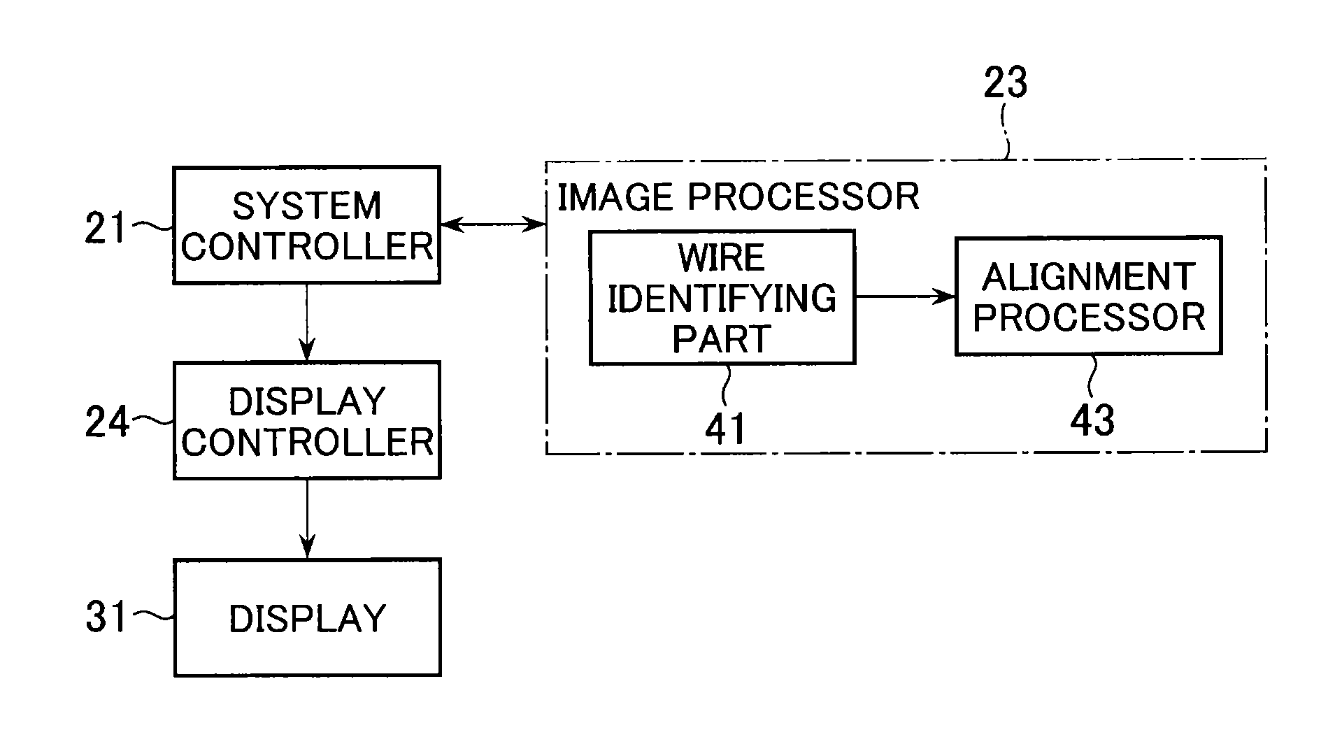
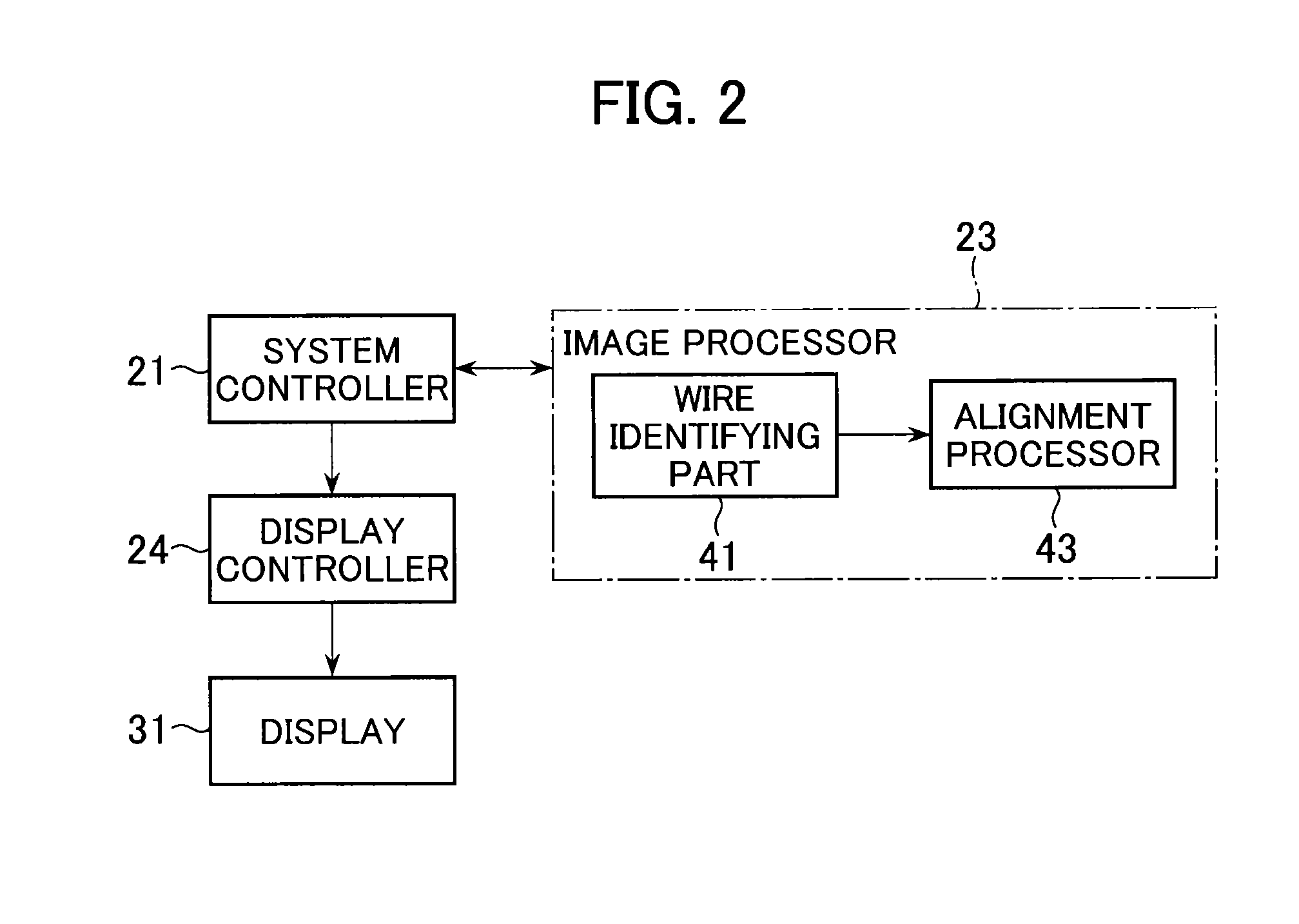
[0064]A first embodiment will be described. First, the configuration of a medical imaging apparatus for performing catheterization under X-ray fluoroscopy will be described. Next, an example will be described in which the amount of change in the shape of a wire is detected as the result of detecting the manipulated state of the wire handled by the operator during catheterization under X-ray fluoroscopy. Then, an example will be described in which the importance levels of X-ray images acquired during catheterization under X-ray fluoroscopy are obtained based on the detection results.
[Device configuration]
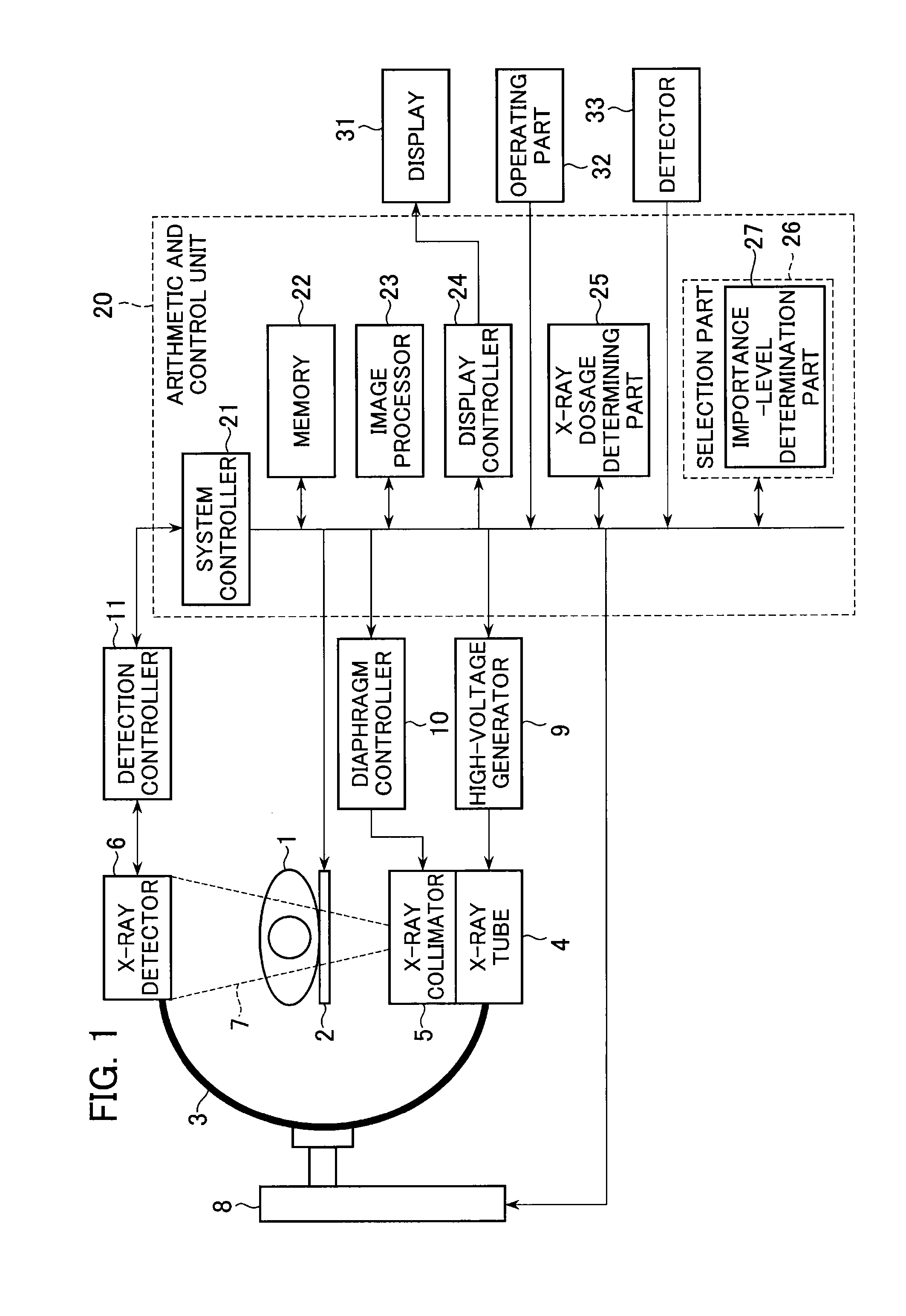
[0065]The configuration of the medical imaging apparatus according to the present embodiment will be described. An example configuration of this medical imaging apparatus (X-ray diagnosis apparatus) is shown in FIG. 1. This medical imaging apparatus has a mechanical composition similar to that of conventional examples.
[0066]The subject 1 represents the patient undergoing catheterizat...
second embodiment
[0168]In the first embodiment, the importance-level determination part 27 deduced each importance level using the size of the X-ray dosage, the acquisition period of the X-ray images, the amount of change in the pixel values of the X-ray images (administration of a contrast agent), and the amount of change in the shape of the wire as information for making a determination, but it is sufficient to use at least one of the manipulated state of an instrument (wire) handled by the operator, biological information of the operator, or the behavior of the operator as information for deducing importance levels. Next, other examples used as information for deducing importance levels will be described.
[0169]In the first embodiment, an example was described in which the results of detecting changes in the shape of the wire were used as information for determining whether or not the operator was manipulating the wire, but the results of detecting sounds or vibrations generated when the wire is m...
third embodiment
[0174]The medical imaging apparatus according to the second embodiment detects the manipulated state of an instrument (wire) handled by the operator, and using the detection results as information for making a determination, the importance-level determination part 27 deduces the importance levels of the X-ray images. In comparison, as other information for making a determination, the results of detecting the posture of the operator may be used. Furthermore, the importance-level determination part 27 may be configured to deduce the importance levels of the X-ray images using a synthesis of the detection results of the manipulated state of the instrument and the detection results of the posture of the operator as information for making a determination. Furthermore, the comprehensive importance levels may be determined by combining these deduced importance levels with other deduced importance levels. In this way, by combining greater numbers of deduced importance levels, the correspond...
PUM
 Abstract
Description
Claims
Application Information
 - R&D
- Intellectual Property
- Life Sciences
- Materials
- Tech Scout
- Unparalleled Data Quality
- Higher Quality Content
- 60% Fewer Hallucinations
Browse by: Latest US Patents, China's latest patents, Technical Efficacy Thesaurus, Application Domain, Technology Topic, Popular Technical Reports.
© 2025 PatSnap. All rights reserved.Legal|Privacy policy|Modern Slavery Act Transparency Statement|Sitemap|About US| Contact US: help@patsnap.com