Drug delivery from rapid gelling polymer composition

a technology of gelling polymer and polymer composition, which is applied in the direction of drug compositions, aerosol delivery, extracellular fluid disorder, etc., can solve the problems of adversely affecting tissue surfaces, worsening tissue inflammation at the site of administration,

Inactive Publication Date: 2012-10-04
ANGIOTECH INT AG (CH)
View PDF6 Cites 5 Cited by
  • Summary
  • Abstract
  • Description
  • Claims
  • Application Information

AI Technical Summary

Problems solved by technology

One difficulty encountered when designing in situ hydrogel forming compositions is that optimizing the composition to enhance gel formation may worsen tissue inflammation at the site of administration.
A possible explanation for this effect is that highly reactive composition components that are capable of rapid gel formation may adversely affect tissue surfaces.

Method used


Image

  • Drug delivery from rapid gelling polymer composition
  • Drug delivery from rapid gelling polymer composition
  • Drug delivery from rapid gelling polymer composition

Examples


example 1

Preparation of a Two-Component Tissue Sealant Composition

[0375]a. First Component

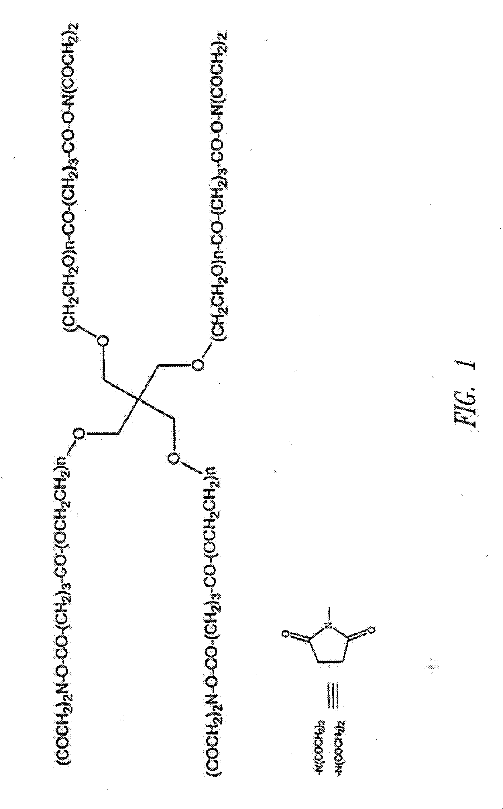
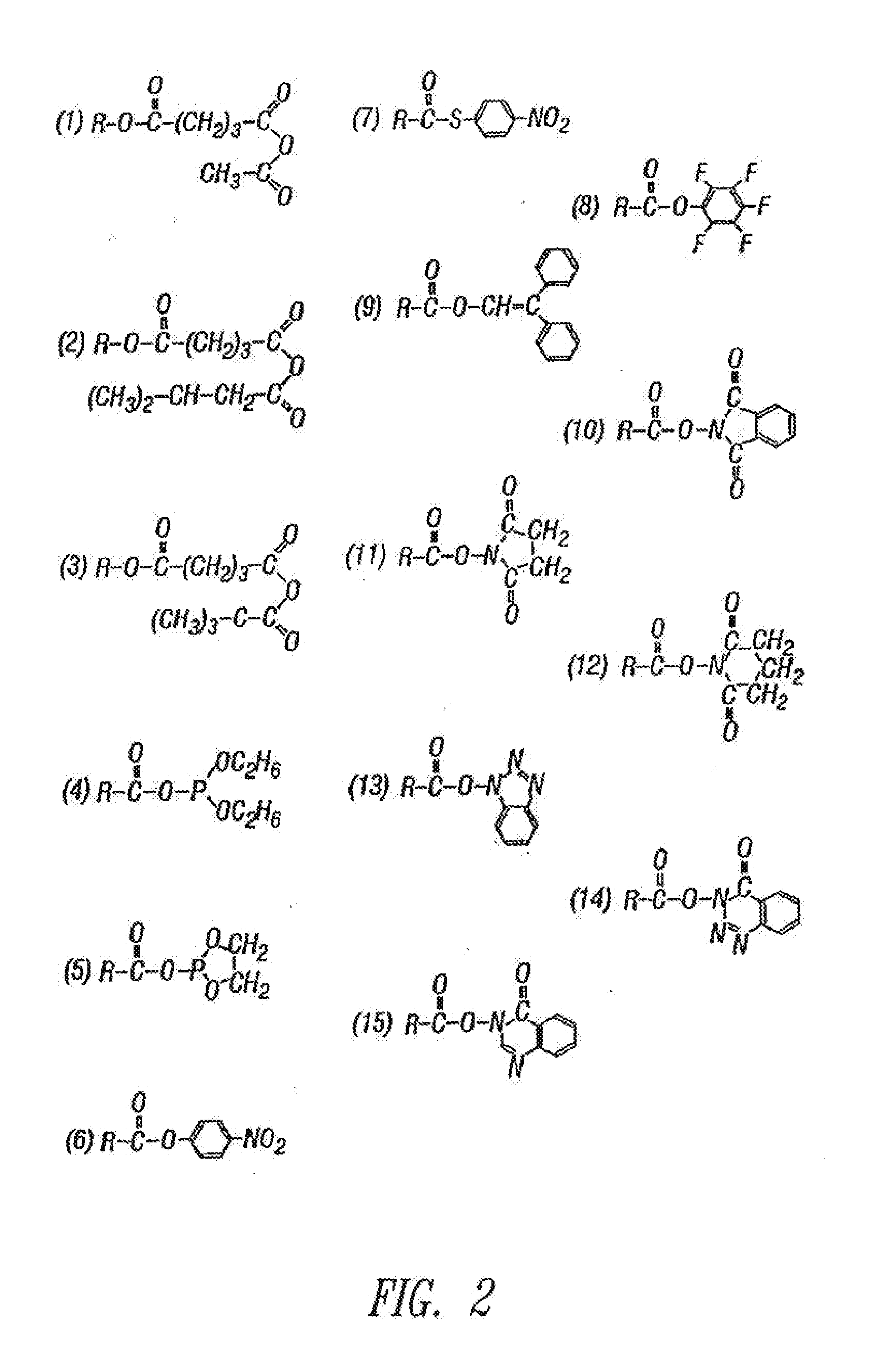
[0376]Pentaerythritol poly(ethylene glycol)ether tetra-succinimidyl glutarate (“SG-PEG”) (mol. wt. 10,000) is dissolved in 0.5 mM sodium phosphate pH 6.0 at a concentration of 20% w / v. (This solution is not stable in aqueous media due to the susceptibility of the active ester to hydrolysis and should be used within one hour of preparation).

[0377]b. Second Component

[0378]Pentaerythritol poly(ethylene glycol)ether tetra-sulfhydryl (mol. wt. 10,000) is dissolved in 300 mM sodium phosphate / sodium carbonate buffer (“P / C buffer”), pH 9.6, at a concentration of 20% w / v. P / C buffer is prepared as follows: 300 mM sodium monobasic phosphate is mixed with 300 mM sodium carbonate to achieve pH 9.6. The final molarity is approximately 117 mm phosphate and 183 mM carbonate. This solution is stable in aqueous media, but care should be taken to prevent the exposure of the solution to oxygen to prevent oxidation to disu...

example 2

Surgical Sealing of Arteries

[0379]The right carotid artery of New Zealand white rabbits is exposed. The rabbits are treated with 200 U / kg of heparin and the vessel is clamped proximally and distally using atraumatic vascular clamps. A puncture hole is made in the carotid artery using a 27G needle. The control rabbits are treated with tamponade until hemostasis is achieved. For the treated rabbits, approximately 0.5 mL of each of the two components of the compositions prepared as described in Example 1 are delivered to the defect site using a two component sprayer (Duo Flow, Hemaedics, Malibu, Calif.). After the material is allowed to set for 30 sec, the clamps are removed and the time to hemostasis and the blood loss are measured. The arteries of the control rabbits also remain clamped for 30 sec for consistency. The results are shown in Table 1.

TABLE 1Blood Loss and Time to Hemostasis as a Function of TreatmentTreatmentBlood Loss (g)Time to Hemostasis (sec)Tamponade (n = 18)5.7 ± 3...

example 3

Surgical Sealing of an ePTFE Graft

[0381]The dogs are treated with heparin to achieve an activated clotting time of greater than 480 sec. The left iliac of the dogs is exposed and isolated using atraumatic vascular clamps placed distally and proximally. A 5 cm segment of the artery is excised and replaced with an ePTFE (polythetrafluoroethylene) graft of the same diameter. Prior to the completion of the anastamosis, the graft was de-aired using a 27G needle. Approximately 3.0 mL of each of the two components of the composition prepared according to Example 1 is delivered to the defect site using a two component sprayer (Cohesion Technologies, Inc., Palo Alto, Calif.). After the material is allowed to set for 30 sec, the clamps are removed and the time to hemostasis and the blood loss are measured. The procedure was repeated on the left iliac, with the exception of material application. The right iliac received only tamponade treatment. The results are shown in Table 2.

TABLE 2Blood Lo...


PUM

PropertyMeasurementUnit
biocompatibleaaaaaaaaaa
hydrophobicaaaaaaaaaa
pHaaaaaaaaaa

Abstract

Compositions are disclosed that afford drug delivery from two-part polymer compositions that rapidly form covalent linkages when mixed together. Such compositions are particularly well suited for use in a variety of tissue related applications when rapid adhesion to the tissue and gel formation is desired along with drug delivery. For example, the compositions are useful as tissue sealants, in promoting hemostasis, in effecting tissue adhesion, in providing tissue augmentation, and in the prevention of surgical adhesions.

Description

CROSS REFERENCE TO RELATED APPLICATIONS[0001]This application is a continuation of U.S. patent application Ser. No. 12 / 259,916, filed Oct. 28, 2008, now pending; which is a continuation of U.S. patent application Ser. No. 10 / 749,117, filed Dec. 30, 2003, now abandoned; which claims the benefit under 35 U.S.C. §119(e) of U.S. Provisional Patent Application No. 60 / 437,471, filed Dec. 30, 2002, and U.S. Provisional Patent Application No. 60 / 440,875, filed Jan. 17, 2003, which applications are included herein by reference in their entirety.BACKGROUND OF THE INVENTION[0002]1. Field of the Invention[0003]This invention relates generally to compositions that afford drug delivery from two-part polymer compositions that rapidly form covalent linkages when mixed together. Such compositions are particularly well suited for use in a variety of tissue related applications when rapid adhesion to the tissue and gel formation is desired along with drug delivery. For example, the compositions are us...

Claims


Application Information

Patent Timeline
Patent Type & Authority Applications(United States)
IPC IPC(8): A61K47/34A61P7/04A61K9/06
CPCA61K9/0024A61K9/06A61L2300/434A61L31/16A61L31/145A61K9/1647A61K31/337A61K31/365A61K31/54A61K47/34A61L31/041C08L71/02C08L89/06A61K47/6903A61P35/00A61P43/00A61P7/04A61P9/00A61K51/1213
Inventor GRAVETT, DAVID M.TAKACS-COX, ANIKOTOLEIKIS, PHILIP M.MAITI, ARPITAEMBREE, LEANNE
Owner ANGIOTECH INT AG (CH)
Why Eureka
Try Eureka