Medical examination apparatus

a medical examination and apparatus technology, applied in the field of medical examination apparatus, can solve problems such as the triggering of emergency calls by patients, and achieve the effects of avoiding unnecessary delays, preventing effective triggering, and facilitating rapid respons

Active Publication Date: 2013-05-02
SIEMENS HEALTHCARE GMBH
View PDF3 Cites 7 Cited by
  • Summary
  • Abstract
  • Description
  • Claims
  • Application Information

AI Technical Summary

Benefits of technology

[0007]The alarm signal emitter also has an alarm button for a patient, the actuation of which triggers the alarm, generating a corresponding alarm signal and routing it to the control unit. During an examination, for example using a computed tomography unit, it is at present normal for the patient to be given a device to hold, which can be used for an “emergency call”. By actuating said device, also referred to as a “call ball”, the operator of the computed tomography unit, who is typically in a different room during the examination, is alerted for example acoustically or visually to the emergency call, so that he / she can take appropriate measures, for example consulting the patient by way of a speaker device or terminating the ongoing examination. However termination then takes place with a time delay that is dependent on the operator, which is particularly undesirable when a contrast agent is used during the examination. Such contrast agents are generally injected into the body of the patient during the examination with the aid of the controllable injection apparatus, which is embodied for example as an injection pump or dosing pump. If the patient is unable to tolerate the contrast agent and side effects occur, the patient will trigger an emergency call. However until the operator terminates the examination and in particular deactivates the injection apparatus, the injection of the contrast agent continues. This undesirable delay can be avoided with the alarm button, which is positioned with the patient at least during the examination and is configured for example in the manner of a switch and in particular according to the principle of an “emergency off” switch. With an inventive automated response of the medical examination apparatus to the alarm triggered by actuation of the alarm button and therefore by the patient, the response is much more rapid and therefore the termination of the injection process is much more rapid, so the resulting dose of contrast agent is much smaller.
[0008]The alarm button and a system according to the principle of the “call ball” can also advantageously be combined, so that the patient can either contact the operator of the medical examination apparatus by “emergency call” or trigger the alarm directly, depending on the situation.
[0009]The alarm button is also integrated in a simple operating element with just a few functions or even just one “trigger alarm” function, which the patient can easily hold during an examination and which is therefore neither part of an operating console for the image-generating modality nor part of an operating console for the controllable injection apparatus nor part of an operating console for the medical examination apparatus. Alternatively such a simple operating element can be positioned in easy reach of the patient during an examination, in other words for example on or next to a patient couch.
[0010]A variant includes an evaluation unit for evaluating the image data generated by means of the image-generating modality is used as an additional alarm signal emitter. The image data is analyzed and compared with stored reference data, with a correspondence between image data and reference data triggering an alarm, whereupon the evaluation unit generates an alarm signal and routes it to the control unit. If the time pattern of an image signal is monitored for example in the region of the heart of the patient, while a contrast agent is injected into a vein at the same time, the contrast agent should reach the heart after a certain time and produce a change in the image signal there. If this change in the image signal does not occur, it is for example possible that the injection is not taking place into the vein as intended but into the surrounding tissue. Such an undesirable injection of the contrast agent into the tissue, referred to as paravasation, is potentially harmful for the patient and should therefore be effectively prevented. Therefore if no image signal change takes place after a predefined time interval in the monitored or examined region of the patient, the evaluation unit triggers the alarm and generates a corresponding alarm signal, which in turn prompts the control unit to deactivate the injection apparatus and thus prevent the further injection of contrast agent. Since the incorrect injection of contrast agent can generally only be perceived after a certain time by both operator and patient, additional automated monitoring by means of the image-generating modality and an evaluation unit, which is also preferably part of the image-generating modality, further improves patient safety.

Problems solved by technology

However termination then takes place with a time delay that is dependent on the operator, which is particularly undesirable when a contrast agent is used during the examination.
If the patient is unable to tolerate the contrast agent and side effects occur, the patient will trigger an emergency call.

Method used


Image

  • Medical examination apparatus
  • Medical examination apparatus

Examples


Embodiment Construction

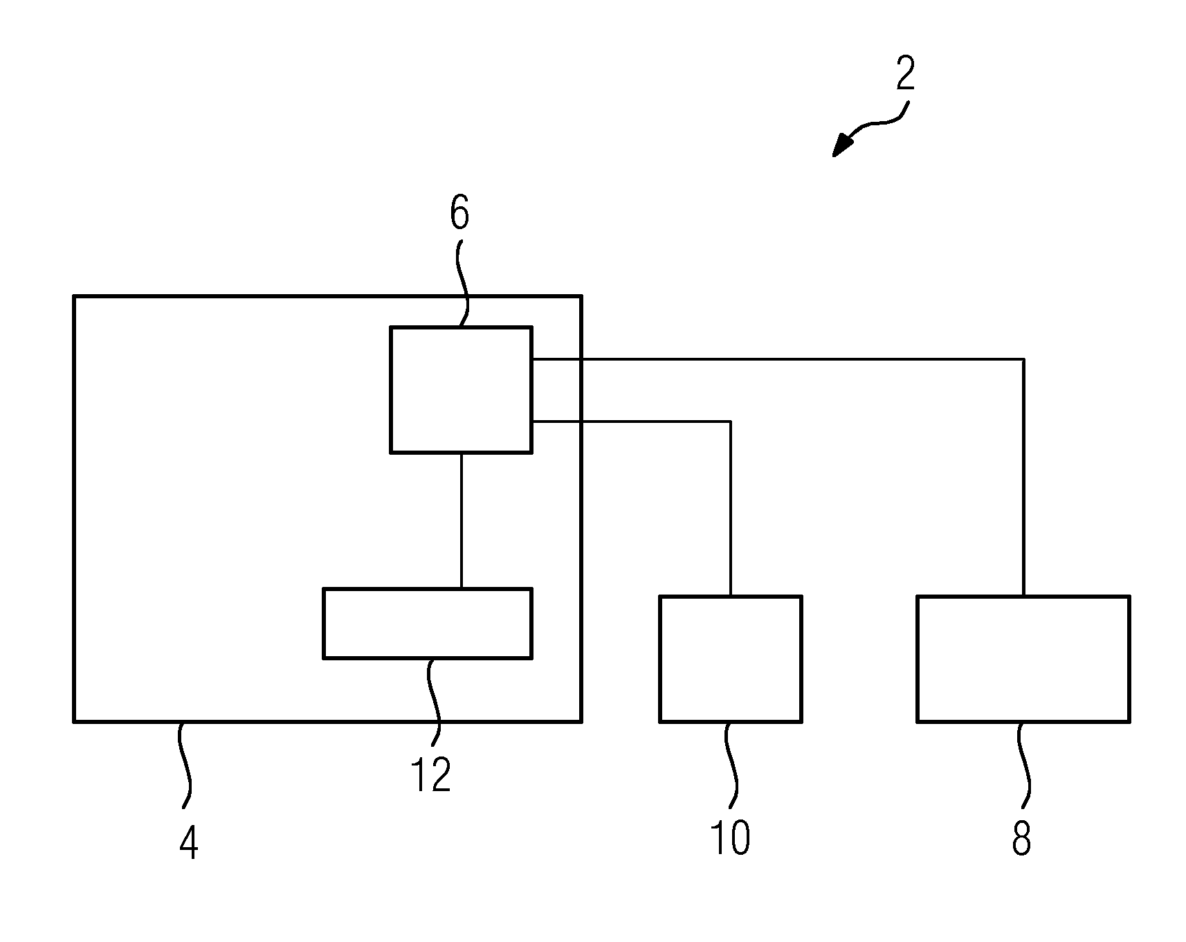
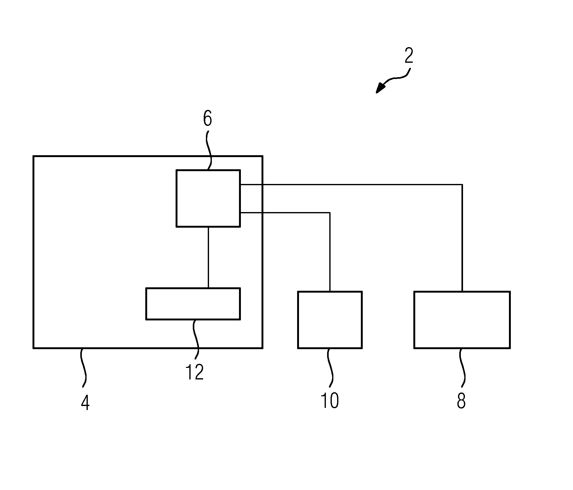
[0021]In the case of the medical examination apparatus 2 described below and illustrated in FIG. 1 a computed tomography unit 4 with a central control unit 6 is used to examine a patient using an imaging method. Depending on the examination method selected, provision is made for a contrast agent to be injected into the patient to assist the imaging method. For this purpose the medical examination apparatus 2 comprises an injection apparatus 8, which is preferably configured as an injection pump, also referred to as a dosing pump.

[0022]The injection apparatus 8 used in the exemplary embodiment is a controllable injection apparatus 8, which is connected for signaling purposes to the control unit 6 of the computed tomography unit 4 and which is activated by way of the control unit 6. This tailors the injection of the contrast agent to the respective selected image-generation method, in order thereby to influence the quality of the image data to be generated in a positive manner.

[0023]B...


PUM


Abstract

A medical examination apparatus including a control unit, an image-generating modality and a controllable injection apparatus for a contrast agent is provided. The control unit is connected to an alarm signal emitter and the control unit switches the modality and the injection apparatus to a safe mode as a function of an alarm signal.

Description

CROSS REFERENCE TO RELATED APPLICATIONS[0001]This application claims priority of European Patent Office application No. 10 2011 085 597.1 DE filed Nov. 2, 2011. All of the applications are incorporated by reference herein in their entirety.FIELD OF INVENTION[0002]The invention relates to a medical examination apparatus having a control unit, having an image-generating modality and having a controllable injection apparatus for a contrast agent.BACKGROUND OF INVENTION[0003]If a problem occurs when a patient is being examined using an imaging modality, it is generally desirable to terminate the ongoing examination. With the modalities currently used provision is generally made for such a termination of the ongoing examination to be performed by an operator, for example a medical technical assistant (MTA), as soon as he / she identifies a problem or judges a situation to be problematic.[0004]A medical imaging apparatus having an image generation unit is also known from WO 00 / 64353, in whi...

Claims


Application Information

Patent Timeline
Patent Type & Authority Applications(United States)
IPC IPC(8): A61B6/00
CPCA61M5/484A61B6/10A61B6/032A61B6/541A61M5/007A61B6/481
Inventor ASSMAN, STEFANHEISMANN, BJORNMERGES, RETOSCHMIDT, MARKUSSCHMIDT, SEBASTIANWESTPHAL, KERA
Owner SIEMENS HEALTHCARE GMBH
Why Eureka
Try Eureka