Image stitching method and camera system

Active Publication Date: 2014-07-17
ALTEK BIOTECH
View PDF10 Cites 26 Cited by
  • Summary
  • Abstract
  • Description
  • Claims
  • Application Information

AI Technical Summary

Benefits of technology

[0006]Accordingly, the invention is directed to an image stitchi

Problems solved by technology

When a vascular hyperplasia or rupture occurs on the fundus (for example, macular degeneration), the cone and rod cells in the retina may die and accordingly the patient may lose his or her eyesight.
However, because these fundus images are not captured all together, they have different exposure values and white balance values.
In addition, the fundus image

Method used


Image

  • Image stitching method and camera system
  • Image stitching method and camera system
  • Image stitching method and camera system

Examples


Embodiment Construction

[0045]Reference will now be made in detail to the present preferred embodiments of the invention, examples of which are illustrated in the accompanying drawings. Wherever possible, the same reference numbers are used in the drawings and the description to refer to the same or like parts.

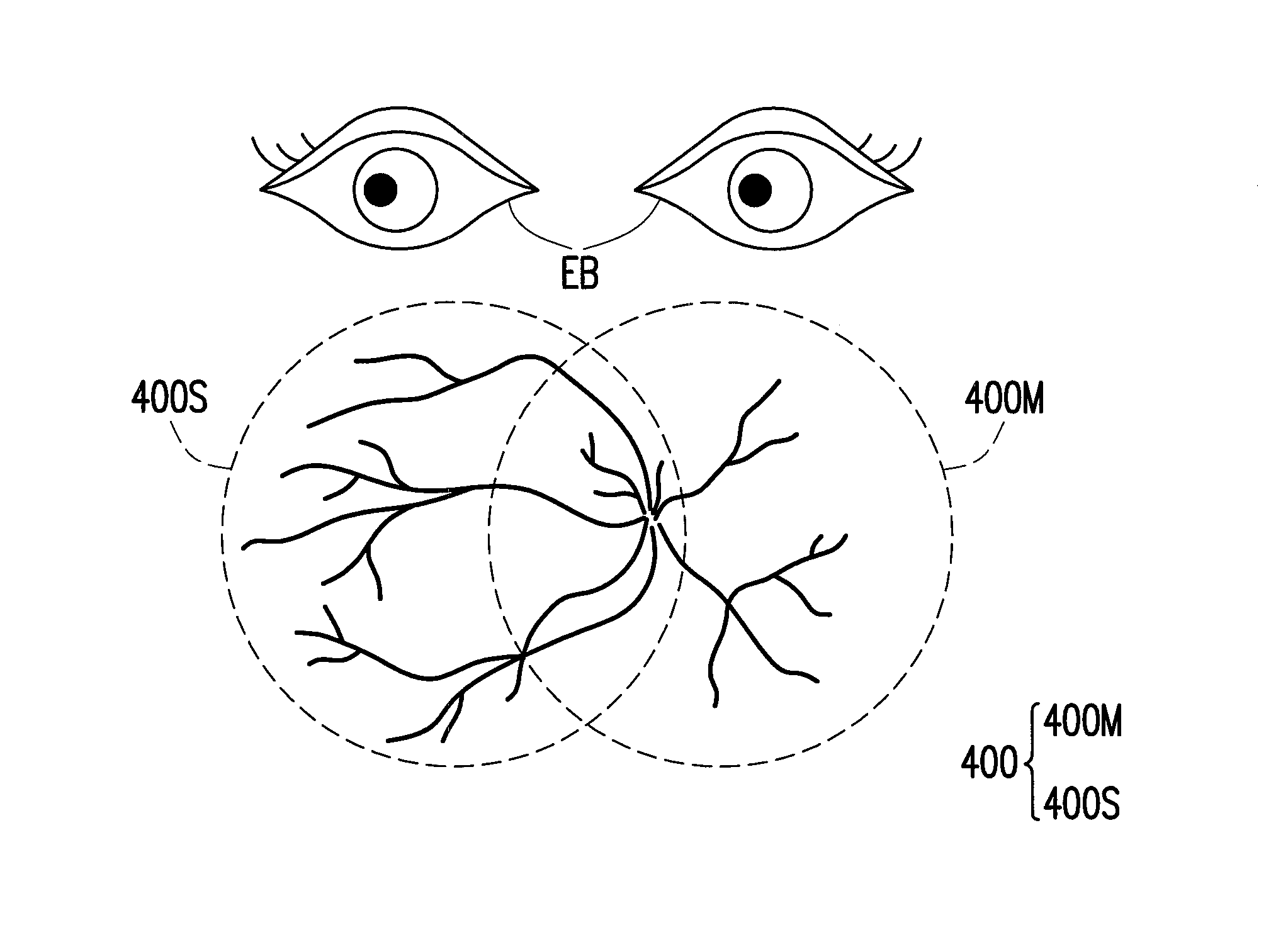
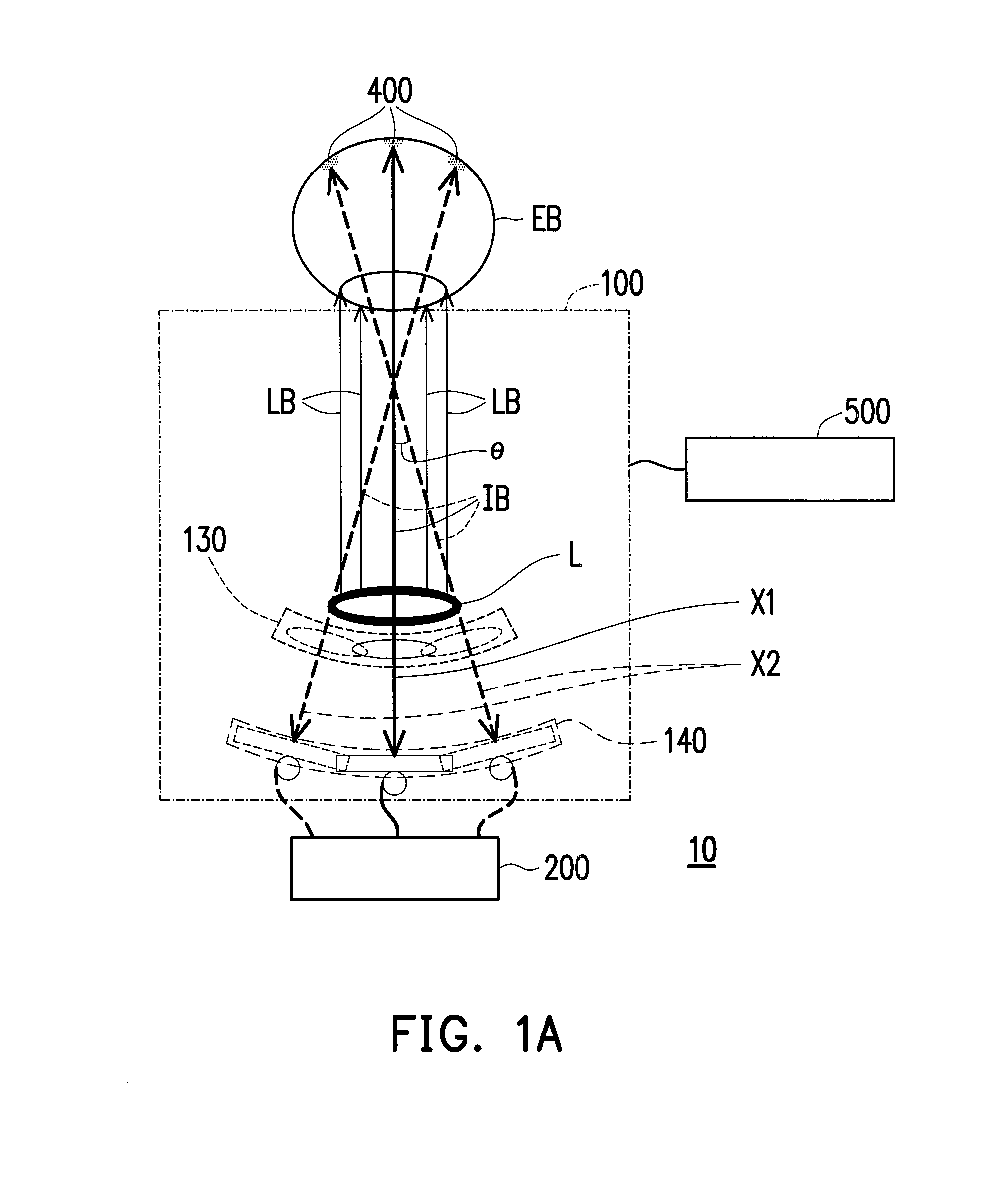
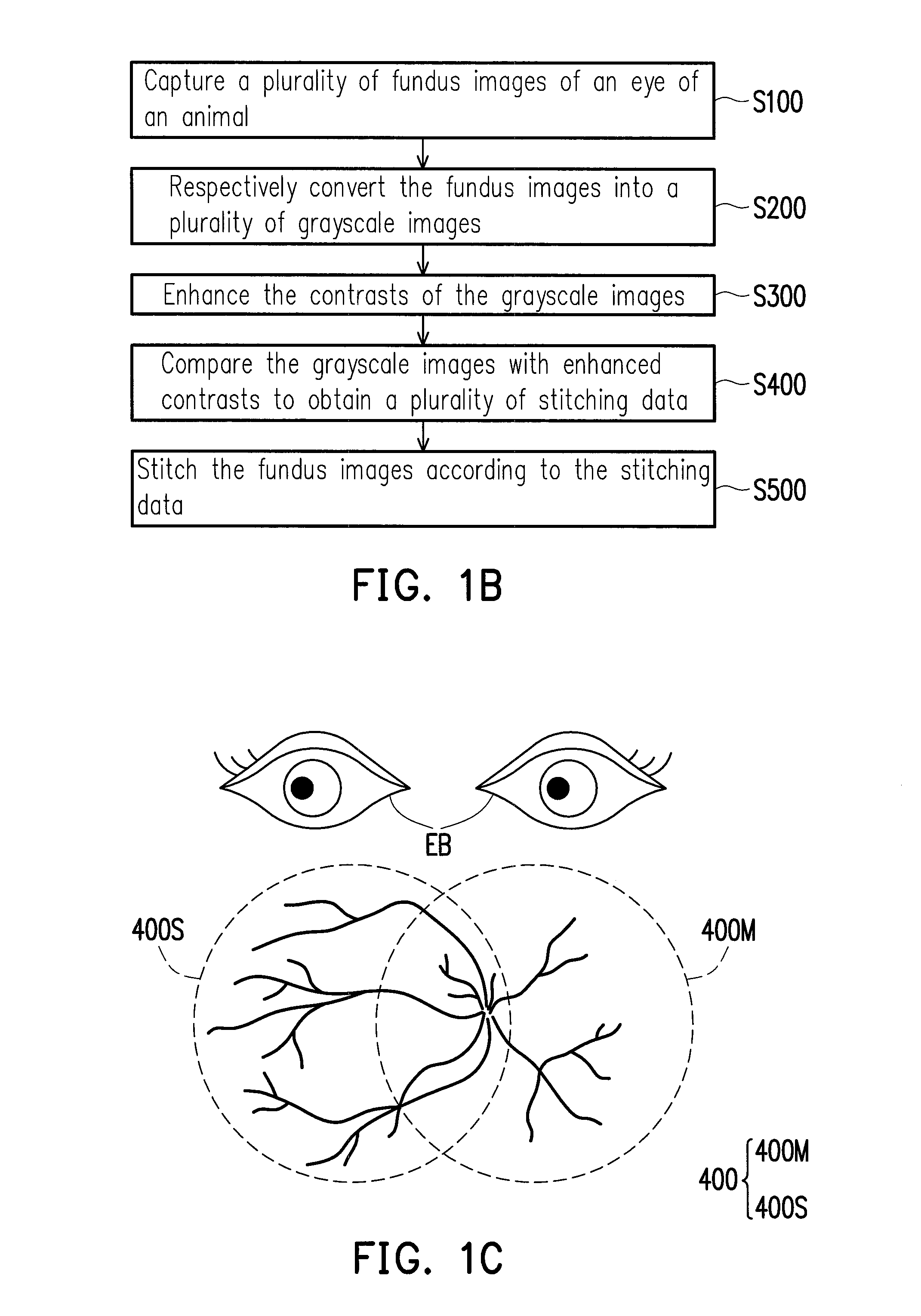
[0046]FIG. 1A is a diagram of a camera system according to an embodiment of the invention. Referring to FIG. 1A, in the present embodiment, the camera system 10 includes a camera unit 100 and a processing unit 200. The camera unit 100 captures a plurality of fundus images 400 of an animal (i.e. images of the fundus of the eye of the animal), where the fundus images 400 partially overlap each other. In the present embodiment, the fundus images 400 are full-color images. However, the invention is not limited thereto, and in other embodiments, the fundus images 400 may also be color or grayscale images obtained through infrared photography or any other technique. The processing unit 200 is electrically ...


PUM


Abstract

An image stitching method adapted to stitch a plurality of fundus images of an animal is provided. The image stitching method includes following steps. A plurality of fundus images of the animal are captured, where the fundus images partially overlap. The fundus images are respectively converted into a plurality of grayscale images. The contrasts of the grayscale images are enhanced. The grayscale images with enhanced contrasts are compared to obtain a plurality of stitching data. The fundus images are stitched according to the stitching data. Additionally, a camera system is also provided.

Description

BACKGROUND OF THE INVENTION[0001]1. Field of the Invention[0002]The invention generally relates to an image processing method and an optical system, and more particularly, to a method of stitching fundus images of an eye of an animal and a camera system.[0003]2. Description of Related Art[0004]Eyes are the window to the soul, and people see the world through their eyes. The cone and rod cells responsible for color and light reception in the eye are located in the retina at the fundus and are the photoreceptors for converting light into physiological electrical signals in the human body. The blood vessels for supplying blood and nutrients to the eye are also located at the fundus. When a vascular hyperplasia or rupture occurs on the fundus (for example, macular degeneration), the cone and rod cells in the retina may die and accordingly the patient may lose his or her eyesight. Thus, it is very important to observe and track fundus images in the diagnosis and prevention of eye disease...

Claims


Application Information

Patent Timeline
IPC IPC(8): A61B3/14
CPCA61B3/14A61B3/0025
Inventor CHENG, SHIN-HAOCHEN, PIN-WEN
Owner ALTEK BIOTECH
Why Eureka
Try Eureka