Method for medical image processing

a medical imaging and processing technology, applied in the field of medical imaging devices, can solve the problems of interruption in concentration, inability to use input peripherals, and inability to meet the needs of practitioners, and achieve the effect of less memory and execution time resources

Active Publication Date: 2016-11-03
GENERAL ELECTRIC CO
View PDF9 Cites 1 Cited by
  • Summary
  • Abstract
  • Description
  • Claims
  • Application Information

AI Technical Summary

Benefits of technology

[0018]Embodiments of the invention allow for the detection of areas which have been examined for a long time by the practitioner, and automatic triggering of the local processing algorithm. Moreover, as this processing is confined to a portion of the medical image, it consumes less memory and execution time resources when it is applied by an image processing module.

Problems solved by technology

However, these input peripherals are not always practical for use, and their use may interrupt the concentration of the practitioner.
Such an interruption in concentration extends the duration of use of the imaging device, and therefore the total duration of a medical intervention in progress made by the practitioner.
Further, such an interruption in concentration may be a problem in the case when the imaging device is used while the practitioner is operating on a patient.
Further, the imaging systems do not traditionally have access to the information indicating what is of interest for the physician, in particular in each of the acquired images or reviewed.
This limits the display optimizations which may result from this.

Method used


Image

  • Method for medical image processing
  • Method for medical image processing
  • Method for medical image processing

Examples


Embodiment Construction

Medical Imaging Device

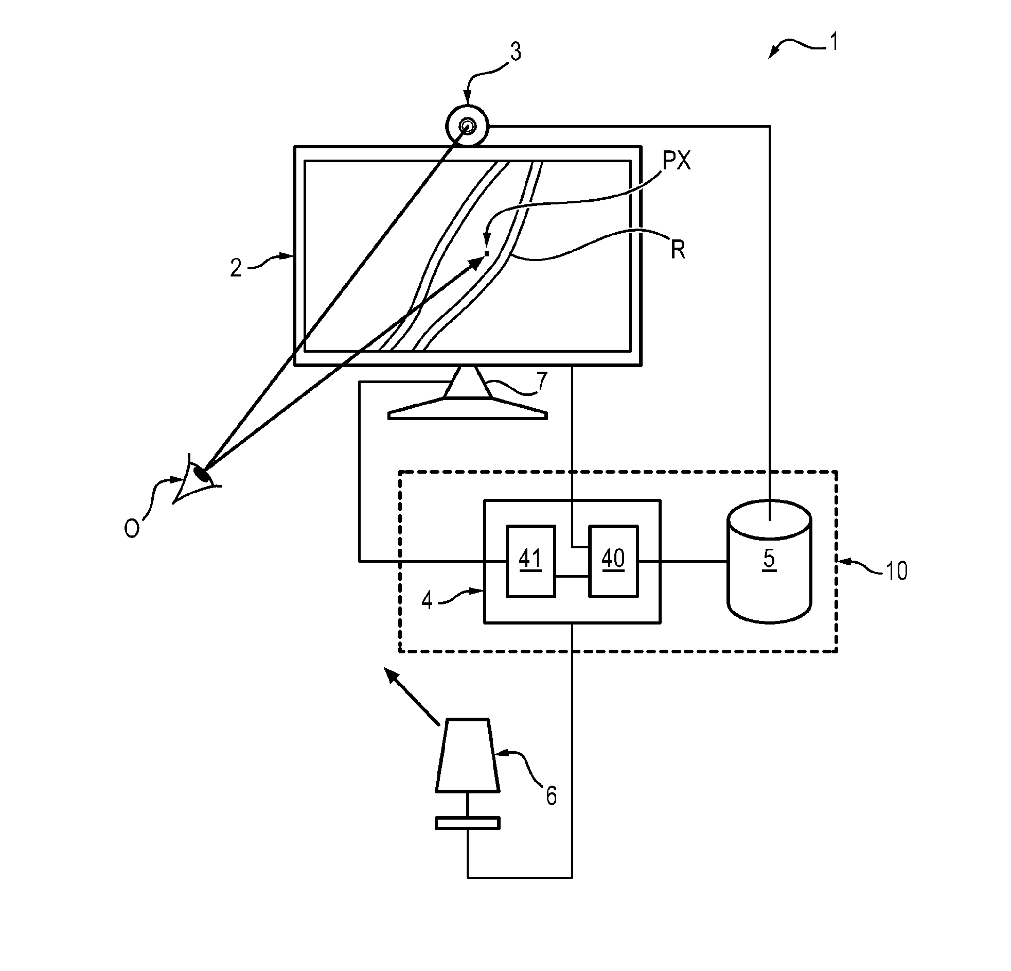
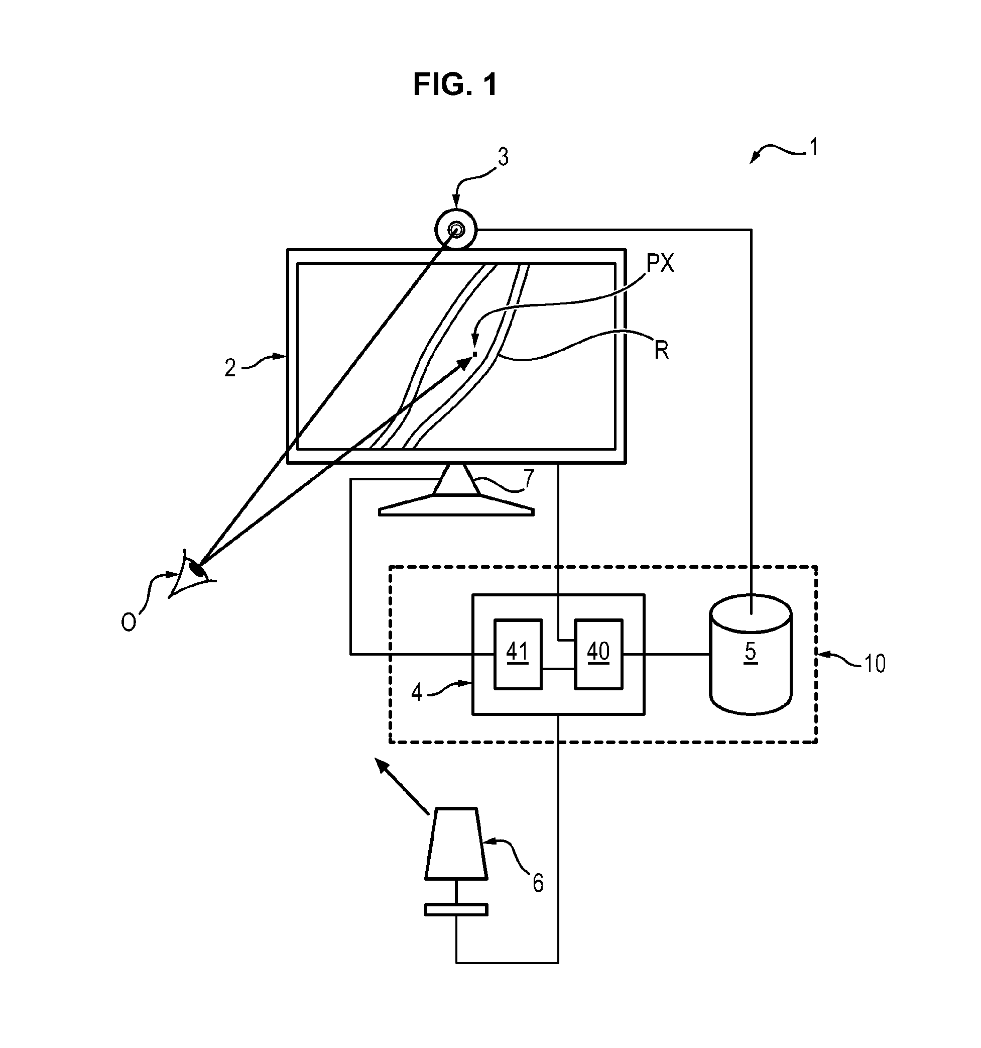
[0031]With reference to FIG. 1, a medical imaging device 1 comprises a display screen 2, a camera 3, an image processing module 4 and a data storage module 5.

[0032]The screen 2 and the camera 3 are laid out so as to be facing a practitioner 0 simultaneously (one eye of whom is schematically illustrated in FIG. 1). The camera 3 may be positioned fixedly relatively to the screen 3, for example above an upper edge of the screen 1.

[0033]The screen 2 is connected to the image processing module 4 and is adapted for displaying images stemming from this image processing module 4.

[0034]The camera 3 is adapted for acquiring images and transferring them to the image processing module 4.

[0035]The image processing module 4 typically comprises at least one processor 40 suitable for applying a gaze tracking algorithm, and at least one memory unit 41 suitable for storing in memory, images to be processed by the processor 40 or already processed by the processor 41, for example...


PUM


Abstract

An image processing method applied by a medical imaging device is proposed, including at least one camera and at least one display screen on which is displayed a medical image showing a region of interest of a patient, the method being characterized in that the region of interest is determined by the following steps, the screen and the camera being laid out so as to be facing a practitioner simultaneously, acquisition by the camera of at least one tracking image containing the eyes of the practitioner, analysis of the tracking image so as to localize in the medical image at least one pixel of interest contemplated by the practitioner on the screen from the orientation of the eyes of the practitioner, processing the medical image so as to select the region of interest from the localized pixel of interest.

Description

TECHNICAL FIELD[0001]Embodiments of the invention relate to the field of medical imaging devices and to that of image processing methods allowing an improvement in the ergonomics of such devices.STATE OF THE ART[0002]The use of imaging devices has become more democratic in the medical field, notably within the scope of interventional radiology.[0003]Medical imaging devices comprise a display screen for viewing an image containing a portion of the body of a patient, and at least one input peripheral intended to be manipulated by a practitioner for selecting an area of interest of the patient, viewed on the screen, or for starting applications requiring interaction with the image such as clicks on particular locations (a mouse, a keyboard, or the actual screen if the latter comprises a touchpad).[0004]However, these input peripherals are not always practical for use, and their use may interrupt the concentration of the practitioner.[0005]Such an interruption in concentration extends t...

Claims


Application Information

Patent Timeline
Patent Type & Authority Applications(United States)
IPC IPC(8): A61B90/00G06T3/40G06T7/00G06T5/00A61B34/00G06F3/01
CPCA61B90/361A61B34/25A61B90/37G06F3/013G06T7/0012G06T7/0044G06T2200/32G06T3/4038G06T2207/20104G06T2207/30201G06T2207/20208G06T2207/20221G06T5/008A61B2017/00216G06T7/74
Inventor BISMUTH, VINCENTVAILLANT, REGISGORGES, SEBASTIENCAZALAS, MAXIMERAMUS, LILIANE
Owner GENERAL ELECTRIC CO
Why Eureka
Try Eureka