Targeting of melanocytes for delivering therapeutic or diagnostic agents using protein nanocages

a technology of protein nanocages and melanocytes, which is applied in the direction of peptide/protein ingredients, peptide sources, drug compositions, etc., can solve the problems of current therapies, pigmentation disorders such as melasma, and suffer from current therapies

Active Publication Date: 2019-01-31
NANYANG TECH UNIV +1
View PDF1 Cites 3 Cited by
  • Summary
  • Abstract
  • Description
  • Claims
  • Application Information

AI Technical Summary

Benefits of technology

[0019]In yet another aspect, encompassed in the invention is a method of diagnosing hyperpigmentation disorders or other melanocyte-related disorders in a subject,

Problems solved by technology

However, hyper-expression of melanin in humans is known to result in pigmentation disorders such as melasma.
However, current therapies suffe

Method used


Image

  • Targeting of melanocytes for delivering therapeutic or diagnostic agents using protein nanocages
  • Targeting of melanocytes for delivering therapeutic or diagnostic agents using protein nanocages
  • Targeting of melanocytes for delivering therapeutic or diagnostic agents using protein nanocages

Examples


example 1

Construction of Plasmids Encoding SPACE1-E2LC2, SPACE2-E2LC2 and αMSH-E2LC2 Fusion Proteins

[0095]The amino acid sequences of SPACE1-E2LC2 (S1LC2; SEQ ID NO:14), SPACE2-E2LC2 (S2LC2; SEQ ID NO:15) and αMSH-E2LC2 (SEQ ID NO:16) fusion proteins were codon optimized using a tool from Integrated DNA Technologies, Inc (http: / / eu.idtdna.com / CodonOpt). The pSMART plasmids encoding said fusion proteins were purchased from the same company and digested by NdeI and BamHI restriction enzymes. Subsequently, the DNA fragments encoding the fusion proteins were purified and subcloned into the pET11a plasmid. Escherichia coli-DH5α and Escherichia coli-BL21(DE3) competent cells were then transformed with the ligated plasmids and plated in LB agar plates supplemented with ampicillin (100 mg / mL) and incubated at 37° C. for 16 hours. Single clones from E. coli BL21 (DE3) agar plates were isolated and inoculated in 20 mL LB medium as the starter culture before being transferred to 1 L cultures for large ...

example 2

Production and Characterization of Protein Nanocages

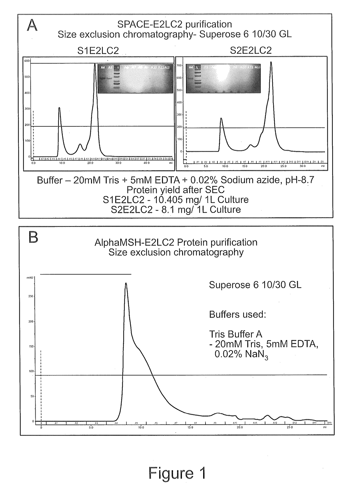
[0096]Protocols for the production and purification of E2 protein used in the present application had been previously reported (Dalmau M, et al. Biotechnol Bioeng. 2008 Nov. 1; 101(4):654-64). E. coli BL21 (DE3) cells were transformed with pET11a plasmids encoding E2LC2, S1LC2, S2LC2 or αMSH-LC2. The 20 ml starter culture of cells were grown overnight and inoculated in 1 L of LB medium. The cells were treated with 1 mM IPTG to overexpress the proteins when the OD value reached 0.6-0.8. After 4 hours cells were harvested by centrifugation at 4,700 g for 30 minutes. Harvested cells were resuspended in a buffer (20 mM Tris, 5 mM EDTA and 0.02% sodium azide) and ultrasonicated by Vibracell VC 750 Ultrasonic Cell Disrupter at 37% amplitude for 20 minutes with pulse on and off for 5 seconds. The sonicated solution was heat-treated at 75° C. for 15 minutes and was ultracentrifuged at 193,011 g for 1 hour. The supernatant was then collecte...

example 3

Conjugation of Protein Nanocages with Alexa Fluor 488 Fluorescent Dye

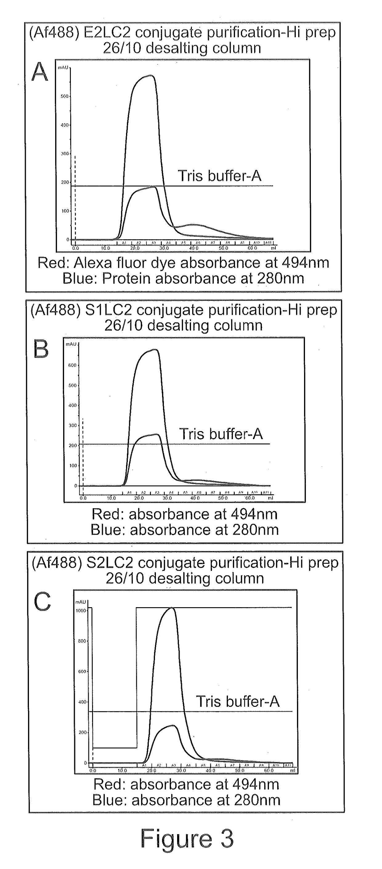
[0097]The purified proteins were conjugated with thiol reactive Alexa Fluor 488 (AF488) dye for convenience of detection and quantification. The E2LC2, S1LC2, S2LC2 and αMSH-LC2 protein nanocages were incubated with 10 mM excess tris carboxy ethyl phosphine (TCEP) for 1 hour to reduce the 120 cysteine residues residing inside the protein nanocages. The reduced proteins were reacted with 50 molar excess of Alexa Fluor 488 dye at room temperature for 2 hours and subsequently at 4° C. for 20 hours at pH 7.0. After the conjugation reaction, the dye-conjugated protein nanocages were purified by gel filtration chromatography using Hi prep 26 / 10 desalting column by FPLC. The samples were spun down at 12,000 g for 15 minutes after the conjugation reaction. The pellet fraction was discarded and the supernatant was collected and loaded onto the desalting column with elution buffer (20 mM Tris, 5 mM EDTA and 0.02% sodium azid...


PUM

PropertyMeasurementUnit
Therapeuticaaaaaaaaaa

Abstract

The present invention relates to protein nanocages comprising a melanocyte-targeting moiety and pharmaceutical compositions comprising the protein cages as well as methods for treating or diagnosing hyperpigmentation disorders or other melanocyte-related disorders using the protein nanocages or pharmaceutical compositions. In the preferred embodiment, the protein nanocage is composed of Bacillus stearothermophilus E2 protein of pyruvate dehydrogenase multi-enzyme complex (E2), with a skin penetrating and cell permeating SPACE moiety, and a melanocyte-targeting moiety of alpha melanocyte stimulating hormone.

Description

FIELD OF THE INVENTION[0001]The present invention relates generally to protein nanocages, pharmaceutical compositions containing them and methods for the treatment or diagnosis of hyperpigmentation disorders or other melanocyte-related disorders using said protein nanocages.BACKGROUND OF THE INVENTION[0002]Melanocytes are unique in that they produce eu- / pheo-melanin pigments in unique membrane-bound organelles termed melanosomes, and are closely related to various diseases and disorders including hyperpigmentation disorders and melanoma.[0003]It is known in the art that melanocytes produce melanin as a protective mechanism in response to the DNA damage induced by exposure to ultra violet (UV) radiation. However, hyper-expression of melanin in humans is known to result in pigmentation disorders such as melasma. Melasma occurs mostly on the face, occasionally on the neck, and rarely on the forearms. It predominantly affects females and has significant effect on psychology and quality ...

Claims


Application Information

Patent Timeline
IPC IPC(8): A61K38/34A61K38/35A61P17/00C07K14/32C07K14/47C07K14/685C07K14/69C07K14/695C07K14/72A61K9/00A61K47/10
CPCA61K38/34A61K38/35A61P17/00C07K14/32C07K14/47C07K14/685C07K14/69C07K14/695C07K14/723A61K9/0014A61K47/10B82Y5/00C12Y102/04001C07K14/195
Inventor LIM, SIERINBHASKAR, SATHYAMOORTHYTHNG, STEVENKUNIYIL, AMBILIMRIDUL, SARKER
Owner NANYANG TECH UNIV
Why Eureka
Try Eureka