Anti-lag3 antibodies and antigen-binding fragments
a technology of antigen binding fragments and antibodies, applied in the field of antilag3 antibodies, can solve the problems of formidable barriers to effective antitumor immunity, and achieve the effect of reducing the number of antibodies
- Summary
- Abstract
- Description
- Claims
- Application Information
AI Technical Summary
Problems solved by technology
Method used
Image

Examples
example 1
Generation of Antibodies Against Human LAG3
[1033]To generate antibodies to human LAG3, Balb / C mice were immunized with 5 ug of human LAG3-hFc tagged recombinant protein using RIBI adjuvant and footpad injection on a biweekly schedule. Immunized mice were bled and serum titers determined for binding to human LAG3 transfected CHOK1 cells using a cell-based ELISA (described below). Mice with the highest titers were given a final boost with recombinant protein and draining popliteal lymph nodes isolated four days later. Hybridomas were generated by electrofusion of isolated lymphocytes with the myeloma fusion partner P3X63-AG8.653 using the Cytopulse Hybrimmune electrofusion system. Fused cells were plated in 96-well plates in DMEM / F12, 15% BCS, HAT, IL-6, OPI supplement, and gentamycin.
[1034]Hybridoma supernatants were assayed for binding to human LAG3 expressing CHOK1 cells and cross-reactivity to cynomolgus LAG3 expressing CHO cells using a cell-based ELISA format. Human LAG3 and cyn...
example 2
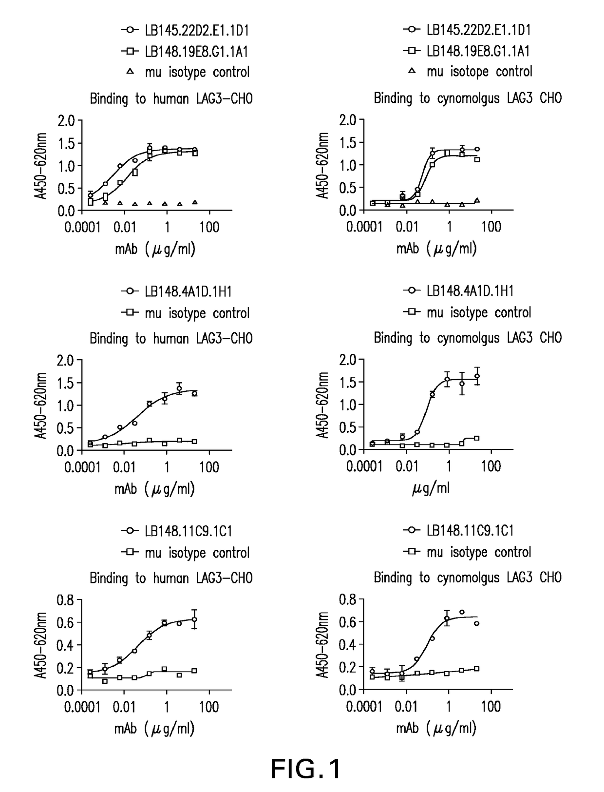
Binding of Mouse Anti-LAG3 Antibodies to Human LAG3 and Cynomolgous Monkey LAG3 Expressed on the Surface of Chinese Hamster Ovary Cells
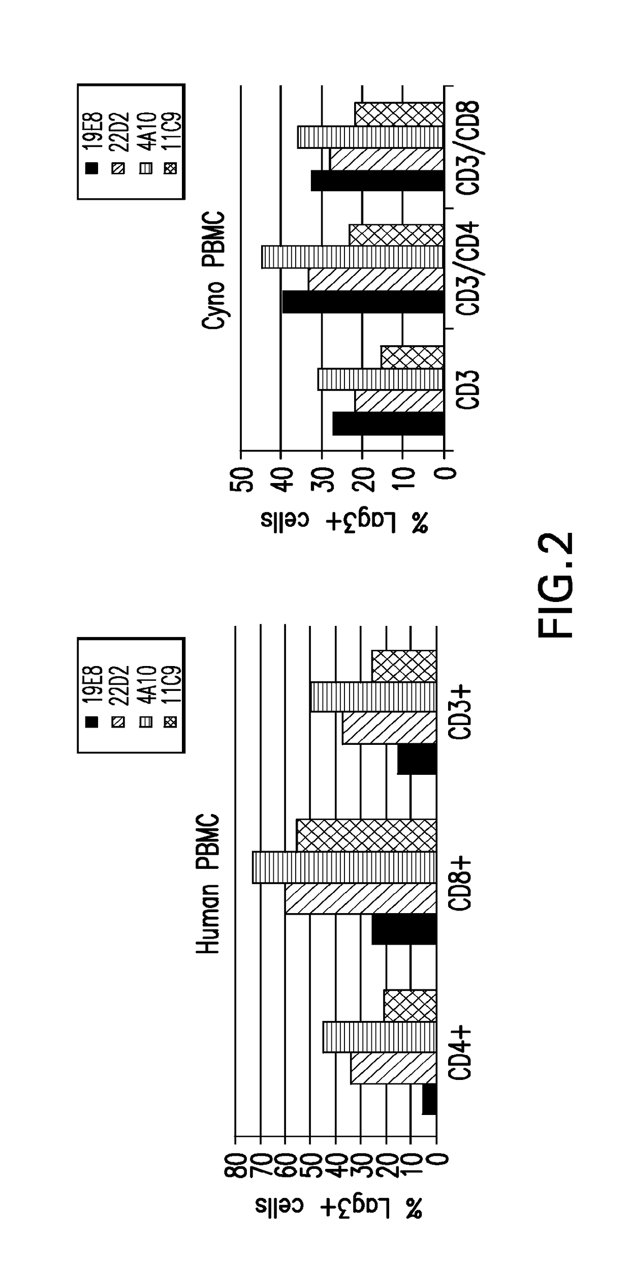
[1036]Mouse anti-human LAG3 antibodies were tested for binding to human LAG3 and cynomolgus LAG3 expressing CHO-K1 cells using a cell-based ELISA. CHO-K1 cells were plated as described above and media removed prior to adding the test samples. Purified antibody from clones LB145.22D2.E1.1D1 (22D2), LB148.19E8.G1.1A1 (19E8), LB148.4A10.1H1 (4A10), and LB148.11C9.1C1 (11C9) were serially-diluted in DMEM / F12, 10% BCS (CHOK1 media) and added to the CHO-K1 plates. The samples were incubated at 37° C. for 30-60 minutes and plates were washed three times with PBS / 0.05% Tween-20 using the cell wash program on the Biotek washer as described above. Binding was detected using an HRP-conjugated goat anti-mouse IgG (Southern Biotech cat# 1031-05) secondary antibody added at a 1:2000 dilution in CHO-K1 media and incubated at 37° C. for 30-60 minutes. Assay plates w...
example 3
Affinity Determination for Binding of Mouse Anti-LAG3 Antibodies to Human LAG3 Recombinant Protein
[1037]The kinetic binding activity of mouse anti-human LAG3 clones LB148.19E8.G1.1A1, LB148.4A10.1H1, LB148.11C9.1Cland LB145.22D2.E1.1D1 using human LAG3-His tagged recombinant protein was measured by surface plasmon resonance using a Biacore T200 system (Biacore, GE Healthcare, Piscataway, N.J.). Approximately 4000 RU of Goat Anti-Mouse IgG Fc gamma, Fragment Specific (Jackson ImmunoResearch Catalog #115-006-071, Lot 81313) was immobilized via amine coupling chemistry onto a Series S CM4 sensor chip, catalog number BR-1005-34. Mouse anti-human LAG3 clones listed above were injected over the immobilized goat anti-mouse surfaces at 1 ug / mL for a capture level of 40 RU. HBS-EP+ buffer (BR-1006-69) was used as the running buffer with a flow rate of 30 μL / min.
[1038]Varying concentrations of human LAG3-His protein ranging from 0.15 nM to 18.8 nM, at a flow rate of 40 μL / min were injected ov...
PUM
 Abstract
Description
Claims
Application Information
 - R&D Engineer
- R&D Manager
- IP Professional
- Industry Leading Data Capabilities
- Powerful AI technology
- Patent DNA Extraction
Browse by: Latest US Patents, China's latest patents, Technical Efficacy Thesaurus, Application Domain, Technology Topic.
© 2024 PatSnap. All rights reserved.Legal|Privacy policy|Modern Slavery Act Transparency Statement|Sitemap