Ophthalmologic device and ophthalmologic system

a technology of ophthalmologic devices and ophthalmologic systems, applied in medical science, diagnostics, skiascopy, etc., can solve the problems of not being able to directly enter, ophthalmologic devices cannot measure the ocular characteristics of subjects eyes, and ophthalmologic devices cannot feedback

Pending Publication Date: 2022-05-19
KK TOPCON
View PDF0 Cites 0 Cited by
  • Summary
  • Abstract
  • Description
  • Claims
  • Application Information

AI Technical Summary

Benefits of technology

[0016]In the ophthalmologic device according to another aspect of the present invention, the second optical element is arranged on an optical axis for each of the right and left optical axes, and the second optical elements are configured to branch the second optical paths from the first optical paths in directions which are parallel to a plane containing both the optical axes and in which second optical paths are away from each other. Thus, it is possible to prevent interference between the right and left of the ophthalmologic device in a case where both eyes of the subject eyes converge.
[0019]The ophthalmologic device according to another aspect of the present invention includes an alignment control unit connected to a relative moving mechanism that moves one of each subject eye and the correcting device relatively to the other, the alignment control unit configured to drive the relative movement mechanism based on a detection result of the alignment detecting unit so as to perform alignment of the ophthalmologic device with respect to the subject eye. This allows the ophthalmologic device to automatically achieve alignment with the subject eye.
[0021]The present invention makes it possible to use the ophthalmologic device integrally with an existing correcting device as necessary.

Problems solved by technology

Incidentally, when an ophthalmologic device that performs objective measurement is not integrated with each phoropter, an examiner is required to enter the result of measurement performed by the ophthalmologic device to a controller of each phoropter, and it is not possible to directly enter the result of the measurement from the ophthalmologic device to the controller.
In this case, the ophthalmologic device cannot measure the ocular characteristics of the subject eye, which views the visual target chart through the examination window, continuously in real time.
Furthermore, the ophthalmologic device cannot feedback the result of the measurement to each phoropter (controller).
Meanwhile, in the case of using a phoropter system in which an ophthalmologic device and each phoropter are integrated as described in Patent Literature 4, existing phoropters need to be replaced with the phoropter system or the phoropter system needs to be introduced separately, which is not practical from a cost perspective, even if the aforementioned problems could be solved.
In addition, although respective users have different needs such as necessity or unnecessity of an ophthalmologic device, the phoropter system described in Patent Literature 4 cannot omit the ophthalmologic device, and therefore it is not possible to meet the needs of users who do not need the ophthalmologic device.

Method used


Image

  • Ophthalmologic device and ophthalmologic system
  • Ophthalmologic device and ophthalmologic system
  • Ophthalmologic device and ophthalmologic system

Examples


first embodiment

Operation of First Embodiment

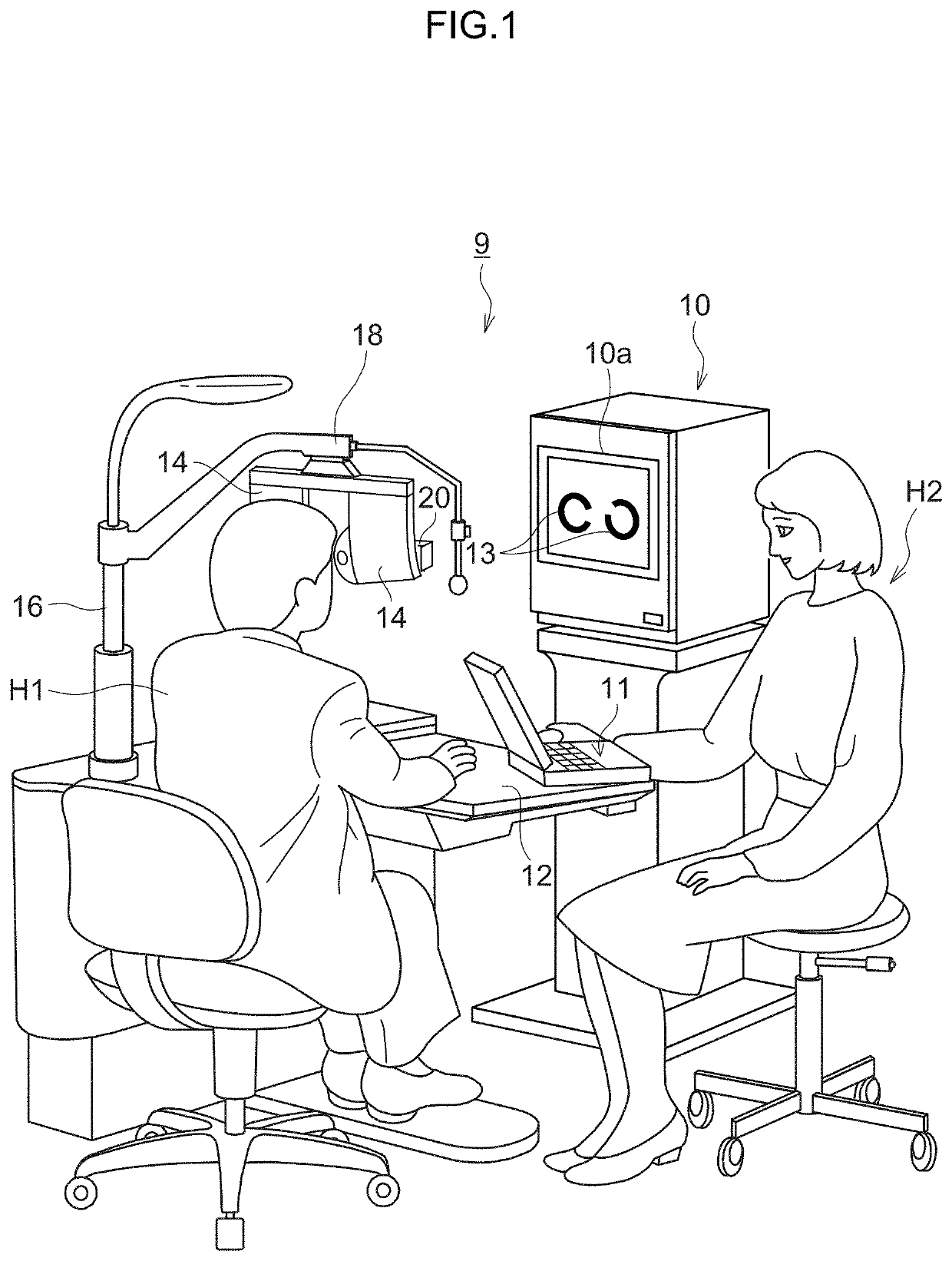
[0097]FIG. 7 is a flowchart showing the flow of determination processing of a prescription value of spectacle lenses for the subject H1 using the ophthalmologic system 9 having the above configuration. As shown in FIG. 7, the examiner H2 first couples the coupling members 27 of the wavefront sensors 20 to the opening parts 23a of the second examination windows 23 of the phoropters 14. Accordingly, the wavefront sensors 20 are coupled to the phoropters 14, respectively.

[0098]Upon activation of each of the wavefront sensors 20, radiation of alignment luminous flux to the subject eye E by the alignment optical system 34, radiation of near-infrared light LA to the subject eye E from the infrared light source 99, acquisition of an observation image of the anterior eye part of the subject eye E by the anterior eye part observing system 29A, and display of the observation image by a display unit not illustrated, are performed. Based on the observation image dis...

second embodiment

[0110]FIG. 8 is a functional block diagram of the controller 11 constituting the ophthalmologic system 9 according to a second embodiment. In the first embodiment, alignment of the wavefront sensors 20 with respect to the right and left subject eyes E is performed manually. In the second embodiment, the alignment is performed automatically.

[0111]As shown in FIG. 8, the ophthalmologic system 9 of the second embodiment is basically similar in configuration to the ophthalmologic system 9 according to the first embodiment except that the controller 11 is connected to a relative moving mechanism 66 and that the wavefront sensor control unit 52 functions as an alignment detecting unit 62 and an alignment control unit 64 in addition to functioning as other units described above (see FIG. 6). Accordingly, the component members identical in function or configuration to those in the first embodiment are designated by identical reference numerals and the description thereof is omitted.

[0112]Th...

third embodiment

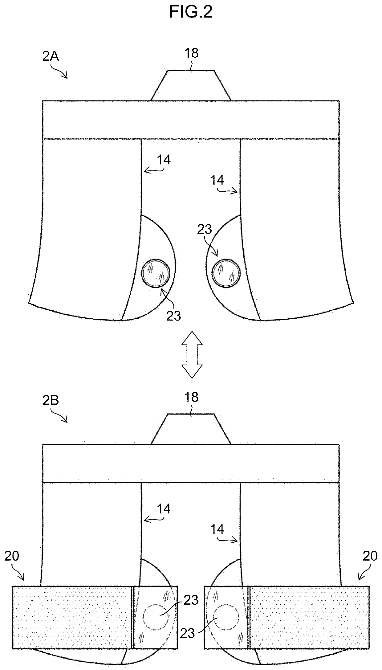
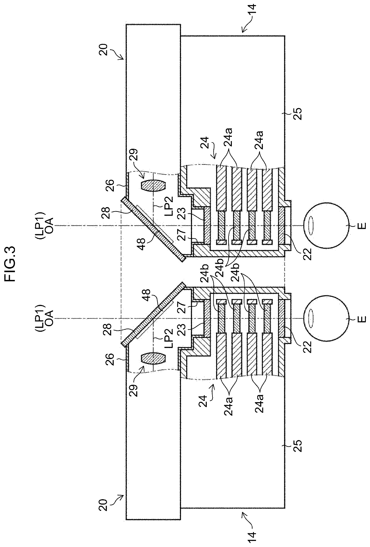
[0115]FIG. 9 is a sectional view of the phoropter 14 and the wavefront sensor 20 provided in the ophthalmologic system 9 according to a third embodiment, in a state before the wavefront sensor 20 is coupled to the phoropter 14. FIG. 10 is a sectional view of the phoropter 14 and the wavefront sensor 20 constituting the ophthalmologic system 9 according to the third embodiment, in a state after the wavefront sensor 20 is coupled to the phoropter 14. Here, since the phoropters 14 and the wavefront sensors 20 have a symmetrical structure, description is given by taking as an example the phoropter 14 and the wavefront sensor 20 corresponding to the right eye of the subject eyes E in FIGS. 9 and 10.

[0116]As shown in FIGS. 9 and 10, in the third embodiment, in a state where the wavefront sensor 20 is coupled to the phoropter 14, the sensor housing 26 of the wavefront sensor 20 is used as the rear side surface of the phoropter housing 25, on the side of the second examination window 23. No...


PUM


Abstract

The ophthalmologic device to be coupled to a correcting device for correcting subject eyes, the correcting device including a right-left pair of two examination windows and a right-left pair of arrangement mechanisms that each arrange first optical elements, includes: a coupling member configured to be detachably coupled to each of coupled members provided on a side surface of the correcting device on a side opposite to an eyepiece side for each subject eye; a second optical element arranged at a position facing a second examination window opposite to a first examination window which is an eyepiece window, when the coupling member is coupled to the coupled member, the second optical element configured to branch a second optical path from a first optical path along the optical axes; and an objective measuring system arranged on the second optical path and configured to acquire ocular characteristics of each subject eye.

Description

CROSS-REFERENCE TO RELATED APPLICATIONS[0001]The present application is a Continuation of PCT International Application No. PCT / JP2020 / 030153 filed on Aug. 6, 2020 claiming priority under 35 U.S.C. § 119(a) to Japanese Patent Application No. 2019-149340 filed on Aug. 16, 2019. Each of the above applications is hereby expressly incorporated by reference, in its entirety, into the present application.BACKGROUND OF THE INVENTION1. Field of the Invention[0002]The present invention relates to an ophthalmologic device that acquires ocular characteristics of a subject eye and an ophthalmologic system including the ophthalmologic device.2. Description of the Related Art[0003]In ophthalmologic clinics and optician's stores, a visual target presenting device and a pair of right and left phoropters (correcting device) are installed for subjective examination of visual functions such as an eyesight of a subject eye or eyes (see Japanese Patent Application Laid-Open No. 2001-346762, hereinafter ...

Claims


Application Information

Patent Timeline
Patent Type & Authority Applications(United States)
IPC IPC(8): A61B3/103A61B3/15A61B3/10A61B3/028
CPCA61B3/103A61B3/0285A61B3/1015A61B3/152A61B3/04A61B3/032A61B3/028A61B3/117
Inventor TATARA, YOKOMIYAGAWA, SUGURUYAMAGUCHI, TATSUOSAIKA, MAKOTO
Owner KK TOPCON
Why Eureka
Try Eureka