Patient specific dosing contrast delivery systems and methods

a technology of contrast delivery system and patient, applied in the direction of instruments, process and machine control, ultrasonic/sonic/infrasonic diagnostics, etc., can solve the problems of not being used on another patient, different results in image contrast and quality, and increasing patient risk

Inactive Publication Date: 2002-05-07
BAYER HEALTHCARE LLC
View PDF123 Cites 224 Cited by
  • Summary
  • Abstract
  • Description
  • Claims
  • Application Information

AI Technical Summary

Benefits of technology

In a principal embodiment, information specific to any given patient is entered into the system and the appropriate concentration and injection parameters are computed before initiating the imaging fluid injection procedure. The system is then ready for injection of a patient. It is important to note that the system is not limited to choosing a particular concentration of contrast media or injection rate for the entire procedure, or even a moderate number of phases with constant velocity as present injectors can now do, but rather is capable of selecting an injection profile which may include a continuously varying injection rate and / or concentration of contrast media. The particular injection profile selected by the system is designed to provide the best image quality for the particular patient based on a variety of factors such as patient weight and circulation system variables.
In a refined version of the system, feedback from at least one sensor is employed by the control system to modify the concentration of the contrast media, injection rate, and / or total volume during the injection procedure. Various types of sensors are disclosed for use with this system, in particular, various electromagnetic sensors or video monitoring devices provide feedback for the system or operator to use. In angiography, where the contrast is injected into the region of interest, the sensor needs to make a measurement in that region of interest. In CT and MR it is sufficient for the sensor to measure a remote area of the body, although measuring within the region of interest (ROI) could be advantageous in some applications. The sensor provides an indication of the actual amount of contrast media in the patient. This is used to calculate the appropriate injection rate or concentration of contrast media to provide a diagnostic image with minimum risk and cost.
A final version of the invention is disclosed which works to further improve doctors confidence by providing tactile feedback to a doctor or operator in addition to visual or other sensed feedback on the amount of contrast media in a patient. This provides the operator with additional information to use in determining injection rate, concentration and pressure for the injection procedure.

Problems solved by technology

Using fixed protocols for delivery simplifies the procedure, however, providing the same amount of contrast media to patients weighing between 100 and 200 pounds, for example, produces very different results in image contrast and quality.
With high contrast costs, this is a waste of money as well as increased patient risk.
Furthermore, in prior art contrast delivery systems, once a bottle of contrast media was opened for use on a patient it could not be used on another patient due to contamination considerations.
Existing contrast delivery systems do not prevent the source of contrast media used for an injection from being contaminated with body fluids of the patient.
However, physically, pressure drives fluid flow.
There are significant drawbacks to fluid delivery systems which are unable to adjust the concentration of contrast media and other injection parameters during an injection procedure.
Many patients may receive more contrast media than is necessary to produce an image of diagnostic quality, while others may receive an amount of contrast media insufficient for producing a satisfactory image.
Existing procedures also frequently result in waste of contrast media as well as the need for repeating the procedure because an image of diagnostic quality could not be produced.

Method used


Image

  • Patient specific dosing contrast delivery systems and methods
  • Patient specific dosing contrast delivery systems and methods
  • Patient specific dosing contrast delivery systems and methods

Examples


Embodiment Construction

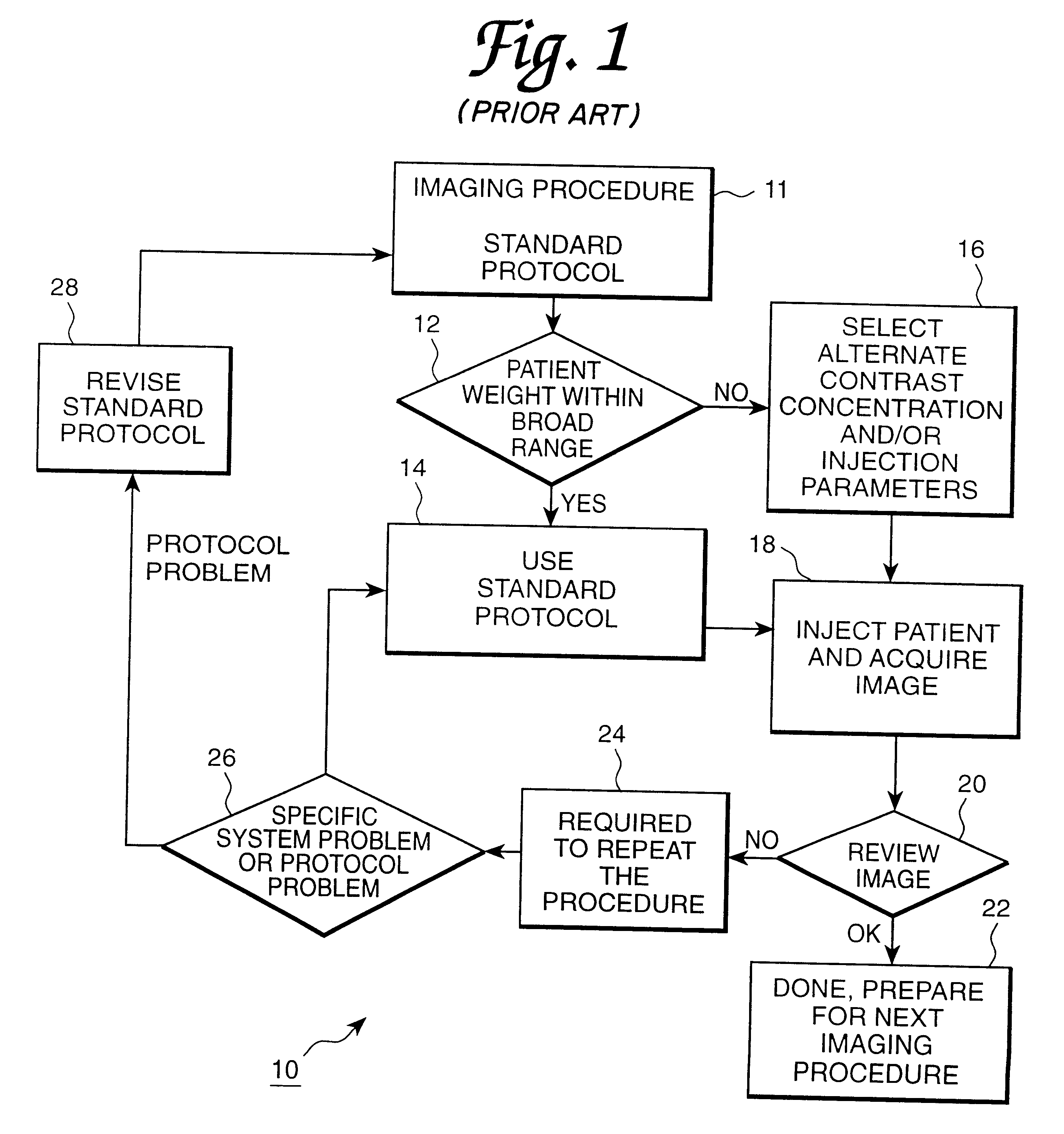
FIG. 1 is a flow diagram showing a conventional medical imaging procedure for implementation of a standard protocol. This diagram is indicated generally by the numeral 10. The imaging procedure standard protocol is selected at first operative step 11, and a decision is made at step 12 as to whether the patient's weight is within a broad range considered to be appropriate for the particular concentration of contrast media and set of injection parameters for the selected protocol. If the patient's weight is within the broad range of weights acceptable for the particular contrast media and set of injection parameters, the standard protocol is determined to be appropriate at step 14. The patient is injected and the image is acquired at step 18. Alternatively, if the weight of the patient is not within the given range, an alternate contrast media concentration and set of injection parameters are chosen at step 16. Once the alternate concentration or set of injection parameters are chosen...


PUM


Abstract

This invention relates generally to the field of medical devices for delivering contrast media during medical diagnostic and therapeutic imaging procedures and more particularly, this invention relates to improved contrast media delivery systems and methods of use which allow adjustment of contrast media concentration and injection parameters either before or during an injection procedure to provide patient specific dosing of contrast media, thus decreasing the waste and cost of these procedures while increasing their efficiency.

Description

This invention relates generally to the field of medical devices for delivering contrast media during medical diagnostic and therapeutic imaging procedures and more particularly, this invention relates to improved contrast media delivery systems and methods of use which allow adjustment of contrast media concentration and injection parameters either before or during an injection procedure to provide patient specific dosing of contrast media, thus decreasing the waste and cost of these procedures while increasing their efficiency.DESCRIPTION OF THE RELATED ARTIt is well recognized that the appropriate dose for many medications is related to the size and weight of the patient being treated. This is readily apparent in the difference between the recommended doses which most medications have for adults and children. The appropriate dose of contrast media for a given medical imaging procedure is equally dependent upon the size and weight of the patient being examined as well as other add...

Claims


Application Information

Patent Timeline
Patent Type & Authority Patents(United States)
IPC IPC(8): A61M5/168A61M5/172A61M5/00
CPCA61B8/06A61B8/481A61M5/007A61M5/1723G06F19/3468A61M2205/80G06F19/321Y10S128/12Y10S128/13A61M2205/581G16H50/20G16H40/63G16H30/20G16H20/17
Inventor UBER, III, ARTHUR E.HIRSCHMAN, ALAN D.WELCH, THOMAS R.ALMON-MARTIN, ROSEMARY
Owner BAYER HEALTHCARE LLC
Why Eureka
Try Eureka