Experimental animals for evaluation of therapeutic effects on corneal epithelial damages

a corneal epithelial damage and experimental animal technology, applied in the field of experimental animals, can solve the problems of unsatisfactory medical needs, dry corneal surface, damage, etc., and achieve the r&d of new medicines withou

Inactive Publication Date: 2005-08-02
BIOCHEM & PHARMACOLOGICAL LAB
View PDF3 Cites 7 Cited by
  • Summary
  • Abstract
  • Description
  • Claims
  • Application Information

AI Technical Summary

Benefits of technology

The present invention provides an experimental animal that can be used to evaluate the therapeutic effect of a medicine on corneal epithelial damages, such as dry eye. This animal can also allow for control or adjustment of the degree of damage and can maintain the damages for a period of time sufficient to evaluate the therapeutic effect of the medicine. Additionally, the invention provides a method for screening or evaluating a medicine for the treatment or improvement of corneal epithelial damages, such as dry eye.

Problems solved by technology

The technical problem addressed in this patent text is the development of a model animal that can be used to evaluate the effect of medicines on corneal epithelial damage, specifically dry eye. The current methods for producing model animals with corneal damage have limitations, such as requiring long periods of time or failing to produce uniform damage on the cornea. There is a need for a new model animal that can be used for this purpose.

Method used


Image

  • Experimental animals for evaluation of therapeutic effects on corneal epithelial damages
  • Experimental animals for evaluation of therapeutic effects on corneal epithelial damages
  • Experimental animals for evaluation of therapeutic effects on corneal epithelial damages

Examples


example 1

Production of the Model Animal Example 1

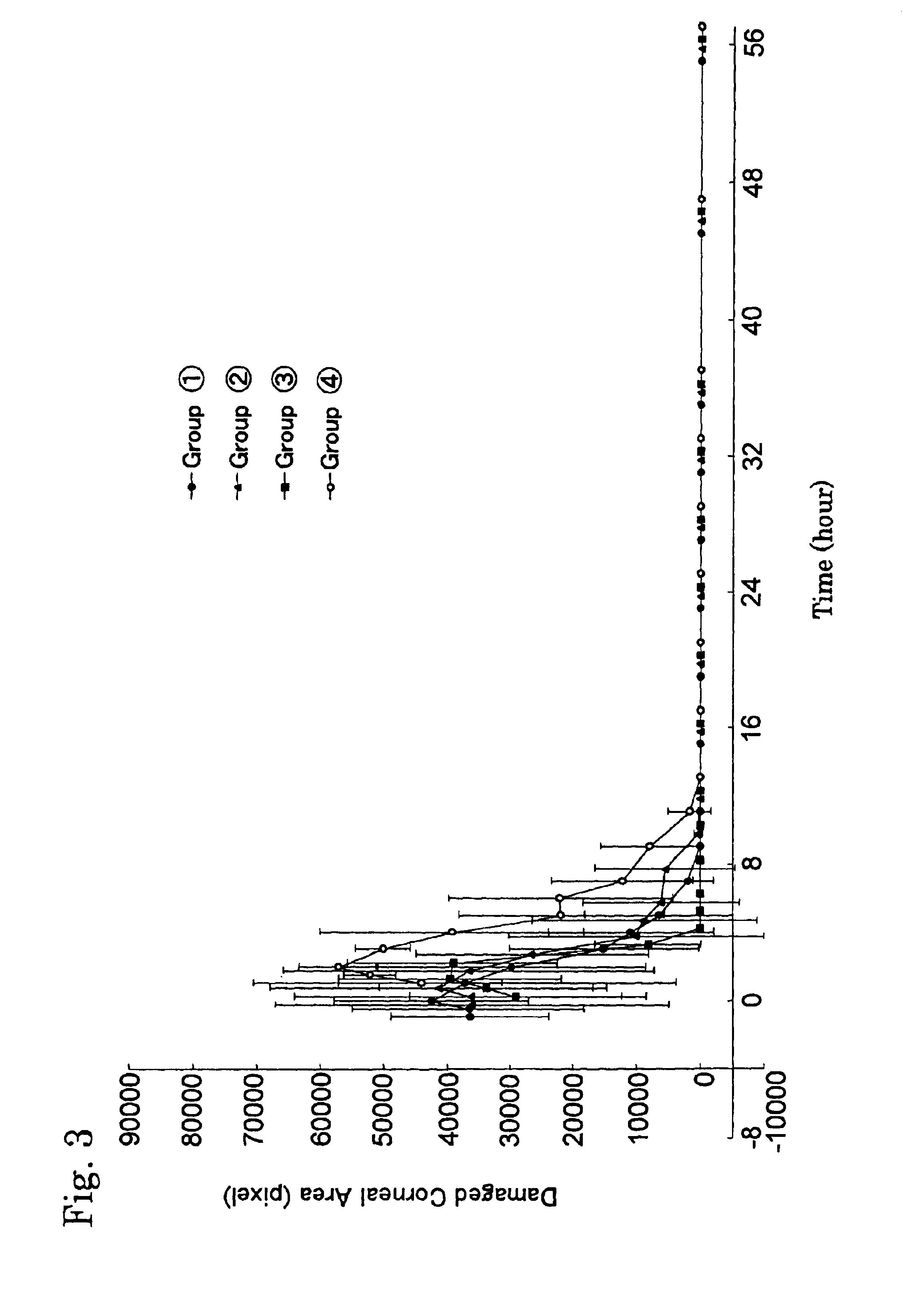
[0116]Rabbits (New Zealand white, male, 13 weeks old, about 3 kg body weight) were quarantined and habituated. They were bred in animal-breeding boxes set in a homothermal and homohumid room (room temperature of 21±3° C.; relative humidity of 50±20%, 12 hours illumination (7 a.m. light-up, 7 p.m. lights-out); and 10-15 times ventilation / hour). They were freely given the commercial feed (manufactured by Oriental Yeast Co., Ltd; RC4) and water (sterilized and filtered through the membrane having pore size of 0.2 μm in diameter). The animals were anaesthetized by intramuscular injection of 20 mg / kg of ketamine hydrochloride (manufactured by Sankyo Co., Ltd under the trade name “Ketaral 50 for intramuscular injection”) and 10 mg / kg of xylazine hydrochloride (manufactured by Bayer under the trade name “Seraktal 2% for injection to dog and cat”). The eyelids of the animal were kept open and glued with the adhesive for surgery (manufactured by Sankyo...

example 2

Production of the Model Animal Example 2

[0119]A model animal was produced by using powder sugar (manufactured by Sankyo Shokuhin Co., Ltd.; a mixture of sucrose and corn starch (97:3; w / w) as the water-absorbing material.

[0120]A quarantined and habituated animal (rabbit) was anaesthetized in the same manner as in Example 1, and further anaesthetized locally by instillation of an eye drop containing a 0.4% oxybuprocaine hydrochloride (manufactured by Senju Pharmaceutical Co., Ltd. under the trade name “Anenocurl”). Then, eyelid retraction was done using a Vanguard eyelid retractor.

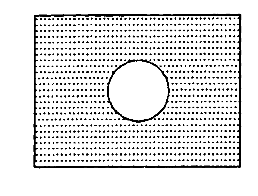
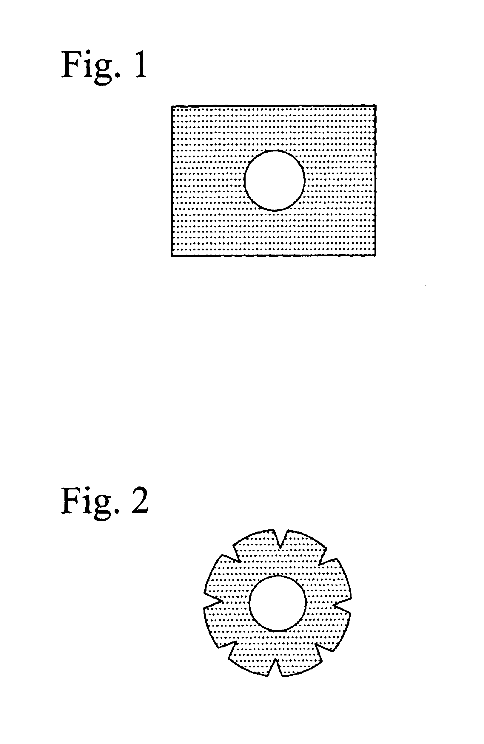
[0121]The eyeball was covered with a polyfluoroethylene sheet film (21 mm ø, 0.2 mm thick; see FIG. 2) having a hole of 8 mm ø in size at around the center thereof. 1 g of the powder sugar was placed on the opening (the hole) of the film covering the eyeball for 20 minutes. Thereafter, the film and sugar were removed from the ocular surface. Said ocular surface was thoroughly washed with a physiological sal...

example 3

Production of the Model Animal Example 3

[0122]A model animal was produced in the same manner as in Example 2 by using the powder of sodium chloride as the water-absorbing material.

[0123]1 g of sodium chloride powder having a particle size of less than 150 μm in diameter (sieved by using a standard sieve (Sieve No. 100) of Japanese Industrial Standard) was placed on the opening (the hole) of the film covering the eyeball. The film and sodium chloride were removed from the ocular surface, and then the ocular surface was thoroughly washed with a physiological saline solution, whereby the model animal example 3 was obtained.


PUM


Abstract

This invention provides experimental animals suffering corneal epithelial damages, such as dry eye, and methods of using the same to assay a variety of compounds for evaluating the therapeutic effect on the disease, and medicine selected using the method, wherein the corneal epithelial damage is induced by the steps of: using a water-absorbing material having a physical state selected from powder, solution, gel, jelly and tablet, and contacting the absorbing materials with the ocular cornea to generate a difference in osmotic pressure between the inside and outside of ocular corneal epithelium cells.

Description


Claims


Application Information

Patent Timeline
Owner BIOCHEM & PHARMACOLOGICAL LAB
Why Eureka
Try Eureka