Human ovarian mesothelial cells and methods of isolation and uses thereof

a technology of ovarian mesothelial cells and methods, applied in the field of development biology and cell biology, can solve the problems of human ovary tissue models, difficult efforts to confirm the ovarian surface epithelium as the source of cancerous growth, and lack of understanding about the development of ovarian surface epithelial cells

Inactive Publication Date: 2005-08-09
MACROGENICS INC
View PDF5 Cites 2 Cited by
  • Summary
  • Abstract
  • Description
  • Claims
  • Application Information

AI Technical Summary

Benefits of technology

[0020]FIG. 5 show the results of immunohistochemical analysis wherein cancerous epithelial cell in ovarian cancer can be detected (brown, indicated by arrows) specifically by anti-ovary monoclonal antibody 5C8.

Problems solved by technology

The lack of human ovary tissue models makes efforts to confirm the ovarian surface epithelium as the source of cancerous growth difficult.
Further, there is a lack of understanding about the development of ovarian surface epithelial cell during oogenesis and follicle formation because culturing conditions in which the OSE cells can be isolated and still retain their original characteristics are still being refined.
However, the ovarian precursor cells, like many precursor cells, are few in number and difficult to isolate.
Once isolated, ovarian precursor cells are difficult to culture to the extent that they retain their precursor traits.
It is postulated that there are precursors to granulosa cells and OSE cells, however, the work accomplished to date only isolates OSE cells and granulosa cells but not a common precursor to both types of cells.
However, the OSE cells are already differentiated and therefore, make studying events related to early OSE development or early cancerous transformation during OSE development difficult.

Method used


Image

  • Human ovarian mesothelial cells and methods of isolation and uses thereof
  • Human ovarian mesothelial cells and methods of isolation and uses thereof
  • Human ovarian mesothelial cells and methods of isolation and uses thereof

Examples


example 1

Isolation and Culturing of Ovarian Mesothelial Cells

[0068]Human fetal ovaries of gestational age between 17 to 25 weeks were obtained from Advanced Bioscience Research at Alameda county, Calif. Ovaries were procured and shipped to the lab in tissue culture medium under wet ice bath. Immediately upon arrival, the ovaries were cleaned of excess connective tissues, carefully separated from fallopian tube, and washed five times with fresh tissue culture medium.

[0069]The ovaries were minced with scissors or cut into small pieces (less than 1 mm thick) with a razor blade. The tissue pieces from each ovary were plated directly in a T75 flask freshly coated with laminin with 10 ml preferred nutrient medium as disclosed herein. Further dissociation of the ovaries with collagenase-dispase (0.5%) for 30 minutes at 37° C. could be done, but the procedure reduced the recovery of ovarian mesothelial cells. The cells were cultured in F12 / DMEM supplemented with 10 μg / ml insulin, 10 μg / ml transferri...

example 2

Characterization of Ovarian Mesothelial Cell Lines

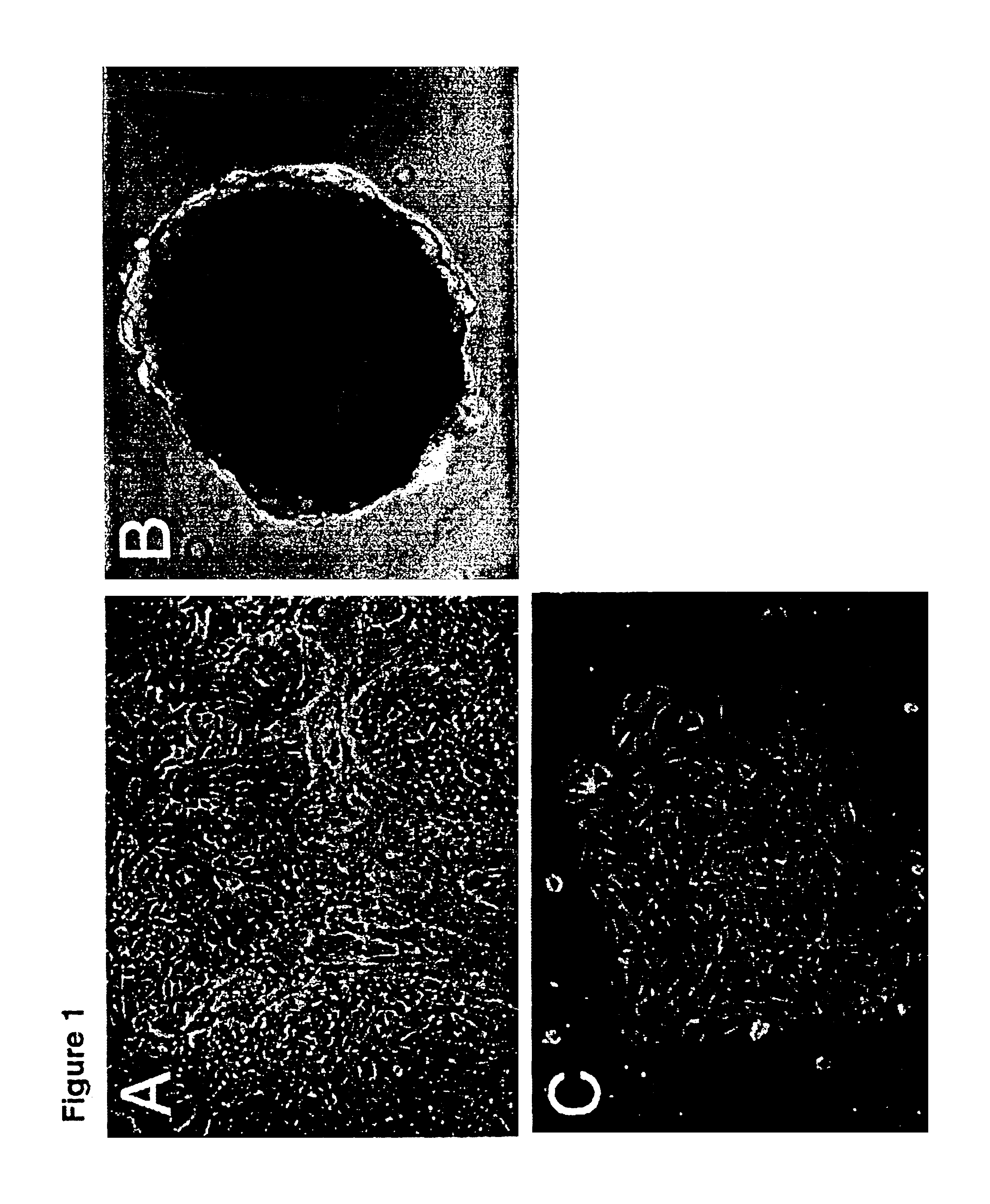
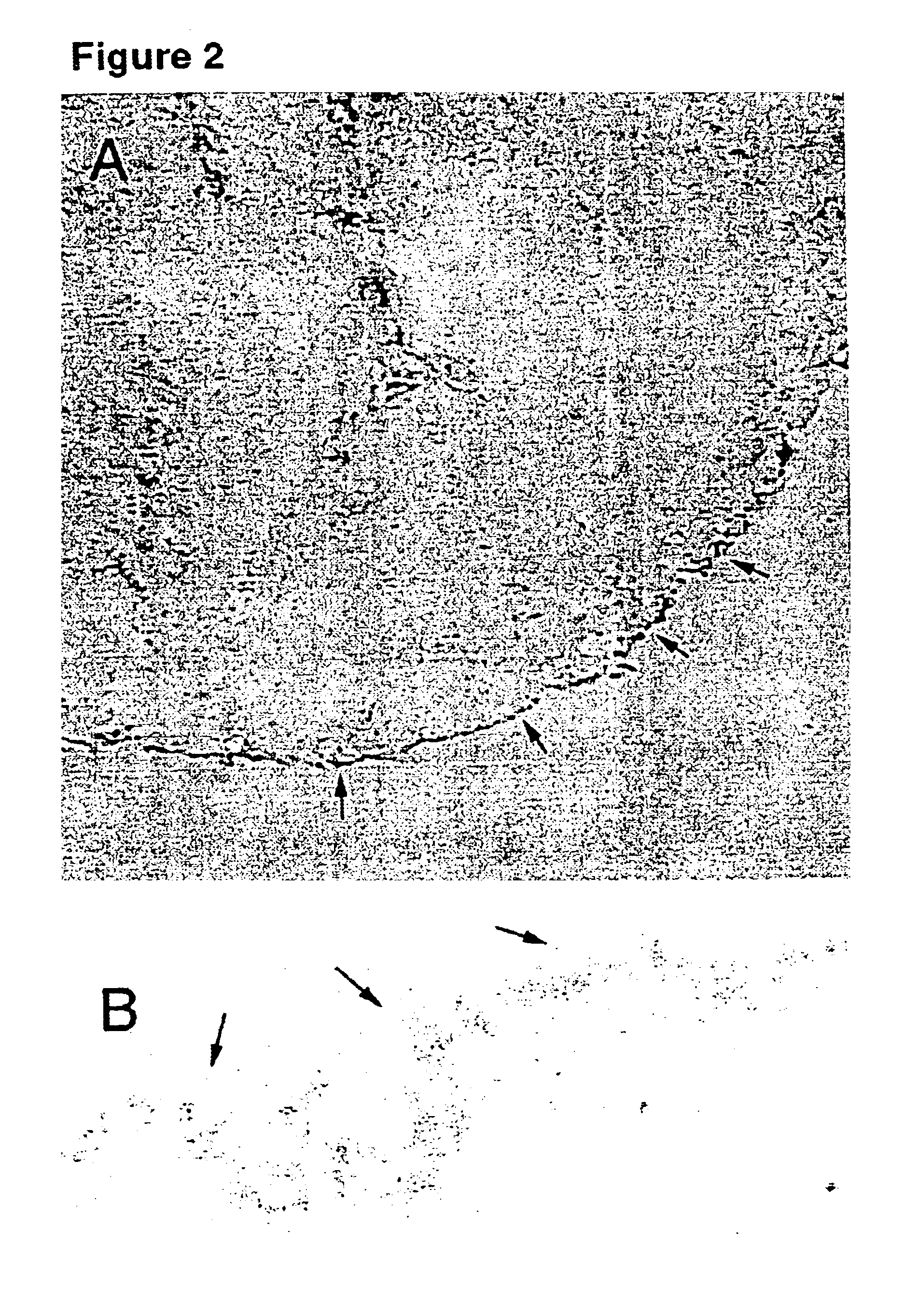
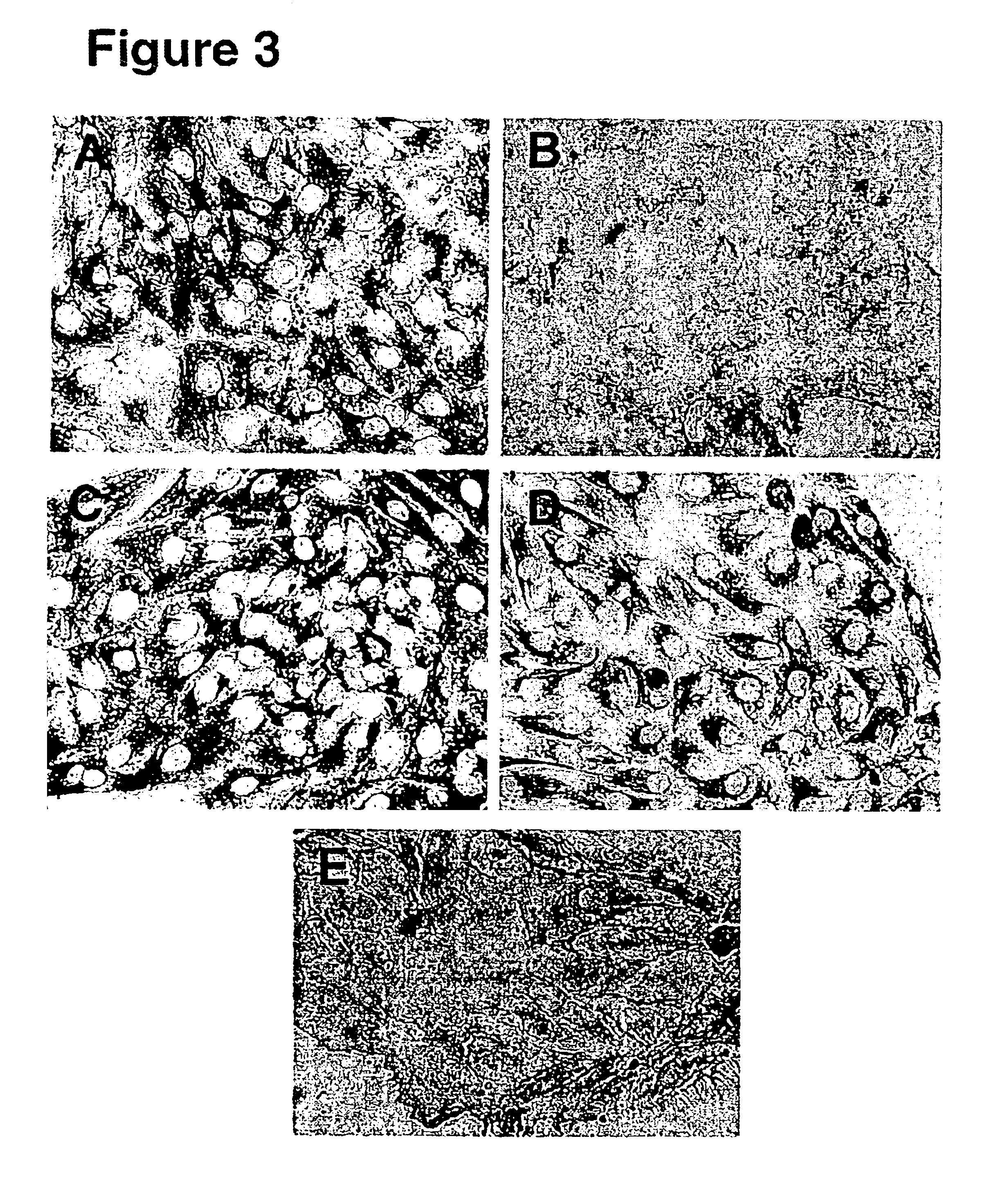
[0070]Ovarian mesothelial cells were harvested from suspension cultures. To directly stain the cells in cluster form, the cell clusters were embedded in optimal cutting temperature (OCT) compound and frozen on dry ice. Cryosections (5-10 μm thick) were cut of the OCT blocks, thaw-mounted onto coverslips, and fixed in 3% paraformaldehyde for 1 hour. A monolayer culture of ovarian mesothelial cells was prepared by plating the cell clusters from the suspension culture on chamber slides in the presence of 2% BSA. The monolayer cell cultures were fixed in situ with 3% paraformaldehyde for 1 hour. After the fixative was washed away with phosphate buffered saline (PBS), the cells were incubated in blocking buffer (5% goat serum and 0.1% Tween 20 in PBS) for 30 minutes, rinsed with PBS, then incubated in primary antibodies for 1 hour, rinsed with PBS, and then incubated with anti-mouse Ig-horseradish peroxidase for 1 hour. The markers and ex...

example 3

Method of Providing a Source of an Immunogen to Produce Monoclonal Antibodies Against Ovarian Mesothelial Cells

[0071]Ovarian mesothelial cells were isolated and cultured as disclosed and then subsequently used as an immunogen to produce a panel of monoclonal antibodies against ovarian mesothelial cells. Mice were each immunized with one injection of about 1×10⁶ cells of ovarian mesothelial cells per week. The immunization was localized into the foot pads of each mice. After 5 injections, the mice were bled to check the titer of ovarian mesothelial cells. When the titer of ovarian mesothelial cells rose above 1:1000 (ovarian cell:total cells) in a mouse, the mouse was sacrificed and lymph nodes were harvested. Lymphocytes were prepared from the lymph nodes and fused to myeloma cell line to produce a hybridoma. Hybridomas were screened by flow cytometry (fluorescence-activated cell sorter or FACS) as well as by immunohistochemistry for monoclonal antibodies that specifically bound to ...


PUM

PropertyMeasurementUnit
thickaaaaaaaaaa
thickaaaaaaaaaa
sizeaaaaaaaaaa

Abstract

The invention discloses a substantially pure population of human ovarian mesothelial cells and methods of isolating and culturing the ovarian mesothelial cells. By carefully manipulating the microenvironment of the ovarian mesothelial cells, multiple passages are attainable wherein the ovarian mesothelial cells are capable of becoming ovary surface epithelial cells or granulosa cells. In addition, several methods of use of human ovarian mesothelial cells are disclosed herein.

Description

CROSS-REFERENCE TO RELATED APPLICATIONS[0001]This application is a divisional of U.S. patent application Ser. No. 09 / 545,659, filed on Apr. 10, 2000, which is incorporated herein by reference in its entirety.TECHNICAL FIELD[0002]This invention is in the field of developmental biology and cell biology. Specifically, this invention relates to a population of ovarian mesothelial cells that are capable of differentiating into ovarian surface epithelial cells and granulosa cells, methods of isolating the ovarian mesothelial cells, characterization of ovarian mesothelial cells, and uses of the ovarian mesothelial cells.BACKGROUND ART[0003]Ovarian cancer is one of the most common causes of death due to cancer in women. Boring C. C. et. al. CA-Cancer J. Clin. 41,19-36, (1991). In about 80-90% of ovarian cancer cases, the ovarian surface epithelium is thought to be the tissue source in which the cancerous transformation occurs. Nicosia S. V. Pathology of Human Neoplasms. New York: Raven Pres...

Claims


Application Information

Patent Timeline
Patent Type & Authority Patents(United States)
IPC IPC(8): C12N5/06G01N33/50G01N33/574A61K35/12C12N5/075A61K35/48A61P35/00C12N5/071
CPCC12N5/0682C12N5/0697G01N33/5011G01N33/57449A61K35/12C12N2500/25C12N2500/36C12N2501/11C12N2501/195C12N2501/70C12N2502/243C12N2502/25C12N2503/02G01N2333/4742A61P35/00C12N5/0609
Inventor LI, RONG-HAOBALD, LAURAMATHER, JENNIE POWELL
Owner MACROGENICS INC
Why Eureka
Try Eureka