Assessment of lesions in an image

a technology for images and lesions, applied in image data processing, eye diagnostics, sensors, etc., can solve problems such as inability to reliably apply methods to images without lesions

Inactive Publication Date: 2009-09-01
RETINALYZE
View PDF10 Cites 31 Cited by
  • Summary
  • Abstract
  • Description
  • Claims
  • Application Information

AI Technical Summary

Benefits of technology

[0010]In order to be able to make automatic detection of various structures in fundus images a reliable method of detecting lesions in fundus images actually containing the lesions, and reliably not detecting lesions in other images not comprising lesions. Current methods may be able to detect the lesions in many images, but the methods are not reliable when applied to images not containing lesions.

Problems solved by technology

Current methods may be able to detect the lesions in many images, but the methods are not reliable when applied to images not containing lesions.

Method used


Image

  • Assessment of lesions in an image
  • Assessment of lesions in an image
  • Assessment of lesions in an image

Examples


Embodiment Construction

Images

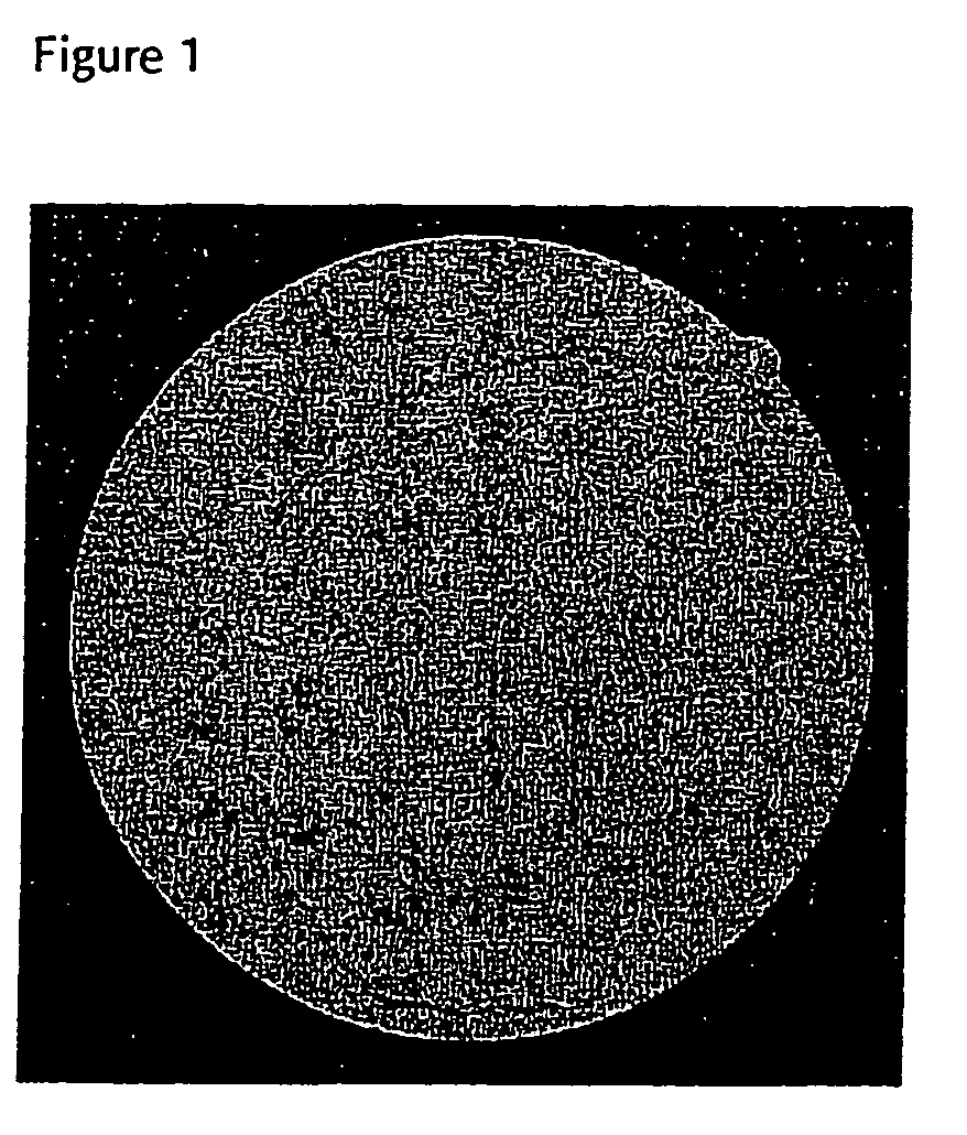
[0065]The images of the present invention may be any sort of images and presentations of the region of interest. Fundus image is a conventional tool for examining retina and may be recorded on any suitable means. In one embodiment the image is presented on a medium selected from dias, paper photos or digital photos. However, the image may be any other kind of representation, such as a presentation on an array of elements, for example a CCD.

[0066]The image may be a grey-toned image or a colour image; in a preferred embodiment the image is a colour image.

Subsets

[0067]At least one subset is established in the image, wherein the subset is a candidate lesion area. The term subset is used in its normal meaning, i.e. one or more pixels.

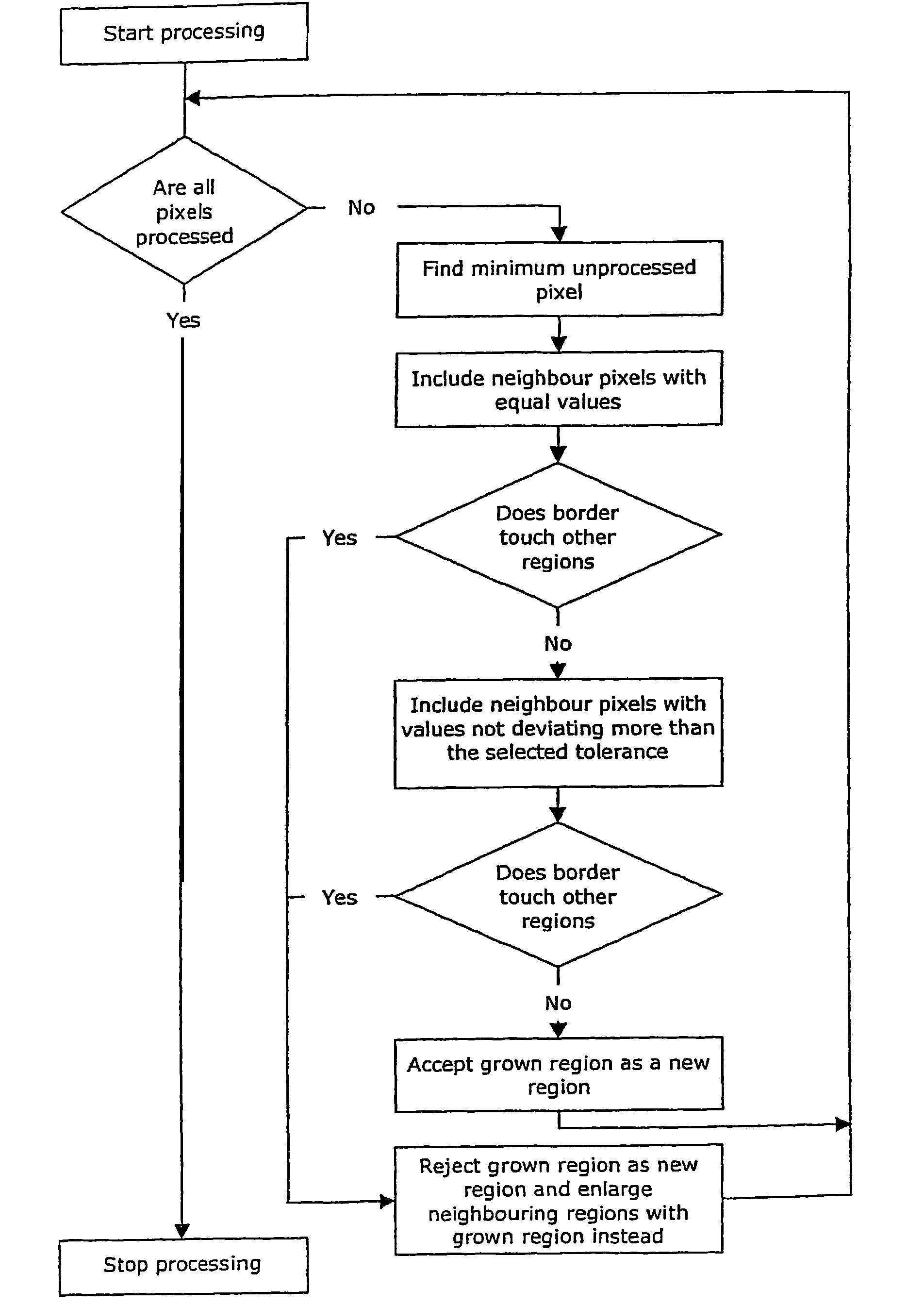
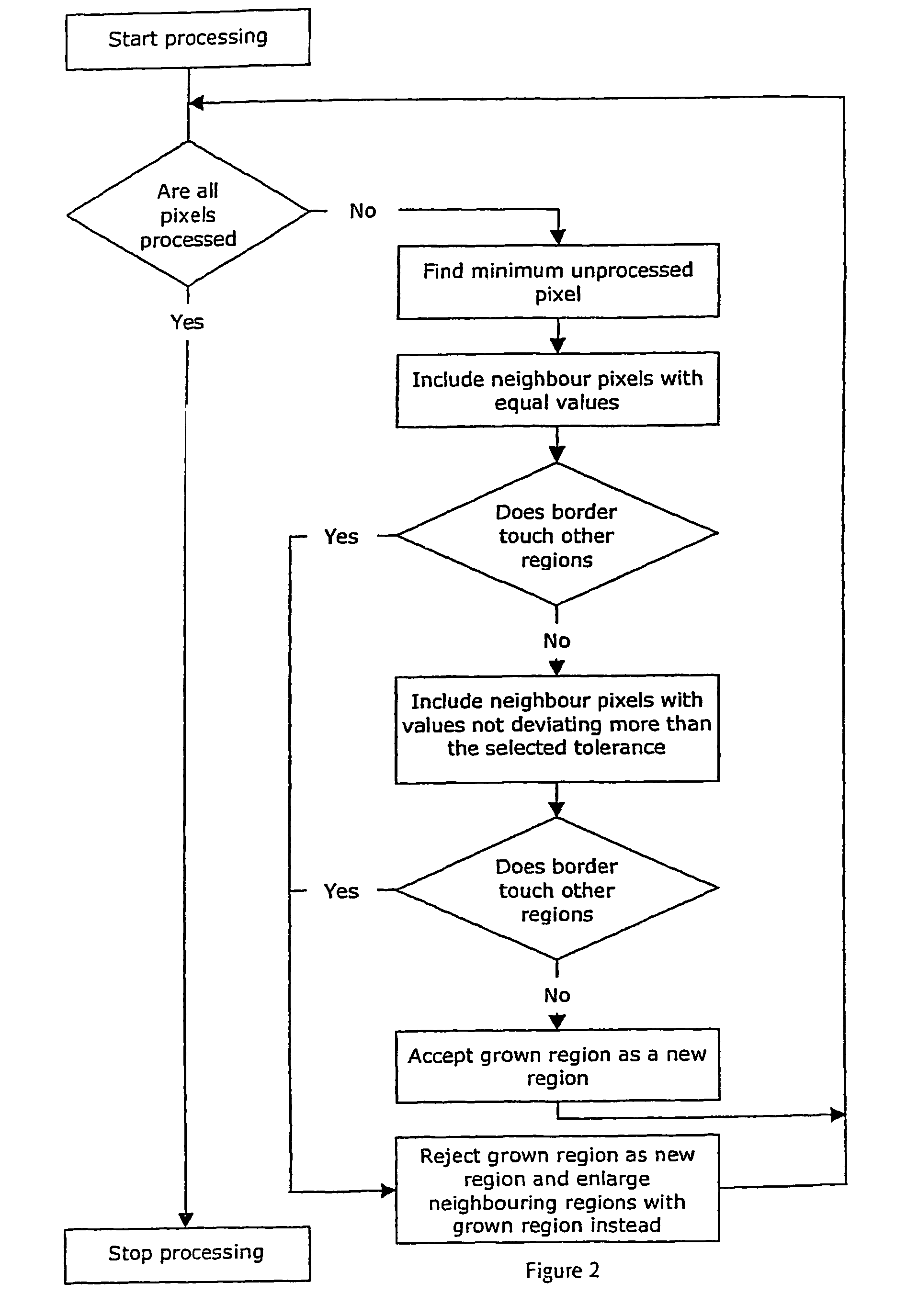
[0068]The subset may be detected establised by any suitable method, for example by filtering, by template matching, by establishing starting points, and from said starting points grow regions and / or by other methods search for candidate areas, and / or c...


PUM


Abstract

The present invention relates to a method for assessing the presence or absence of lesion(s) in an image and a system therefor, wherein said image may be any image potentially comprising lesions, in particular an image from medical image diagnostics, and more particularly an ocular fundus image. The lesions are identified from starting points being candidate lesion areas and validated with respect to their visibility as compared to the local surroundings.

Description

[0001]The present invention relates to a method for assessing the presence or absence of lesion(s) in an image and a system therefor, wherein said image may be any image potentially comprising lesions, in particular an image from medical image diagnostics, and more particularly an ocular fundus image.BACKGROUND[0002]Fundus image analysis presents several challenges, such as high image variability, the need for reliable processing in the face of nonideal imaging conditions and short computation deadlines. Large variability is observed between different patients—even if healthy, with the situation worsening when pathologies exist. For the same patient, variability is observed under differing imaging conditions and during the course of a treatment or simply a long period of time. Besides, fundus images are often characterized by having a limited quality, being subject to improper illumination, glare, fadeout, loss of focus and artifacts arising from reflection, refraction, and dispersi...

Claims


Application Information

Patent Timeline
Patent Type & Authority Patents(United States)
IPC IPC(8): G06K9/00A61B3/14A61B5/00G06K9/62G06Q50/00G06T1/00
CPCG06T7/0012G06T2207/30041Y02A90/10
Inventor HANSEN, JOHAN DOREGRUNKIN, MICHAELHARTVIG, NIELS VAEVERGODT, JANNIKANDRESEN, PER RONSHOLTSORENSEN, EBBESMITH, SOFFIA BJORK
Owner RETINALYZE
Why Eureka
Try Eureka