Method and apparatus for evaluating a 3D image of a laterally-symmetric organ system

a three-dimensional image and organ system technology, applied in the field of three-dimensional image evaluation of a laterally-symmetric organ system, can solve the problem that both halves of the organ system cannot be subjected to direct comparison, and achieve the effect of saving computation tim

Inactive Publication Date: 2010-05-25
SIEMENS HEALTHCARE GMBH
View PDF17 Cites 17 Cited by
  • Summary
  • Abstract
  • Description
  • Claims
  • Application Information

AI Technical Summary

Benefits of technology

[0014]Since, after the deformation, the organ system has two halves that are mirror-symmetrical with regard to shape, in a third method step a comparison of both halves of the deformed organ system can be implemented in a simple manner. Differences between both halves are identified by the comparison of both halves and these differences are presented to a user. The user can now direct his or her attention to the determined differences such that this method supports the user in the assessment of the image.
[0015]In an embodiment, the deformation of the segmented organ system ensues by mirroring and deforming one of the two halves of the segmented organ system such that it corresponds to the other half with regard to its external shape. In another embodiment, the deformation of the segmented organ system ensues by mirroring one of the two halves of the segmented organ system and deforming the other half such that it corresponds to the mirrored half with regard to its external shape. Both embodiments have the advantage that only one half of the segmented organ system is deformed, such that computation time is spared.
[0018]This embodiment has the advantage that the organ system exhibits a predominant symmetry with regard to the mirror plane so determined. The step in which one of the two halves is deformed such that it coincided in terms of its shape with the other half can be fashioned in a simpler manner because possible rotations and translations no longer have to be taken into account (or at least must only be taken into account to a very small degree) before a deformation.
[0021]In this embodiment the organ system itself is not deformed but instead a mirror-symmetrical normal model is deformed to the organ system. In that the undeformed normal model is mirror-symmetric, regions of the normal model corresponding with regard to mirror symmetry are known. These corresponding regions are likewise deformed by the deformation of the normal model and now serve to determine corresponding regions in the organ system. The differences between the halves of the organ system thus can be determined in a simple manner.

Problems solved by technology

The segmented, laterally-symmetric organ system is in fact already largely mirror-symmetric; but, both halves of the organ system cannot be subjected to a direct comparison since, due to natural lateral variations, both halves are congruent with regard to their shape only in rare cases.

Method used


Image

  • Method and apparatus for evaluating a 3D image of a laterally-symmetric organ system
  • Method and apparatus for evaluating a 3D image of a laterally-symmetric organ system
  • Method and apparatus for evaluating a 3D image of a laterally-symmetric organ system

Examples


Embodiment Construction

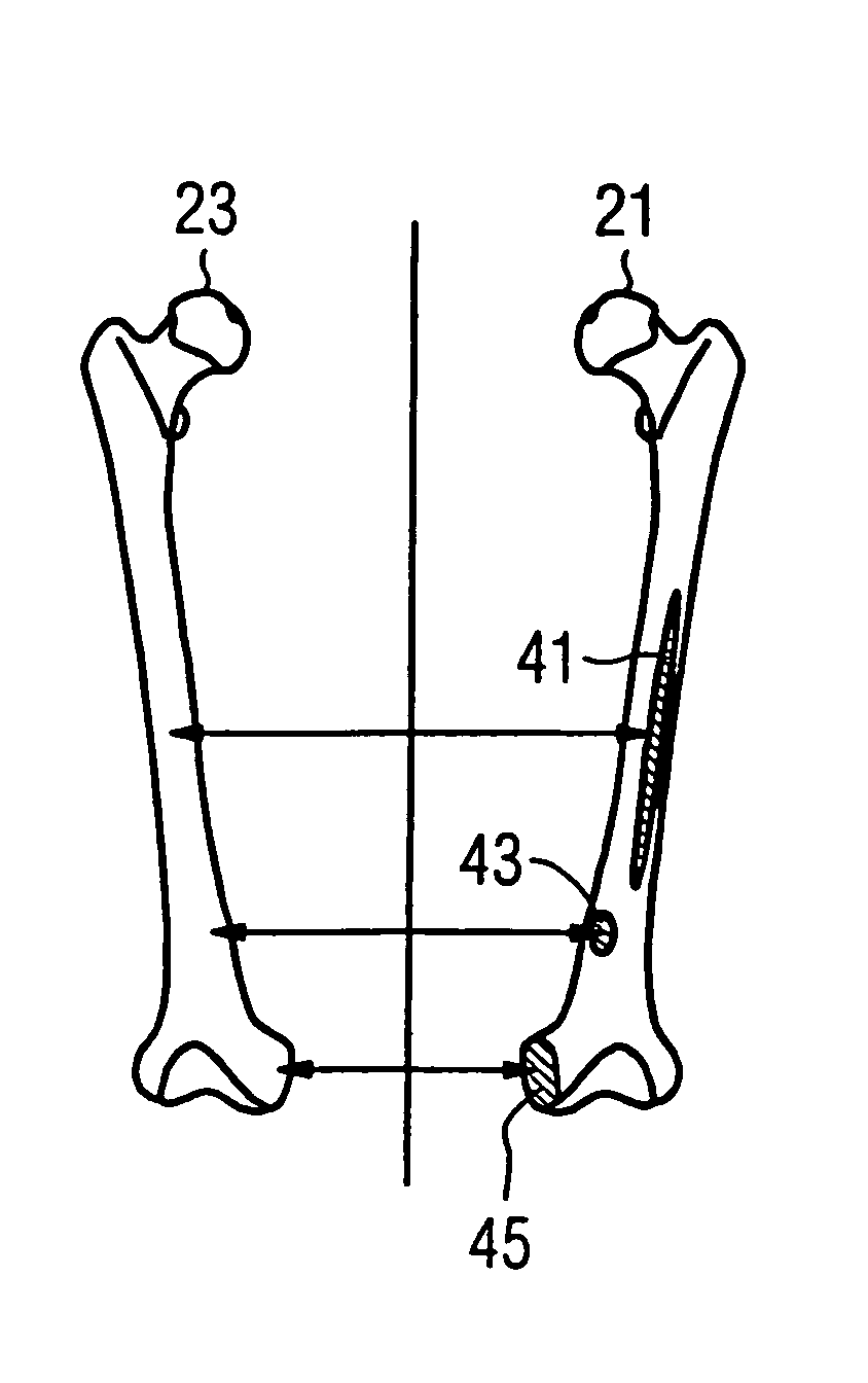
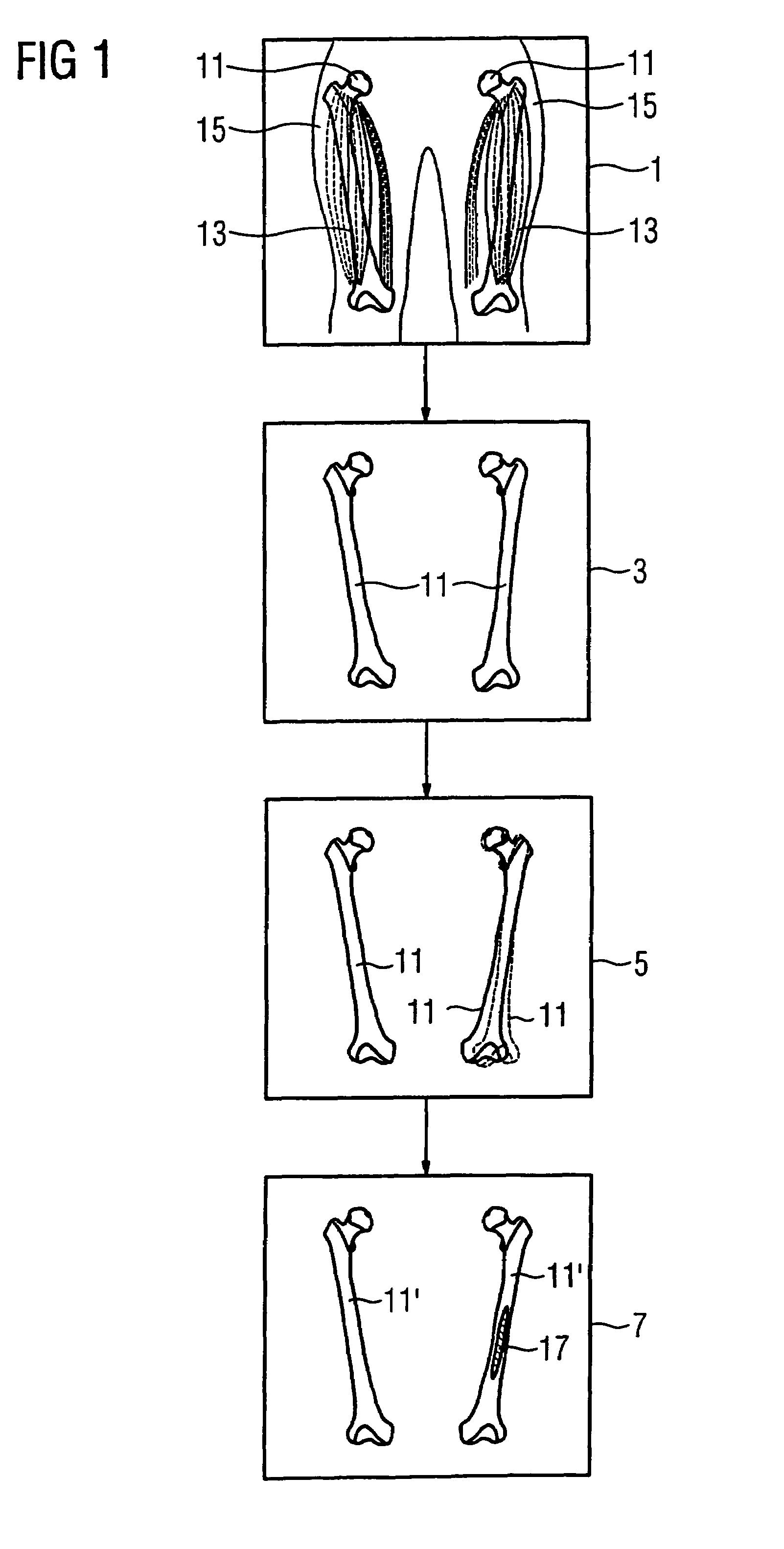
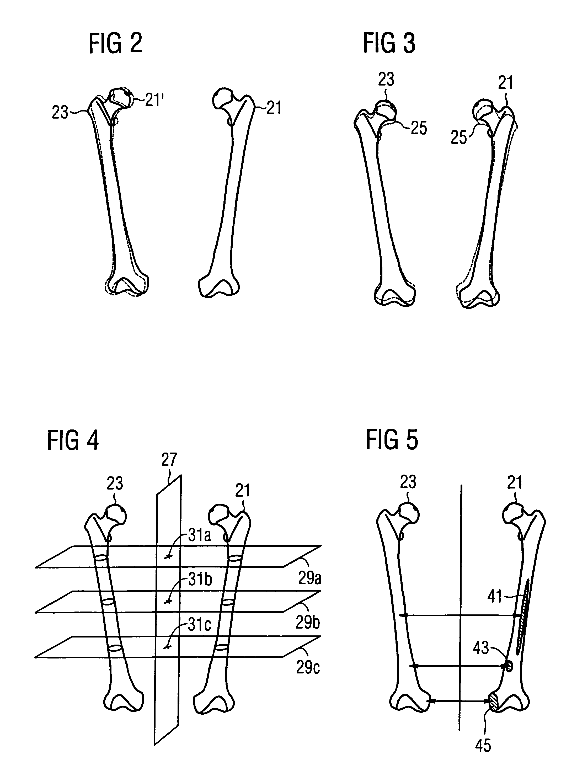
[0038]FIG. 1 schematically shows the basic method steps that are used in the evaluation of laterally-symmetrically situated organ systems. The exemplary embodiment explained in FIG. 1 is demonstrated on a portion of the skeletal system (more precisely on the femur 11), but functions in an analogous manner for all laterally-symmetrically situated organ systems such as, for example, the brain or musculature, and even when the organ is in fact laterally symmetrical but is not paired (such as, for example, the spinal column). It is the goal of the method to implement an automatic evaluation of the organ system with regard to laterally-different findings. Laterally-different findings are of central importance, for example, in the context of oncological questions since they provide indications of possible metastasization of a tumor and thus indicate possible progress of the illness necessitating therapy.

[0039]The starting point of the method is a three-dimensional image 1 of the laterally...


PUM


Abstract

In a method for automated evaluation of a three-dimensional map of a laterally-symmetric organ system as well as medical image acquisition system and a computer program for this purpose, a three-dimensional image of the organ system is segmented, and the segmented organ system is deformed such that the deformed organ system has two mirror-symmetric halves with regard to its external shape. The differences between both mirror-symmetrical halves of the deformed organ system are determined by a comparison of one half with the other half. Alternatively, instead of the deformation of the segmented organ system a mirror-symmetric normal model of the organ system can be deformed, relative to the organ system, or vice versa, such that differences of corresponding regions of both halves of the organ system can be determined using the normal model. The determined differences are presented to a user.

Description

BACKGROUND OF THE INVENTION[0001]1. Field of the Invention[0002]The present invention concerns a method for automated evaluation of a three-dimensional image of a laterally-symmetric organ system as well as a medical image data acquisition system and a computer program for this purpose.[0003]2. Description of the Prior Art[0004]Data acquisition methods in which a three-dimensional data set or image of organ systems of a patient to be examined is produced are well known in medical imaging due to the exceptional diagnostic possibilities. The methods employed (such as, for example, computed tomography or magnetic resonance tomography) generate large data sets. For example, modern computed tomography systems produce dozens of slice image exposures in a few hundred milliseconds. The large number of exposures that arise can often be evaluated by a radiologist only with a very large expenditure of time. The risk of overlooking findings increases given the evaluation of large data sets.[000...

Claims


Application Information

Patent Timeline
Patent Type & Authority Patents(United States)
IPC IPC(8): G06K9/00A61B5/05G06K9/46
CPCG06K9/6206G06T7/0012G06T2207/30008G06V10/754
Inventor KUTH, RAINERSCHMIDT, SEBASTIAN
Owner SIEMENS HEALTHCARE GMBH
Why Eureka
Try Eureka