System for Processing Medical Image data to Provide Vascular Function Information

a technology of medical image data and information about vascular function, which is applied in the field of system for combining 3d (three dimensional) medical image data with vessel blood flow information, can solve problems such as complex vascular anatomy

Inactive Publication Date: 2010-03-04
SIEMENS MEDICAL SOLUTIONS USA INC
View PDF11 Cites 178 Cited by
  • Summary
  • Abstract
  • Description
  • Claims
  • Application Information

AI Technical Summary

Benefits of technology

[0005]A system generates a visually coded 3D image that depicts both vascular morphology and function by visually (e.g. color) coding functional information (e.g. transit time of blood flow through the anatomy) directly on a 3D morphologic image of the vasculature. The functional information is acquired by iteratively computing and scaling transit time curves for individual voxels and minimizing the difference between transit time curves of pixels in a 2D (two dimensional) image to the calculated transit time curves of corresponding projections through a 3D volume. A system combines 3D medical image data with vessel blood flow information. The system uses at least one repository for storing, 3D image data representing a 3D imaging volume including vessels, in the presence of a contrast agent and 2D image data representing a 2D X-ray image through the imaging volume in the presence of a contrast agent. An image data processor uses the 3D image data and the 2D image data in deriving blood flow related information for the vessels. A display processor provides data representing a composite single displayed image including a vessel structure provided by the 3D image data and the derived blood flow related information.

Problems solved by technology

Vascular anatomy can be complex, especially in sick patients, with vessels overlapping, branching, and running in directions perpendicular to standard angiographic viewing orientations.

Method used


Image

  • System for Processing Medical Image data to Provide Vascular Function Information
  • System for Processing Medical Image data to Provide Vascular Function Information
  • System for Processing Medical Image data to Provide Vascular Function Information

Examples


Embodiment Construction

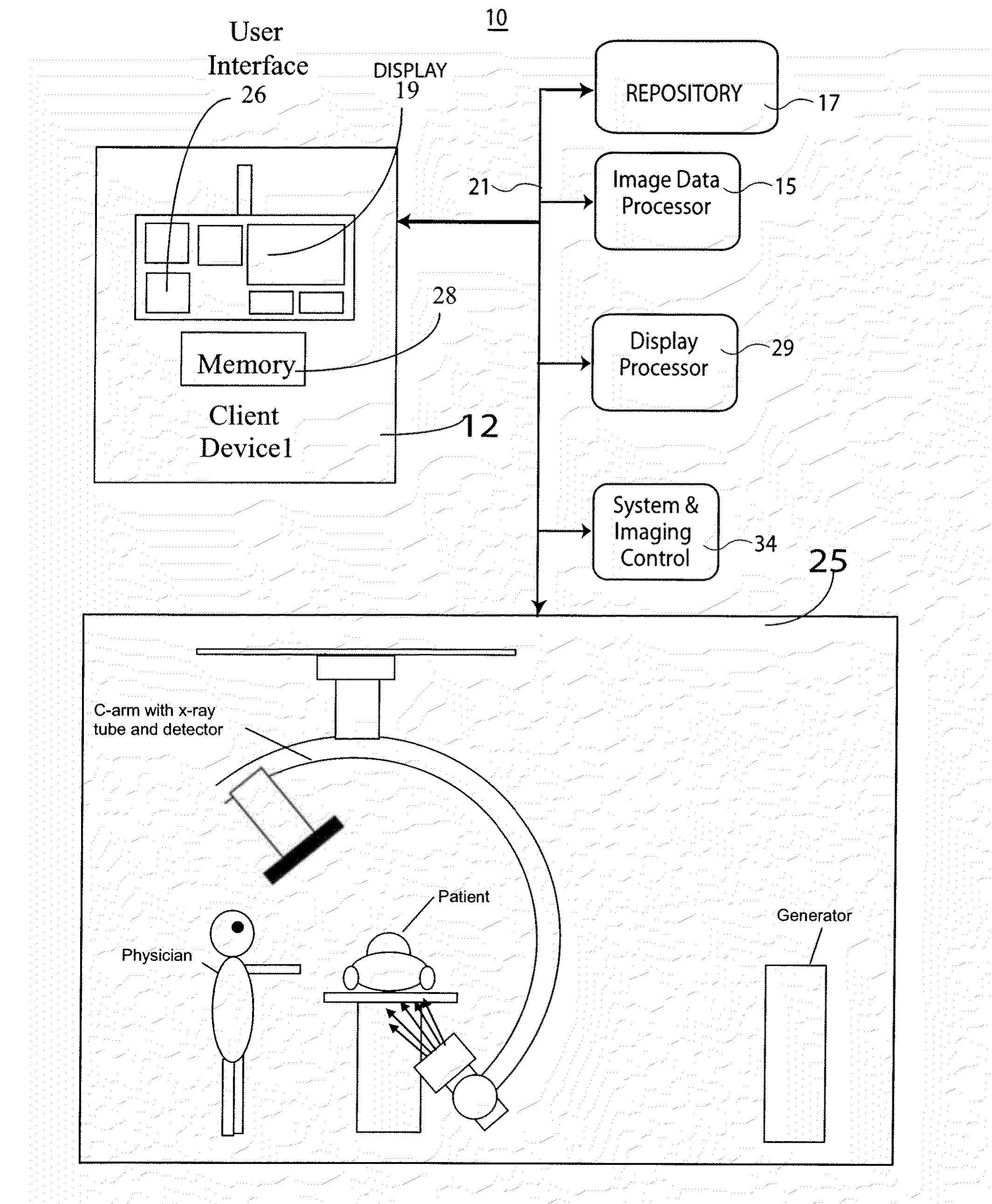
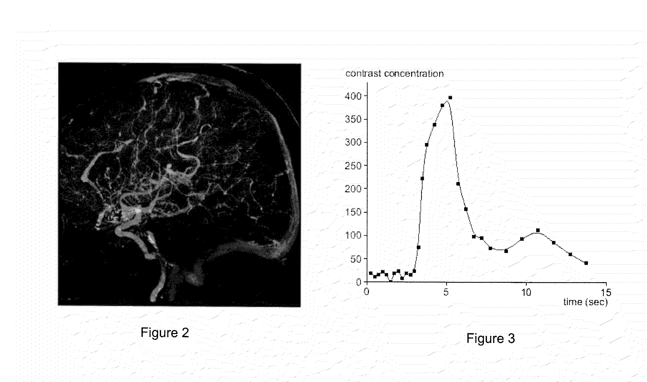
[0016]A system generates a visually (e.g., color) coded 3D image that depicts 3D vascular function information including transit time of blood flow through the anatomy. A transit time curve identifies blood flow by tracking the flow of contrast agent through a region of the anatomy (tissue or vessel). The transit time curve itself plots the x-ray luminance of a pixel or region of pixels in a DSA sequence over the length of the DSA sequence: the amount of contrast in the region of interest over time. Since the blood is carrying the contrast agent, it is possible to obtain a functional measure of the time required for blood to flow through the vessel by examining the time to peak value or time to leading edge of the transit time curves at different locations in the vessel. The functional information is provided using multiple subtracted angiography acquisitions of patient anatomy, while a 3D image of the vasculature provides the morphology of the vascular anatomy. The functional infor...


PUM


Abstract

A system creates a visually (e.g., color) coded 3D image that depicts 3D vascular function information including transit time of blood flow through the anatomy. A system combines 3D medical image data with vessel blood flow information. The system uses at least one repository for storing, 3D image data representing a 3D imaging volume including vessels, in the presence of a contrast agent and 2D image data representing a 2D X-ray image through the imaging volume in the presence of a contrast agent. An image data processor uses the 3D image data and the 2D image data in deriving blood flow related information for the vessels. A display processor provides data representing a composite single displayed image including a vessel structure provided by the 3D image data and the derived blood flow related information.

Description

[0001]This is a non-provisional application of provisional application Ser. No. 61 / 093,002 filed Aug. 29, 2008 and of provisional application Ser. No. 61 / 092,997 filed Aug. 29, 2008, by J. Baumgart et al.FIELD OF THE INVENTION[0002]This invention concerns a system for combining 3D (three dimensional) medical image data with vessel blood flow information and providing a composite single displayed image including a vessel structure provided by the 3D image data and derived blood flow related information.BACKGROUND OF THE INVENTION[0003]In diagnosing and treating patients with vascular problems or deficiencies, it is often necessary to examine both the morphologic and functional characteristics of vasculature. Morphologic information includes the size, geometry, number and placement of the vessels in the anatomy. For vascular anatomy, functional information pertains mainly to the flow of blood including transit times, blood flow, and perfusion. In a conventional angiography laboratory,...

Claims


Application Information

Patent Timeline
Patent Type & Authority Applications(United States)
IPC IPC(8): G09G5/00A61B6/00
CPCA61B6/4441A61B6/504G06T5/50A61B6/507G06T15/08G06T19/20G06T2219/2012G06T2210/41
Inventor RAUCH, JOHN CHRISTOPHERBAUMGART, JOHN
Owner SIEMENS MEDICAL SOLUTIONS USA INC
Why Eureka
Try Eureka