
Precision sensing and treatment delivery device for promoting healing in living tissue

a technology of living tissue and delivery device, which is applied in the field of micro-needles, can solve the problems of inefficiency of systemic distribution of medication within the patient by the arteries and veins, lack of precise volume delivery of one or more therapeutic fluids to tumors or numerous groups of diseased cells, and lack of needles, etc., to facilitate the healing of diseased and/or damaged cell tissue, facilitate cellular healing or poisoning of malignant cells, and facilitate the effect of healing

Inactive Publication Date: 2010-11-23
UNITED STATES OF AMERICA THE AS REPRESENTED BY THE SEC OF THE ARMY
View PDF6 Cites 9 Cited by
  • Summary
  • Abstract
  • Description
  • Claims
  • Application Information

AI Technical Summary

Benefits of technology

[0011]The microneedle includes one or a plurality of microtubes disposed in a bundled configuration within the needle body. The microtubes have distal ends grouped proximal to and interior of the microneedle insertion end. The plurality of microtubes include at least one fluid flow microtube extending to a distal end disposed proximal of the microneedle insertion end, thereby allowing repetitive delivery of a primary treatment fluid into, or removal of cell fluids from the target cell tissue. A second fluid flow microtube is readily incorporated within the plurality of microtubes, with the second fluid flow microtube extending to a second flow end disposed at the microneedle insertion end, thereby allowing repetitive delivery of secondary treatment fluid into or removal of cell fluids treated with the primary treatment fluid delivered by the first fluid flow microtube. Each fluid flow microtube is coupled with the means for fluid flow source such as a microfluidic pump capable of fluid delivery rates of about five microliters / minute.
[0013]Implementation of the microneedle includes the insertion end being positioned within or adjacent to the target cell tissue, thereby positioning the plurality of microtubes distal ends and associated sensing fibers and fluid flow microtubes within the target cell tissue or adjacent to the target tissue mass. The sensing fibers provide evaluation of the cell tissue internal conditions while the fluid flow microtubes provide flow paths for delivery of one or more treatment fluids to the target cell tissue, thereby adjusting the cell tissue internal conditions to preferred levels of pH, oxygen content, temperature, and osmotic balance to facilitate healing of diseased and / or damaged cell tissue. Upon the advice by medical personnel after monitoring of the cell tissue internal conditions with the microneedle, an exact dose of therapeutic medicine, oxygen, vibration, and / or thermal transfer is dispensed through the microneedle, with resulting promotion of cellular healing or poisoning of malignant cells. The microneedle is readily removed and discarded, or reused after sterilization.

Problems solved by technology

If the disease is diagnosed early and is localized in one organ or a group of cells within a patient, then systemic distribution of the medication within the patient by the arteries and veins is not efficient when compared to delivery of medication by hypodermic needle inserted proximal of the diseased cells.
The system of the '184 patent provides for systemic infusion of a relatively large volume of pre-mixed fluid and lacks the ability to deliver one or more therapeutic fluids in precise volumes to a tumor or numerous groups of diseased cells.
The apparatus of the '281 patent does not provide for one needle which remains positioned in an incision during a surgical procedure, with one needle having multiple channels therein for positioning of optic fibers for viewing while concurrently positioning one or more treatment instruments against the joint tissue undergoing repair.
The laser probe diameter may not allow insertion through numerous layers and densities of tissues disposed between a dermal surface and internal organs disposed medially within a patient.
The diameter and configuration of the dual cannula needle may limit precise insertion into a specific tumor in an organ after needle insertion through multiple layers and tissue densities within the patient.

Method used


Image

  • Precision sensing and treatment delivery device for promoting healing in living tissue
  • Precision sensing and treatment delivery device for promoting healing in living tissue
  • Precision sensing and treatment delivery device for promoting healing in living tissue

Examples


Embodiment Construction

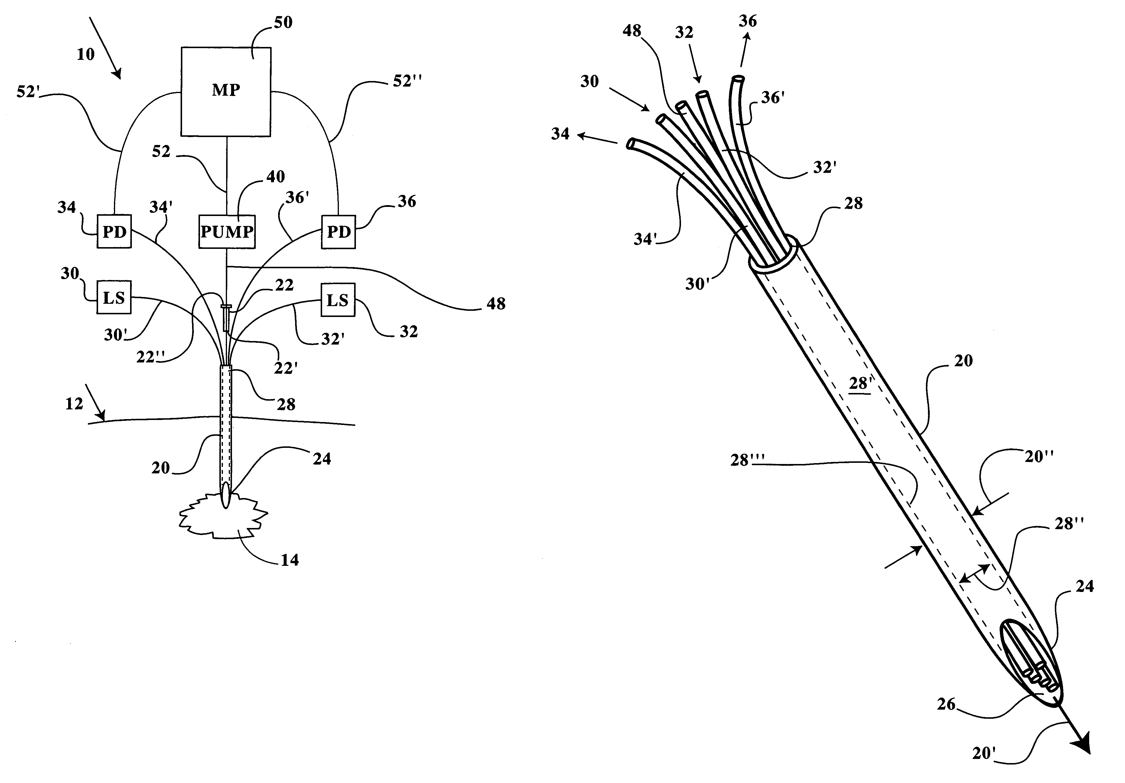
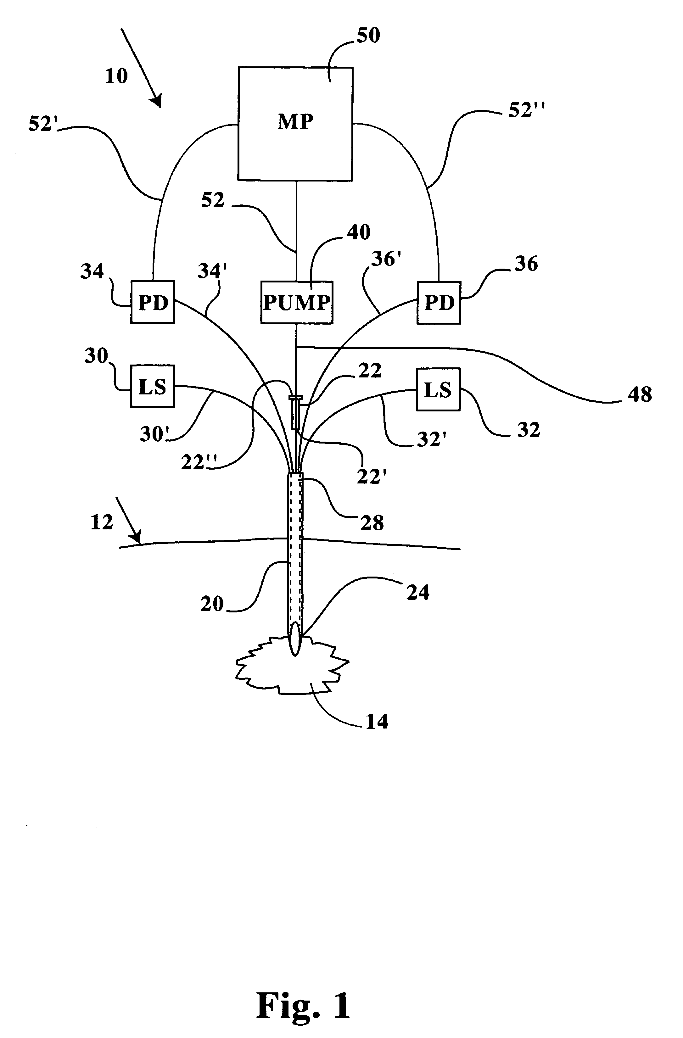
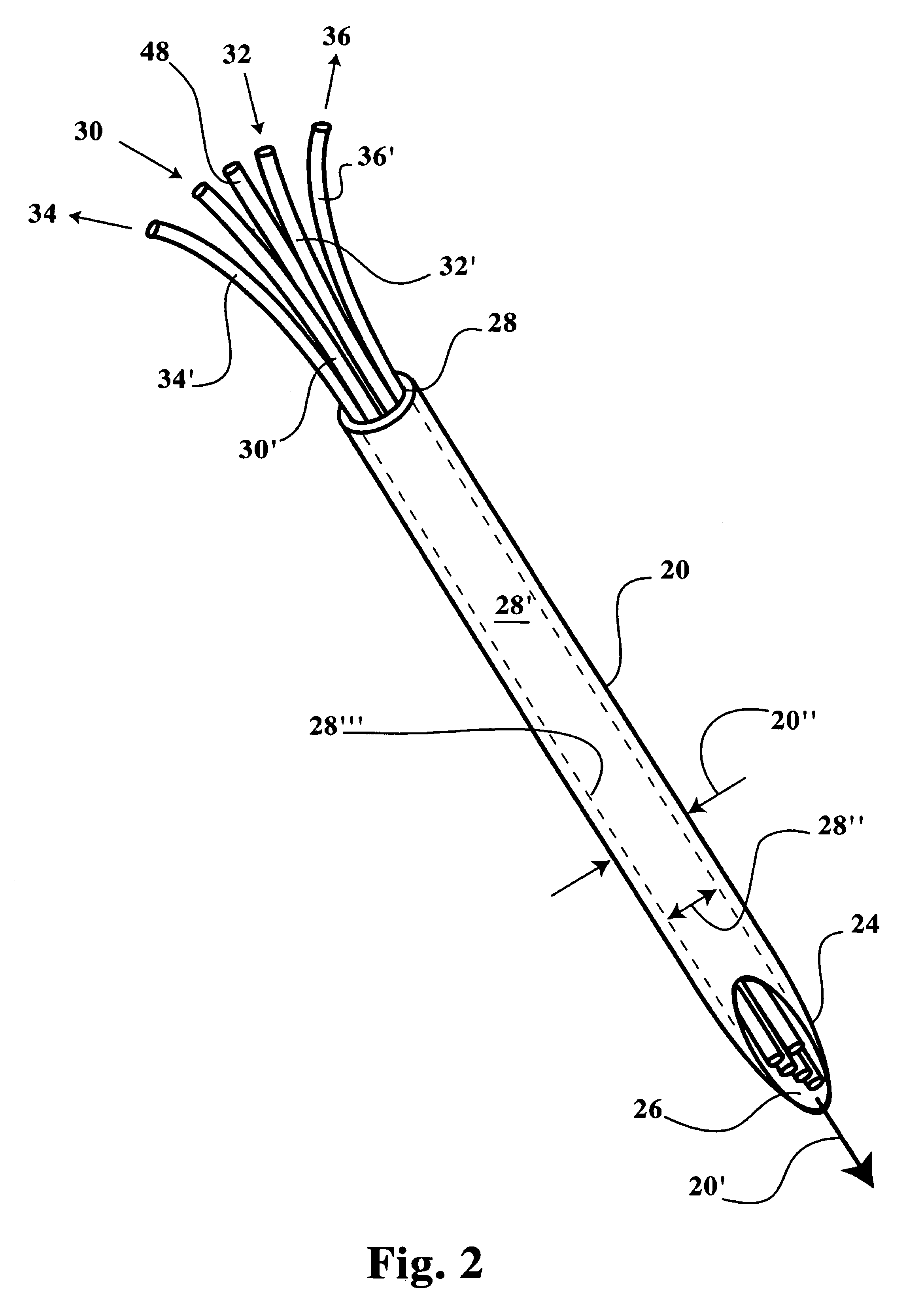
[0027]Referring now to FIGS. 1-8B, a multiple channel needle and delivery system 10 is disclosed, including a microneedle 20 sized for insertion into cell tissue within a patient without significantly disrupting a patient's dermal surface 12 or underlying tissue layers proximal of a target cell tissue 14 or a target tumor mass 16. The microneedle 20 includes a distal insertion end 24 having an elongated and tapered end opening 26 (see FIGS. 2, 3A and 3B), thereby readily allowing positioning of the insertion end 24 and end opening 26 within the target cell tissue 14, a tumor mass 16, or an internal organ or joint (see FIGS. 6-8B). The microneedle 20 includes a proximal end, identified herein as a manipulative end 28, which is maintained exterior of the target cell tissue 14 during positioning of the insertion end 24. The manipulative end 28 is sized in internal diameter (ID) to receive therein one or more microtubes 48, 48′, and / or one or more microfibers 30′, 32′, 34′, 36′. The man...


PUM


Abstract

A microneedle insertable in a target cell tissue, including a manipulative end maintained exterior of cell tissue and an insertion end positionable in or adjacent of target cell tissue. A plurality of microtubes are bundled to pass through the needle body and extend to respective distal ends grouped proximally interior of the insertion end. A sensing fiber is extendable from means for sensing for passage through the needle body to a distal end capable of sensing cell tissue parameters. The insertion end and the bundled microtube and sensing fiber distal ends are positionable in or adjacent of cell tissue thereby providing rapid evaluation of cell parameters by optic fiber sensing, fiber sampling of cell parameters, and precise delivery of therapeutic fluids or additional treatment measures. A method is also disclosed of precisely positioning a microneedle having a plurality of microtubes and sensing fibers therein for evaluating and treating cell tissue.

Description

STATEMENT REGARDING FEDERALLY SPONSORED RESEARCH OR DEVELOPMENT[0001]The invention described herein may be manufactured, used and licensed by or for the Government for governmental purposes without the payment to the inventors and / or the assignee of any royalties thereon.CROSS-REFERENCE TO RELATED APPLICATIONS[0002]Not Applicable.BACKGROUND OF THE INVENTION[0003]1. Field of the Invention[0004]The present invention relates to a micro-sized needle which is insertable through a patient's dermal layer for sampling of tissue in vivo. More specifically, the present invention relates to a needle having a micrometer internal diameter through which a plurality of sensing probes and delivery tubules are extended for monitoring cell parameters at the needle distal end and for delivering therapeutic fluids directly into tissue cells.[0005]2. Description of the Related Art[0006]Prior medical procedures for delivery of therapeutic fluids to treat systemic diseases include utilizing a hypodermic n...

Claims


Application Information

Patent Timeline
Patent Type & Authority Patents(United States)
IPC IPC(8): A61M5/00
CPCA61B5/01A61B5/145A61B5/14539A61B5/4839A61B5/685A61M5/16836A61B17/22012A61M5/141A61M2005/1404A61M2005/1726
Inventor SHUMATE, DANIEL L.RUFFIN, PAUL B.FULDA, JOHN CURTIS
Owner UNITED STATES OF AMERICA THE AS REPRESENTED BY THE SEC OF THE ARMY
Learn More