ECG lead misplacement detection and correction

a lead misplacement and correction technology, applied in the field of physiological information processing systems and methods, can solve the problems of inability to detect unrealistic constraints, and inability to achieve the effect of detecting other electrode wire interchanges, and reducing the difficulty of detection

Active Publication Date: 2010-12-28
KONINKLIJKE PHILIPS ELECTRONICS NV
View PDF8 Cites 28 Cited by
  • Summary
  • Abstract
  • Description
  • Claims
  • Application Information

AI Technical Summary

Problems solved by technology

However, such constraints are unrealistic, if not impossible.
However, detection of other electrode wire interchanges can be relatively difficult.
This approach can be time consuming and resource intensive.

Method used


Image

  • ECG lead misplacement detection and correction
  • ECG lead misplacement detection and correction
  • ECG lead misplacement detection and correction

Examples


Embodiment Construction

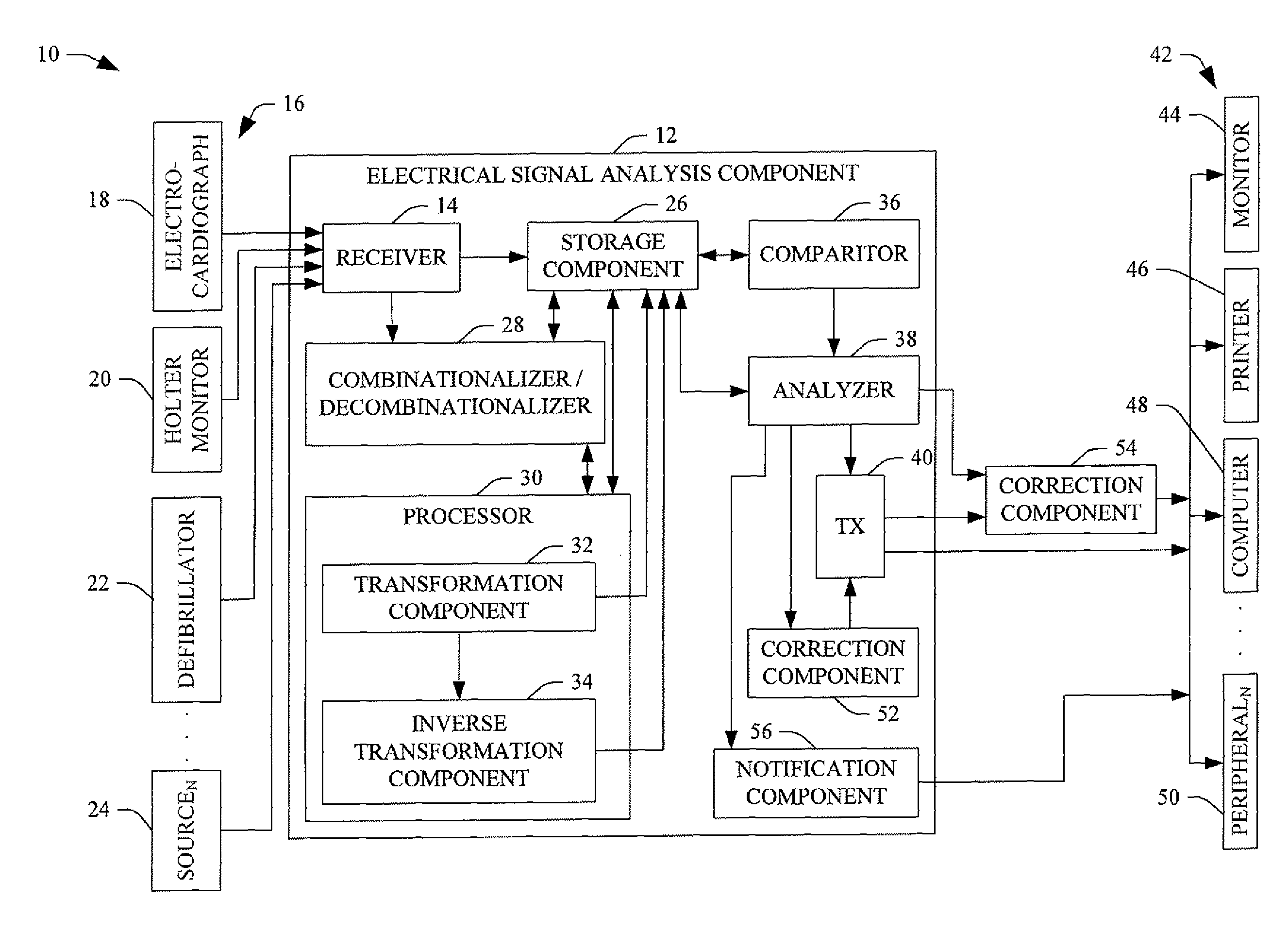
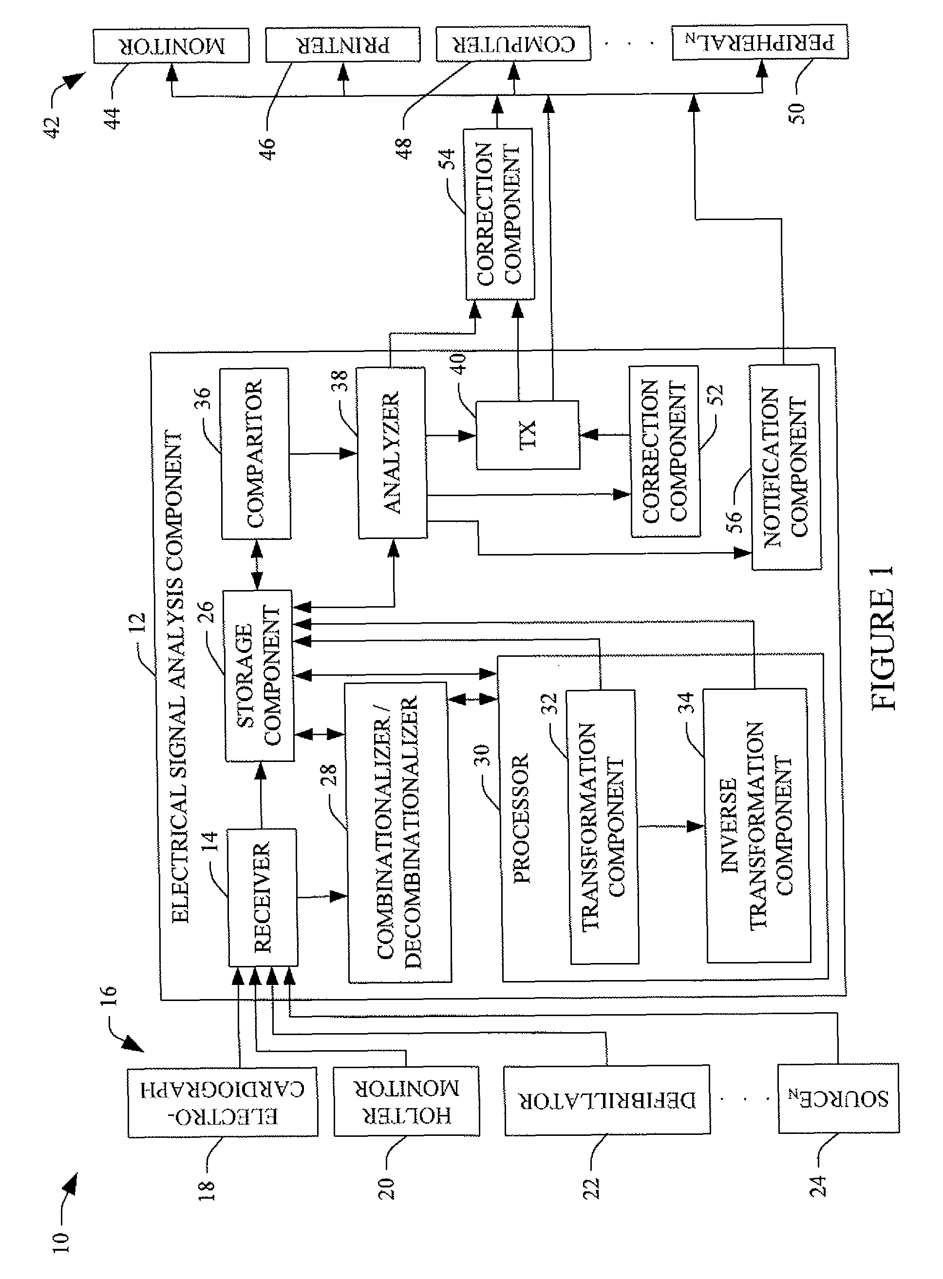
[0018]FIG. 1 illustrates a physiological parameter analysis system 10 (hereafter “system 10”) that at least detects ECG electrode misplacement, including interchanged electrodes, from ECG signals. Optionally, the system 10 additionally corrects detected ECG electrode misplacement by determining which leads are reversed in the saved ECG data, swapping the lead signals, and re-calculating the ECG. The system 10 can also be used to identify electrodes (e.g., from randomly placed electrodes) and / or determine which lead system from a plurality of known lead systems has been used to process the ECG information.

[0019]The system 10 includes an electrical signal analysis component (ESAC) 12 having a receiver 14 that receives ECG information from N sources 16 (wherein N is positive integer). Examples of suitable sources include an electrocardiograph 18, a Holter monitor 20, a defibrillator 22, and other sources 24, including, but not limited to, a computer (not shown), a database (not shown),...


PUM


Abstract

A physiological parameter analysis system (10) detects ECG electrode wire misplacement. The system (10) includes a transformation component (32) that transforms original ECG information and combinations thereof from a first ECG lead system to a second ECG lead system and an inverse transformation component (34) that derives ECG information in the first ECG lead system from the transformed ECG information and the combinations thereof. The system (10) further includes an analysis component (38) that determines a correct ECG lead configuration in the first ECG lead system from among the original ECG information and the combinations thereof based on the derived ECG information.

Description

CROSS REFERENCE TO RELATED APPLICATIONS[0001]This application claims the benefit of U.S. provisional application Ser. No. 60 / 787,827 filed Mar. 31, 2006, which is incorporated herein by reference.BACKGROUND[0002]The following relates to systems and methods for processing physiological information. It finds particular application to detecting interchanged or misplaced ECG electrode wires from ECG information, and also contemplates correcting the ECG information.[0003]An electrocardiogram (ECG or EKG) is a graphical representation of the electrical activity (e.g., electrical potentials / signals) of the heart used for applications such as screening and diagnosing cardiovascular disease. Such electrical activity is non-invasively measured by an electrocardiograph or other suitable electrical transducer, and the graphical representation is generated therefrom and / or by another device such as a computer. Conventional twelve lead (I, II, III, aVR, aVL, aVF, and V1-6) electrocardiographs inc...

Claims


Application Information

Patent Timeline
Patent Type & Authority Patents(United States)
IPC IPC(8): A61B5/0402
CPCA61B5/0424A61B5/0428A61B5/276A61B5/30
Inventor FEILD, DIRK Q.GREGG, RICHARD E.
Owner KONINKLIJKE PHILIPS ELECTRONICS NV
Why Eureka
Try Eureka