

Line structure detection and analysis for mammography CAD
a line structure and mammography technology, applied in the field of computer-aided cancer detection, can solve the problems of significant differences between the different approaches, high rate of unnecessary biopsies, and limited efficacy and efficiency of the above-mentioned methods
- Summary
- Abstract
- Description
- Claims
- Application Information
AI Technical Summary
Problems solved by technology
Method used
Image

Examples
Embodiment Construction
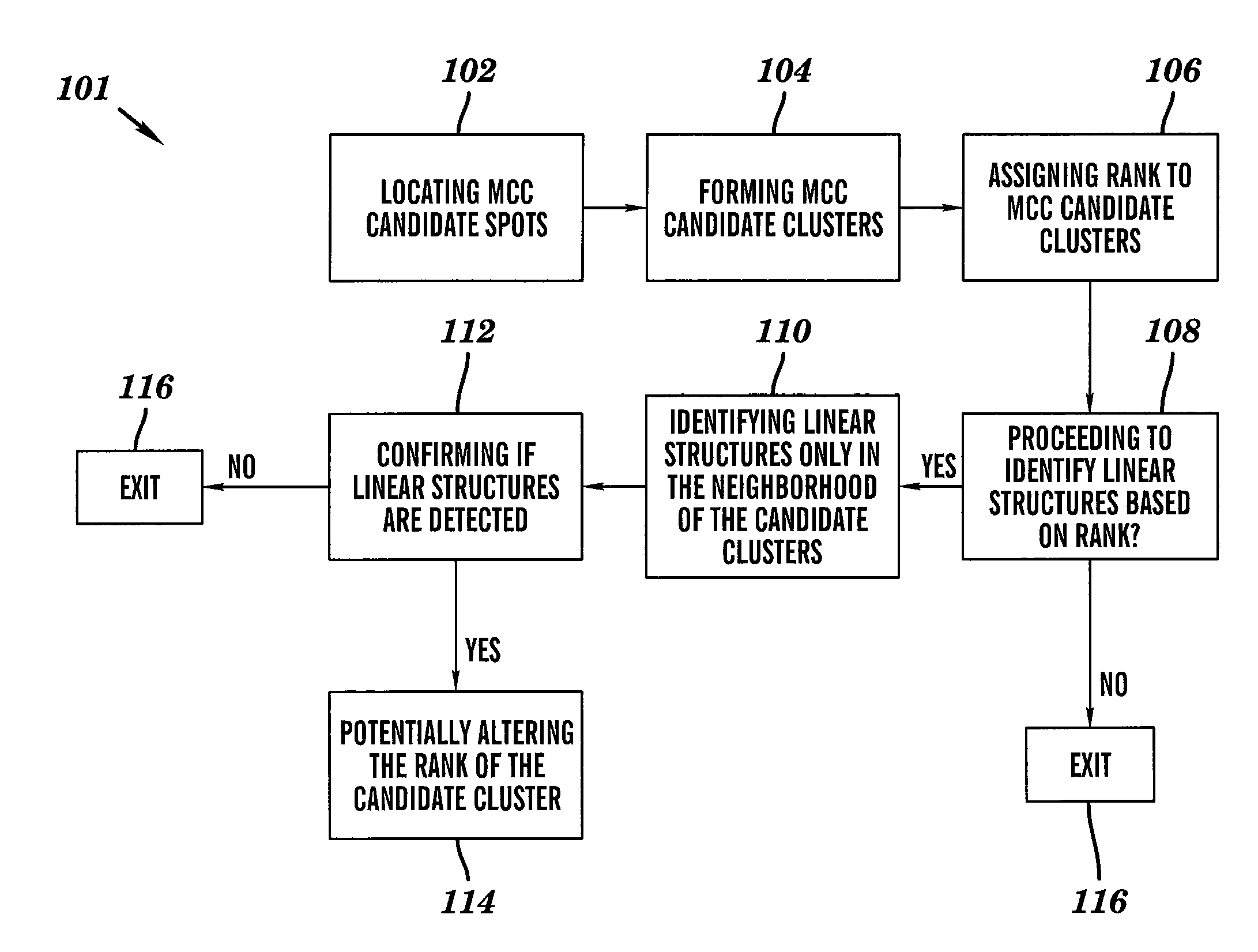
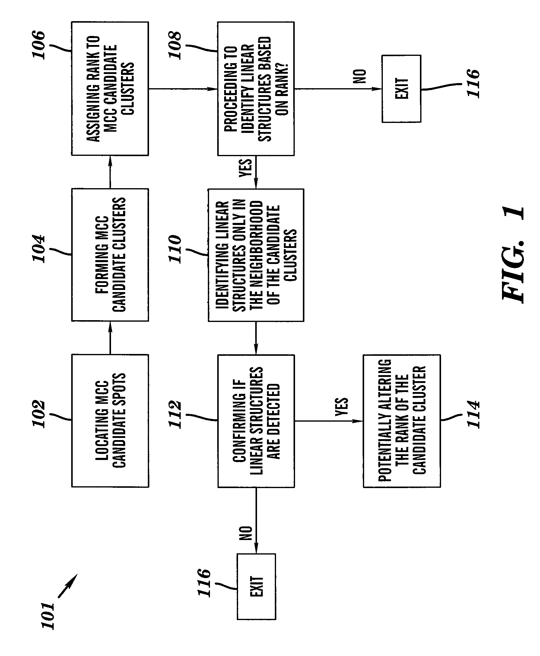
[0021]In one embodiment of the method of image linear structure detection of the present invention, the mammographic image is a digitized X-ray film mammogram in the present invention; the mammographic image is a digital mammogram captured with a computerized radiography system in the present invention; the mammographic image is a digital mammogram captured with a digital radiography system in the present invention.
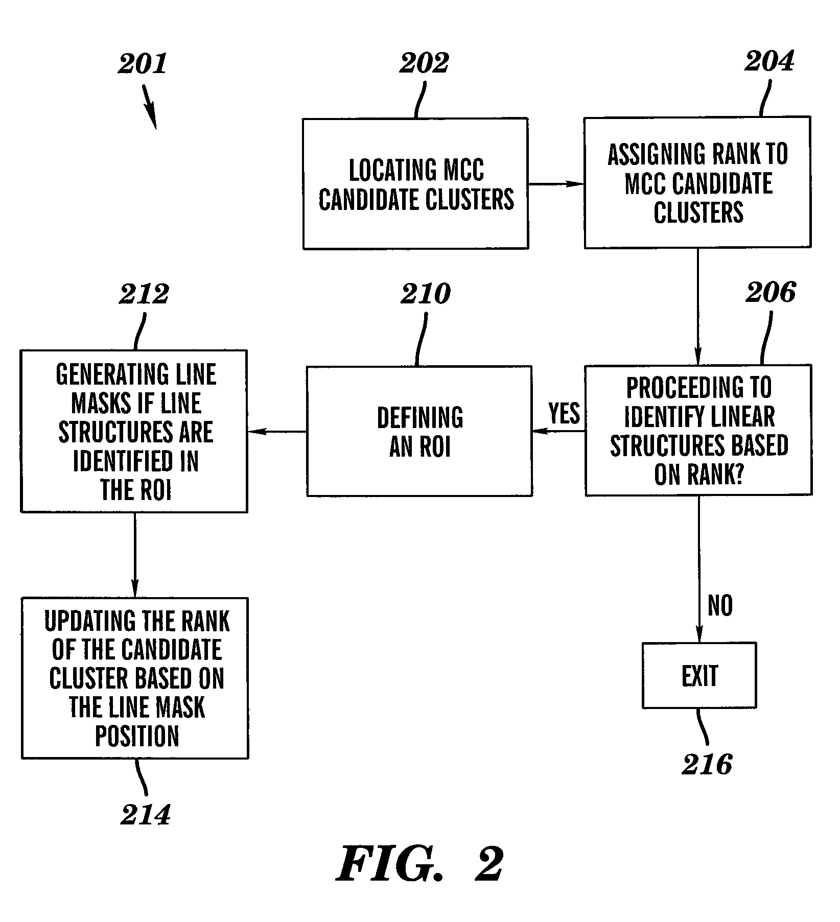
[0022]The step of locating microcalcification candidate spots in a mammographic image consists of a plurality of image processing and computer vision procedures that find clusters of connected pixels that present characteristics which are similar to that of microcalcification in mammogram.
[0023]The step of forming candidate clusters each of which has a plurality of microcalcification candidate spots groups a plurality of microcalcification candidate spots (mammogram image pixels) that are close to each other within a certain distance into a cluster. For each cluster, atta...
PUM
 Abstract
Description
Claims
Application Information