
Apparatus for use in fascial cleft surgery for opening an anatomic space

a technology for anatomic space and surgical equipment, applied in the field of surgical equipment for opening an anatomic space, can solve the problems of long scar, unnecessary damage, and cover the entire chest, and achieve the effect of convenient and efficient manufacture and marketing

Inactive Publication Date: 2011-06-28
TYCO HEALTHCARE GRP LP
View PDF199 Cites 4 Cited by
  • Summary
  • Abstract
  • Description
  • Claims
  • Application Information

AI Technical Summary

Benefits of technology

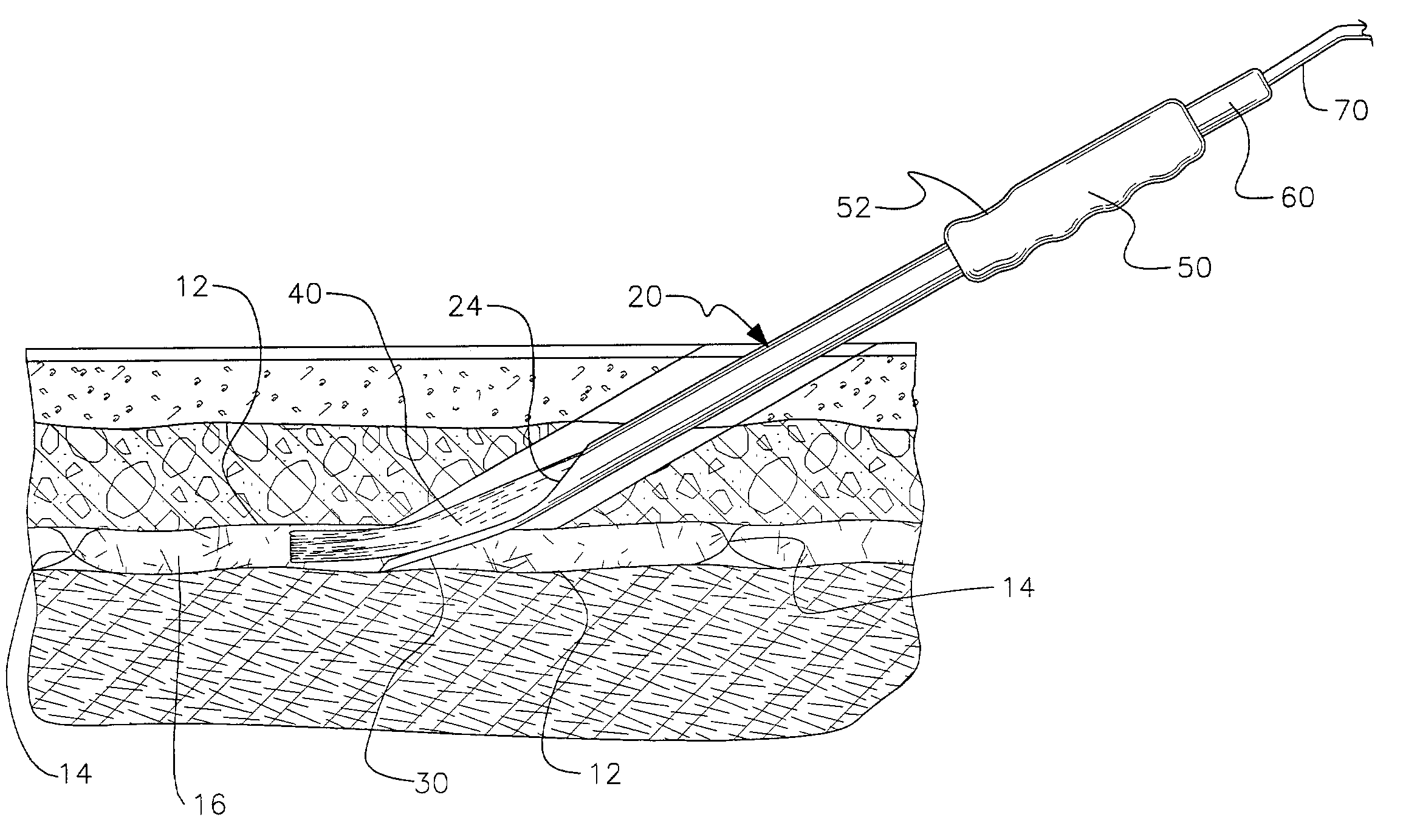
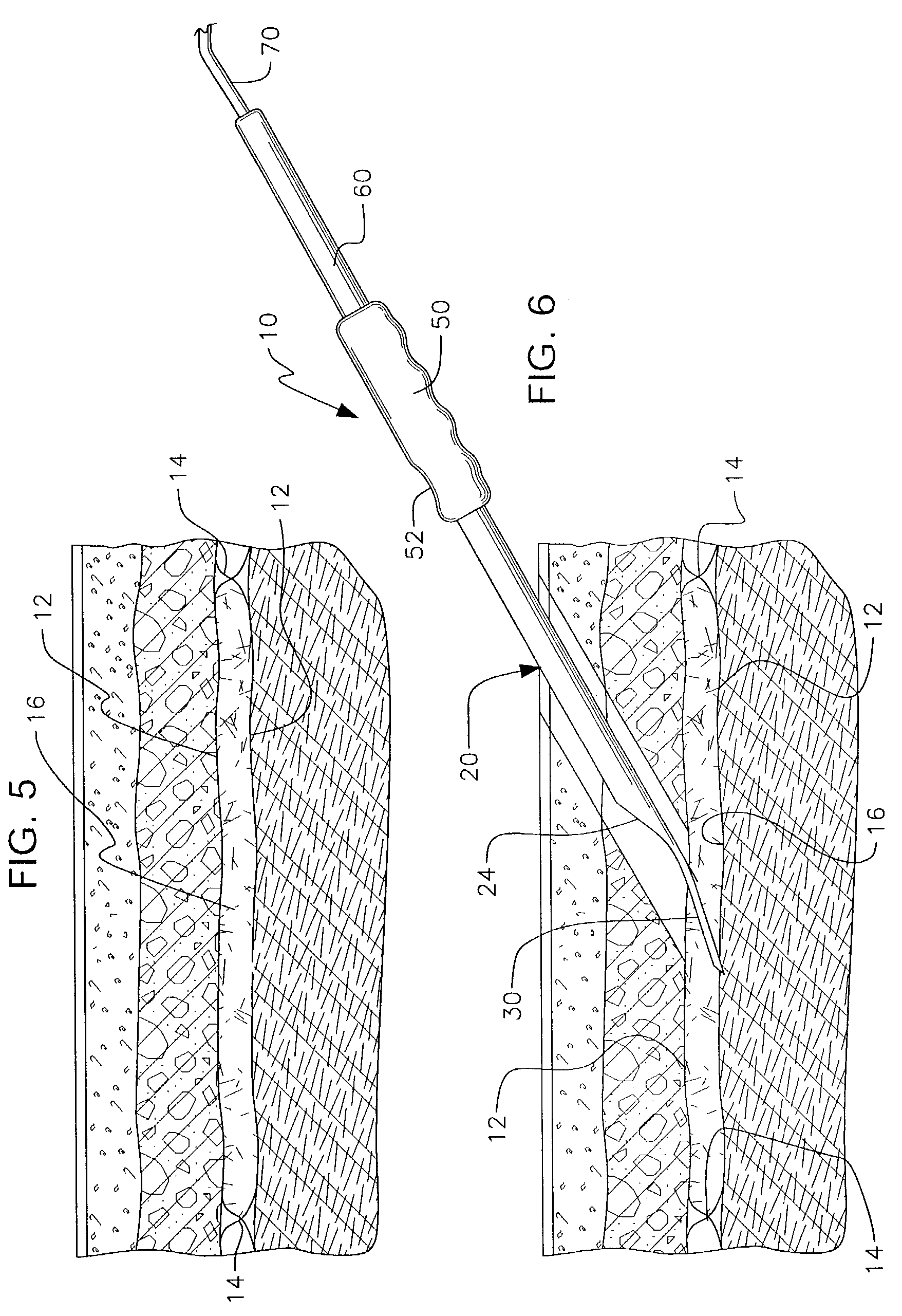
[0040]A second application in the abdominal wall makes use of fascial cleft surgery in performing endoscopically assisted ventral hernia repair. A technique known as components separation technique requires separation of the outer layer of the abdominal wall from the two inner layers to allow advancement of the attached rectus muscle for closure of the midline without tension. This technique repairs ventral hernias without the use of mesh on lays or inlays. Traditional open techniques require extensive undermining of skin to expose the anterior abdominal wall so that open dissection of the fascial cleft, between the lateral abdominal wall muscles, can be bluntly dissected to their anatomic boundaries at the lumbar region. This destroys the circulation of blood to the skin edges of the open wound and leads to a high incidence of wound complications. A minimally invasive approach uses a small incision over the lateral abdominal wall and performs a minimal open dissection down to the fascial cleft between the lateral abdominal wall muscles. The present invention provides for introduction of an elastic inflatable dissector into this fascial cleft which is inflated until dissection reaches the anatomic limits of the cleft. The balloon can be left in place in the expanded condition for a period of time during the procedure to perform immediate tissue expansion prior to release of the fascia, lateral to the rectus muscles, prior to advancement to the midline for closure. This illustrates the ability to perform complex soft tissue surgery without unnecessary collateral damage to the abdominal wall skin and its circulation.
[0045]It is another object of the present invention to provide a new and improved balloon dissection apparatus which may be easily and efficiently manufactured and marketed.

Problems solved by technology

In addition it leaves a long scar, which covers the entire chest.
This creates unnecessary damage that is difficult to reconstruct.
However, most patients are either poor candidate for this invasive reconstructive surgery or do not desire the lengthy recovery that is associated with it.
This however fails to restore functional strength of the abdominal wall and is still associated with an unacceptably high rate of recurrence (20 to 25% in most series).
Mesh repairs have the additional drawback of potential devastating entero-cutaneous fistulas and infections.
Despite dramatic reduction in recurrence rates (to single digits in multiple published reports) several factors have prevented widespread adoption of this procedure.
Second, the wide undermining of abdominal skin and management of resulting skin excess is outside the comfort zone of most general surgeons.
In fact, ischemia of the edges of these skin flaps with resultant wound separation and infection has been the major criticism of the Ramirez technique.
Finally, the components separation technique requires more effort and time, than the mesh repair, due to the need for open exposure of the anterior abdominal wall.
Attempts by Lowe and others to use existing balloon dissectors based on the Kieturakis prior art failed to dissect the fascial cleft to its anatomic borders due to limitations of its design and the inelastic nature of the balloon.
It also failed to fully dissect the entire anatomic space since dissection was limited to the dimensions of the device.
Perhaps this incomplete dissection resulted in insufficient release of tension when the midline was closed and accounts for the higher recurrence of hernia formation in Lowe's endoscopic series when compared to his open components separation repairs.
Minimally invasive procedures outside the peritoneum have defied exploration because of the lack of an easily accessible and inflatable working space.
Johnson is still limited to creating spaces based on the dimensions of the device, even when the device was “overfilled by 50%”.
For instance, it is not possible to perform minimally invasive blunt dissection with an inflatable device within the subcutaneous fat as Johnson claims.
This method does nothing to delineate the size or boundaries of an underlying anatomic space.
This is undesirable in the '989 Rehnke method of fascial cleft surgery.
This sort of straight rigid shaft with a bullet shaped obturator is highly undesirable if one wants to keep the device within a specific anatomic cleft during introduction of the inflatable member.
The Kieturakis device with its rigid shaft and obturator is prone to tunneling out of a desired fascial cleft.
This intrusive approach to invasive dissection generally disregards human anatomy.
The use of a penetrating obturator for tunneling has the same design drawbacks of the Kieturakis device.
This sort of device clearly has no use in fascial cleft surgery.
While these devices fulfill their respective, particular objectives and requirements, the aforementioned patents do not describe a surgical apparatus for use in fascial cleft surgery for tissue dissection wherein a balloon device performs the function of tissue dissection in a minimally invasive manner.

Method used


Image

  • Apparatus for use in fascial cleft surgery for opening an anatomic space
  • Apparatus for use in fascial cleft surgery for opening an anatomic space
  • Apparatus for use in fascial cleft surgery for opening an anatomic space

Examples


Embodiment Construction

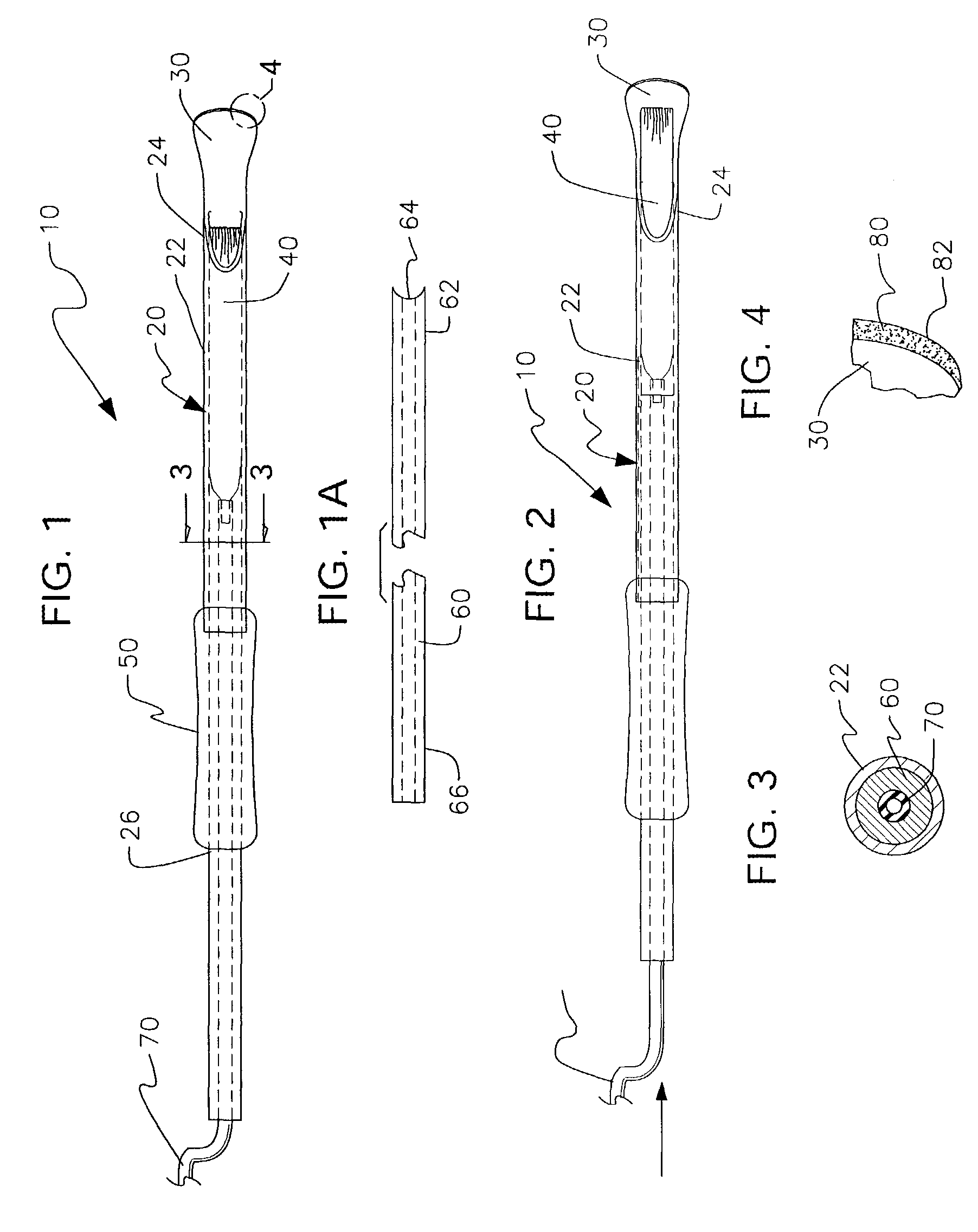
[0059]With reference now to the drawings thereof, the preferred embodiment of the new and improved surgical apparatus for use in fascial cleft surgery for tissue dissection wherein a balloon device performs the function of tissue dissection in a minimally invasive manner embodying the principles and concepts of the present invention and generally designated by the reference numeral 10 will be described.

[0060]The present invention, the surgical apparatus 10, is comprised of a plurality of components. See FIG. 1. Such components in their broadest context include an applicator, an elastic dissection balloon movably positioned within said applicator and a hollow introducing rod slideably positioned within said applicator for positioning said dissection balloon exterior said applicator to within an anatomic space for subsequent inflation and deflation. Such components are individually configured and correlated with respect to each other so as to attain the desired objective.

[0061]The sur...


PUM


Abstract

An apparatus for use in fascial cleft surgery adapted to perform blunt dissection between two layers of anatomically named fascia. The dissection performed by the apparatus extends to the limits of anatomic space defined by fusion of said two layers of fascia in a minimally invasive manner. The apparatus is formed of a hollow tube body member including a malleable introducing flange having a spoonbill-like shape. Further, the apparatus includes an elastic dissection balloon movably positioned within the applicator. The dissection balloon is reversibly expandable between a deflated condition and an expanded condition and is movable from a first storage position within the hollow tube body of the applicator to a position exterior thereof. The dissection balloon is formed of a chosen elastic material having a tensile strength less than the tensile strength of the points of fusion between two layers of fascia such that the dissection balloon fails prior to achieving pressures that would destroy the anatomic boundaries of the fascial cleft such that a working space is demonstrated not created. The apparatus also includes a gripping handle and introducing rod slideably positioned within said applicator for positioning said dissection balloon exterior said applicator to within an anatomic space for subsequent inflation and deflation. Finally, a fill tube extends through the hollow introducing rod to the dissection balloon and operably associated therewith for inflating and deflating the dissection balloon.

Description

BACKGROUND OF THE INVENTION[0001]1. Field of the Invention[0002]The present invention pertains generally to the field of surgical apparatus for opening an anatomic space. More particularly, the present invention pertains to surgical apparatus for use in fascial cleft surgery for tissue dissection wherein a balloon device performs the function of tissue dissection in a minimally invasive manner.[0003]2. Description of the Prior Art[0004]The American Cancer Society estimates that in 2001, 190,000 women were diagnosed with breast cancer. Approximately one in eight women in the U.S. will develop breast cancer in their lifetime. This makes breast cancer the most common cancer among women. These statistics have made breast cancer one of the most pressing issues in women's healthcare today.[0005]The American Society of Plastic Surgeons reports a 174% increase in breast reconstruction procedures between 1992 and 2001, with 81,089 women having reconstructive procedures last year. This trend ...

Claims


Application Information

Patent Timeline
Patent Type & Authority Patents(United States)
IPC IPC(8): A61M29/02A61B17/00A61B17/02A61B17/32A61B19/00
CPCA61B17/02A61B2017/00557A61B2019/307A61B2017/320048A61B2019/302A61B2017/320044A61B2090/032A61B2090/037
Inventor REHNKE, ROBERT D.
Owner TYCO HEALTHCARE GRP LP
Learn More