Portable Digital Medical Camera for Capturing Images of the Retina or the External Auditory Canal, and Methods of Use

a digital medical camera and camera body technology, applied in the field of digital cameras, can solve the problems of limited electrical distribution and unreliability, and achieve the effects of low cost screening, convenient use and simple devices, and rapid growth of diabetes inciden

Inactive Publication Date: 2008-11-11
CHINNOCK RANDAL B
View PDF3 Cites 60 Cited by
  • Summary
  • Abstract
  • Description
  • Claims
  • Application Information

AI Technical Summary

Benefits of technology

[0011]This invention features a simple, compact, low-cost screening device targeted toward medical practitioners with little or no prior experience with retinal imaging or imaging of the external auditory canal. The market for such devices is likely to grow in size as the shift toward lower-cost medical providers continues, driven by the managed care industry. An aging population, and rapidly growing incidence of diabetes also drive this market. Primary care physicians are called on more frequently to do the work of specialists, and nurse practitioners are taking on more of the duties of doctors, including seeing patients. Doctors will continue to surrender more of their traditional duties to less-skilled staff and will assume more of a care manager and advisory role for their patients. It is important for medical device developers to keep this trend in mind in order to place the proper emphasis on ease of use and simplicity of their devices, especially those intended for use by non-specialists. Since it is clear in the case of diabetic retinopathy screening that the best chance of maximizing the diagnosis of retinal disorders is through screening within the primary care setting, the invention provides for increased access to screening for the populations that need it the most. In rural and underserved areas where the lack of specialists coincides with the lowest rate of screening, highest risk of undetected diabetic eye disease, and the least amount of resources to buy large and expensive equipment, the small, low-cost inventive digital opthalmoscope provides a promising means to decrease the incidence of blindness and vision loss among diabetics.
[0012]The inventive handheld, battery-powered device acquires images of the retina. The images can be transmitted from the examination site to an expert reading center via a standard telephone line, cellular link, or Internet connection. An expert consultant can then view the images on a high-resolution color monitor. The images may be compared to previous images stored for that particular patient, or to a library of standard diagnostic images. A diagnostic response may be made back to the referring party within minutes, either by telephone, fax, pager, or e-mail. If evidence of retinal abnormality is seen, the patient may be referred to an ophthalmologist for a complete mydriatic exam and follow-up treatment. For the primary care physician, routine screening of high-risk patients may be offered economically. The periodic monitoring of known diabetic patients will improve their standard of care and reduce medical costs by detecting ocular problems at an early, treatable stage. The device may also be used as part of a routine physical exam, and may be able to identify previously undiagnosed diabetics by means of diabetic retinopathy. Some patients already have detectable diabetic retinopathy by the time a diagnosis of diabetes is made. An eye exam included as part of a routine physical may also allow other eye diseases to be detected earlier and referred to an ophthalmologist, since patients with no perceived visual problems are not as likely to have regular eye exams.
[0013]In addition to the telemedicine service, image data can also be output from the inventive device in standard file formats for storage in any personal computer. Data formats can be compatible with emerging Picture Archiving and Communication Systems (PACS) standards such as the Digital hnaging and Communication in Medicine (DICOM) standard, enabling integration with the user's medical management software. Optional color printers may be used to produce hard copy records of the images for inclusion in paper records, to give to the patient, or to provide to another consulting practitioner. The ability to store images is important in order for a healthcare worker to quickly assess disease progression. There is a great opportunity for the advancement of scientific knowledge by having access to larger numbers of retinal images for research purposes.

Problems solved by technology

The inventive device is greatly needed in developing countries where electrical distribution is limited and unreliable.

Method used


Image

  • Portable Digital Medical Camera for Capturing Images of the Retina or the External Auditory Canal, and Methods of Use
  • Portable Digital Medical Camera for Capturing Images of the Retina or the External Auditory Canal, and Methods of Use
  • Portable Digital Medical Camera for Capturing Images of the Retina or the External Auditory Canal, and Methods of Use

Examples


Embodiment Construction

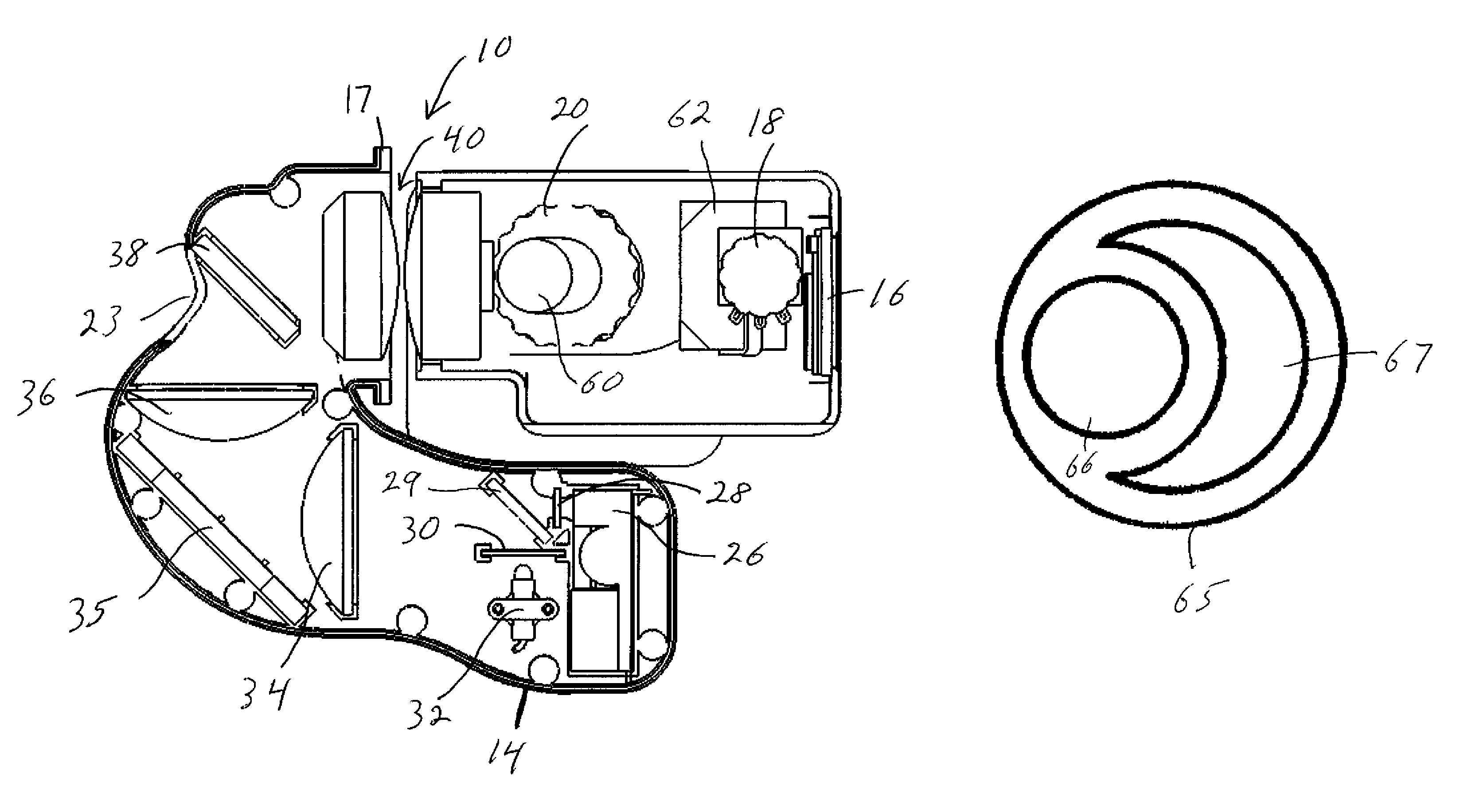
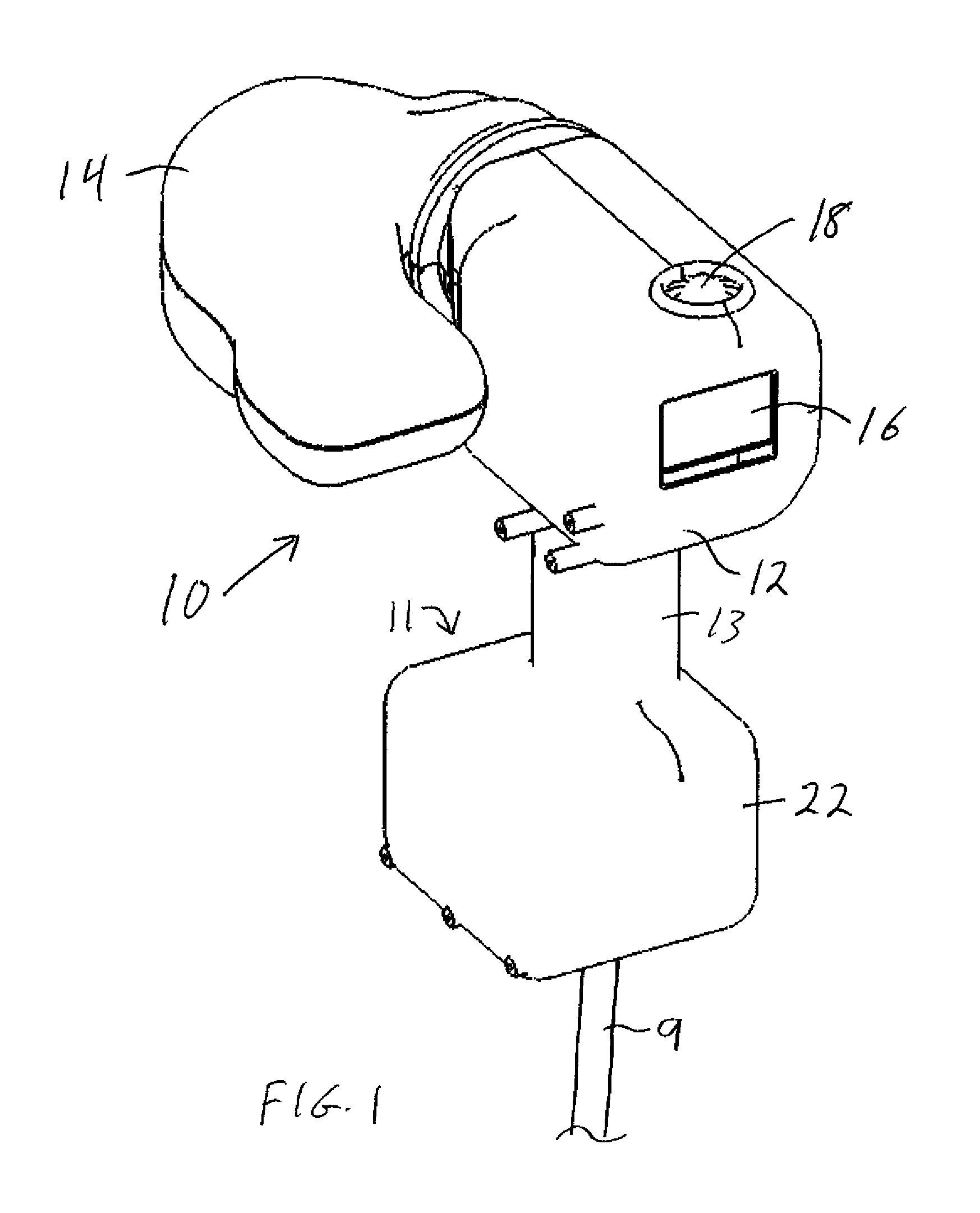
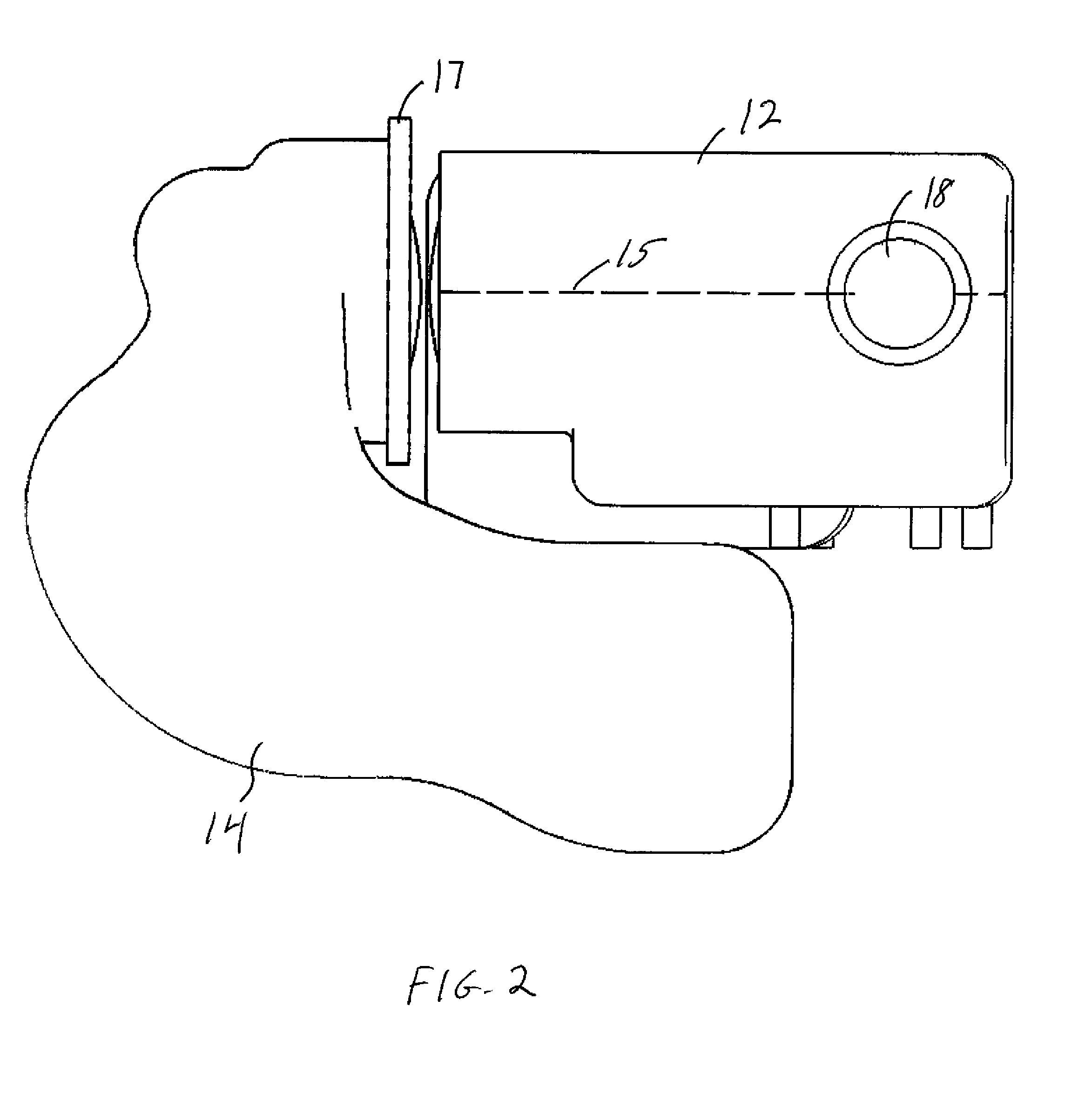
[0058]The preferred embodiment of the inventive device, shown in FIGS. 1-7, offers the following features and benefits:[0059]Handheld device for maximum portability.[0060]Overall size of about 7W″×9L″×10H″ (180×230×250 mm).[0061]Overall weight of about 2 lbs (1 kg).[0062]Powered by a rechargeable battery with a small wall charger.[0063]Designed for non-mydriatic use (no pupil dilating drugs required).[0064]40° Field of View.[0065]A near infrared light source used for aiming and focusing. This prevents pupil constriction that is caused by exposure to visible light.[0066]When the user sees that an image is lined up and focused, a trigger is squeezed. This initiates a flash and a white light image is automatically captured.[0067]The captured image is automatically presented on the screen, and then the camera is ready for the next shot.[0068]Zoom and pan features let the user view the image on a small screen to determine whether image quality is acceptable.[0069]Images are stored on rem...


PUM


Abstract

A hand-held digital camera for obtaining images of a portion of a patient's body and having a hand-held housing, a visible light source located within the housing for providing light along an illumination path from the housing aperture to the patient's body, an image sensor located within the housing that detects light returning from the patient's body along an imaging path that passes into the housing aperture, an optical system located within the housing with separate illumination and imaging paths, an external optical aperture common to the illumination and imaging systems, wherein the illumination and imaging sub-apertures are wholly contained within the common external aperture, are longitudinally coincident, and are laterally separated and non-overlapping, a digital memory device for storing captured images, an output display carried by the housing, and the ability to electronically transmit stored images. The camera can be used for retinal imaging and for otoscopy.

Description

CROSS-REFERENCE TO RELATED APPLICATION[0001]This application claims priority of Provisional application Ser. No. 60 / 700,513 filed on Jul. 19, 2005, the disclosure of which is incorporated herein by reference.GOVERNMENT RIGHTS[0002]This invention was made with Government support under (Grant Number 2 R44 EY 13492-02) awarded by the National Bye Institute. The Government has certain rights in the invention. The inventor is an employee of the assignee, Optimum Technologies, Inc. of Southbridge, Mass.FIELD OF THE INVENTION[0003]This invention relates to a digital camera that is adapted to capture high-quality images of the retina and / or the external auditory canal of the ear, and transmit the images to an expert at a remote location, and to methods by which such a camera is used.BACKGROUND OF THE INVENTION[0004]Diabetic Retinopathy is the leading cause of blindness of adults aged 20-74 years, causing up to 14,000 new cases of blindness each year. 15.7 million Americans, or 8.2% of the a...

Claims


Application Information

Patent Timeline
Patent Type & Authority Patents(United States)
IPC IPC(8): A61B3/14
CPCA61B3/14A61B1/00108A61B1/227A61B3/0083A61B2560/0431
Inventor CHINNOCK, RANDAL B.
Owner CHINNOCK RANDAL B
Why Eureka
Try Eureka