Automated assessment of optic nerve head with spectral domain optical coherence tomography

an optic nerve head and spectral domain technology, applied in image analysis, medical science, diagnostics, etc., can solve problems such as loss of visual field, eventual irreversible blindness, and substantial inter-observer variability

Active Publication Date: 2011-08-09
UNIVERSITY OF PITTSBURGH
View PDF3 Cites 14 Cited by
  • Summary
  • Abstract
  • Description
  • Claims
  • Application Information

AI Technical Summary

Benefits of technology

[0009]In view of the above-mentioned problems, the present inventors developed, pursuant to the invention described herein, a fully automated ONH assessment system based on SD-OCT, thereby providing important disc parameters for clinical analysis, early detection, and monitoring of progression. The system of the invention can provide subjective and reliable results automatically. The input data are from at least one 3D cube image, as provided by SD-OCT.

Problems solved by technology

Glaucoma is a group of diseases of the eye characterized by progressive damage of the optic nerve that may result in visual field loss and eventual irreversible blindness if inadequately controlled.
This subjective definition leads to substantial inter-observer variability.
Some studies have suggested automated methods to extract the disc parameters for analysis, but none of these methods has been commercialized.
Moreover, the existing stereo disc photography could not get high resolution in depth due to the optical properties of cameras.
The aforementioned detection / analysis cannot be implemented for stereo disc photos, because the nature of the information captured by SD-OCT is quite different from that of stereo disc photos.

Method used


Image

  • Automated assessment of optic nerve head with spectral domain optical coherence tomography
  • Automated assessment of optic nerve head with spectral domain optical coherence tomography
  • Automated assessment of optic nerve head with spectral domain optical coherence tomography

Examples


Embodiment Construction

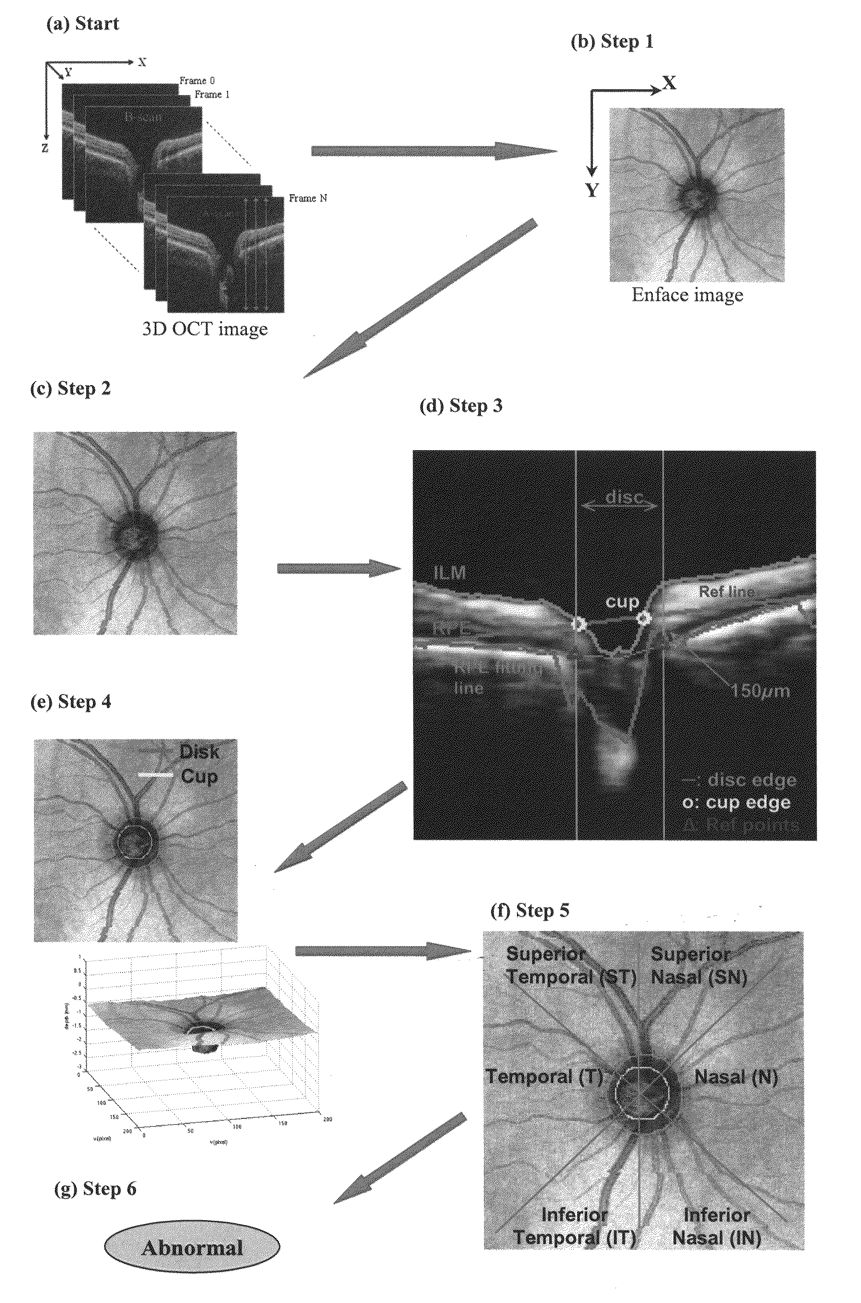
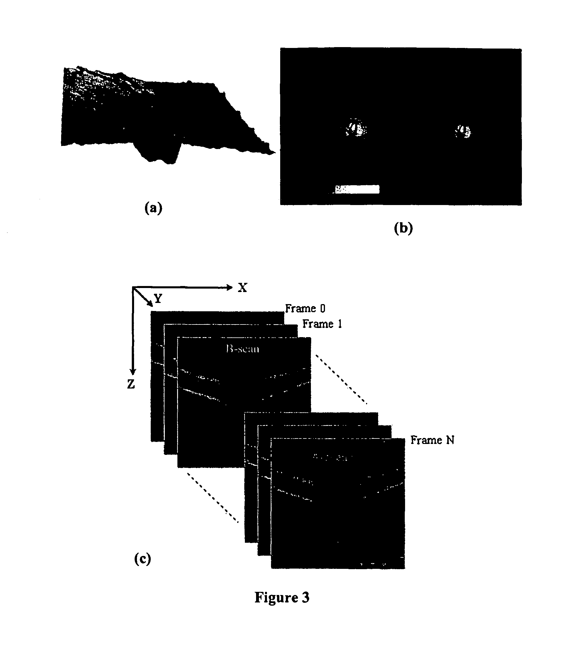
[0042]All the references mentioned herein are incorporated by reference in their entirety. As noted above, the overall system of the present invention is diagramed schematically in FIGS. 6(a)-6(g). A detailed description of the procedures is given as follows:[0043]1. Generate enface image (2D ONH image) from the 3D OCT image taken from SD-OCT, as shown in FIG. 6(a) and / or FIG. 3(c):

[0044](a) The enface image is generated by averaging the intensity values of each A-scan line.

[0045](b) The intensity values is normalized to obtain a high contract enface image.

[0046]Step 1, generation of enface image, comports with existing methodology (FIG. 6(b)). The current SD-OCT machines of several different brands already have implemented enface image generation, based on this basic idea [5]. Data can be utilized from different modes of scanning protocols, such as A-scan, B-scan, and C-scan.[0047]2. Detect the disc margin on 2D ONH image (FIG. 6(c))

[0048]Automated detection of disc margin is a cha...


PUM


Abstract

A fully automated optic nerve head assessment system, based on spectral domain optical coherence tomography, provides essential disc parameters for clinical analysis, early detection, and monitoring of progression.

Description

RELATED PATENT APPLICATIONS[0001]This application claims priority to U.S. Provisional Application Ser. No. 61 / 071,347, filed Apr. 23, 2008, which is incorporated by reference in its entirety.STATEMENT REGARDING FEDERALLY SPONSORED RESEARCH[0002]This invention was made with Government support under grant number RO1-EY013178-8, awarded by the National Institutes of Health. The government has certain rights in the invention.BACKGROUND OF THE INVENTION[0003]Glaucoma is a group of diseases of the eye characterized by progressive damage of the optic nerve that may result in visual field loss and eventual irreversible blindness if inadequately controlled. The World Health Organization currently ranks glaucoma as the second most common cause of blindness. As majority of patients do not experience any symptoms until the disease reaches an advanced stage, early detection and treatment is of utmost importance.[0004]The diagnosis of glaucoma is based on the appearance of the optic nerve head (O...

Claims


Application Information

Patent Timeline
Patent Type & Authority Patents(United States)
IPC IPC(8): A61B3/14
CPCA61B3/102A61B3/1233A61B3/14G06T7/0012G06T7/0083G06T7/402G06T2207/10016G06T2207/10072G06T2207/20081G06T2207/30041G06T7/12G06T7/42
Inventor XU, JUANWOLLSTEIN, CHAIM-GADIISHIKAWA, HIROSHISCHUMAN, JOEL STEVEN
Owner UNIVERSITY OF PITTSBURGH
Why Eureka
Try Eureka