
Endoscopic imaging system in bulk optics biopsy spectral coverage OCT

An optical biopsy and imaging system technology, applied in the field of spectral domain OCT endoscopic imaging system, can solve the problems of limited space scale and restriction of high-quality imaging methods, and achieve the effect of good implementability, simple structure and enhanced anti-interference ability

Inactive Publication Date: 2007-09-12
ZHEJIANG UNIV
View PDF0 Cites 47 Cited by
  • Summary
  • Abstract
  • Description
  • Claims
  • Application Information

AI Technical Summary

Problems solved by technology

However, the problem brought about by miniaturization is that the spatial scale is severely limited, which seriously restricts the alternative high-quality imaging methods

Method used


Image

  • Endoscopic imaging system in bulk optics biopsy spectral coverage OCT
  • Endoscopic imaging system in bulk optics biopsy spectral coverage OCT
  • Endoscopic imaging system in bulk optics biopsy spectral coverage OCT

Examples


Embodiment Construction

[0023] The present invention will be further described below in conjunction with the accompanying drawings and embodiments.

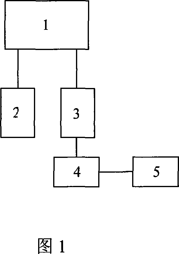
[0024] FIG. 1 shows the framework of a spectral domain OCT endoscopic imaging system, including an optical fiber interferometer 1 , an imaging probe 2 , a detection unit 3 , an image acquisition card 4 and a computer 5 . One end of the fiber optic interferometer 1 is connected to the imaging probe 2 to realize the circular scanning of the endoscope. The light received by the imaging probe 2 returns to the fiber optic interferometer 1, and the generated interference signal enters the detection unit 3 at the other end, and the detection signal is quickly transmitted to the image acquisition card 4, and then the computer 5 performs subsequent processing and image reconstruction and display.

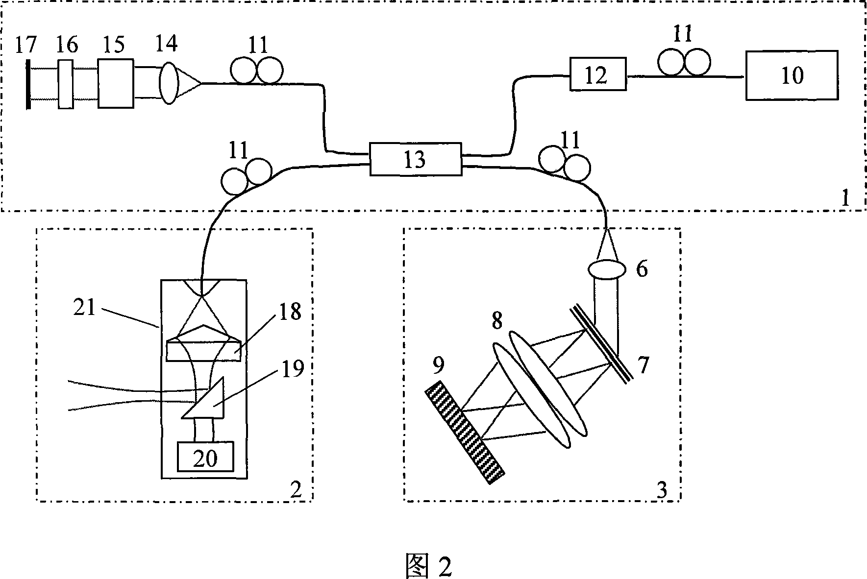
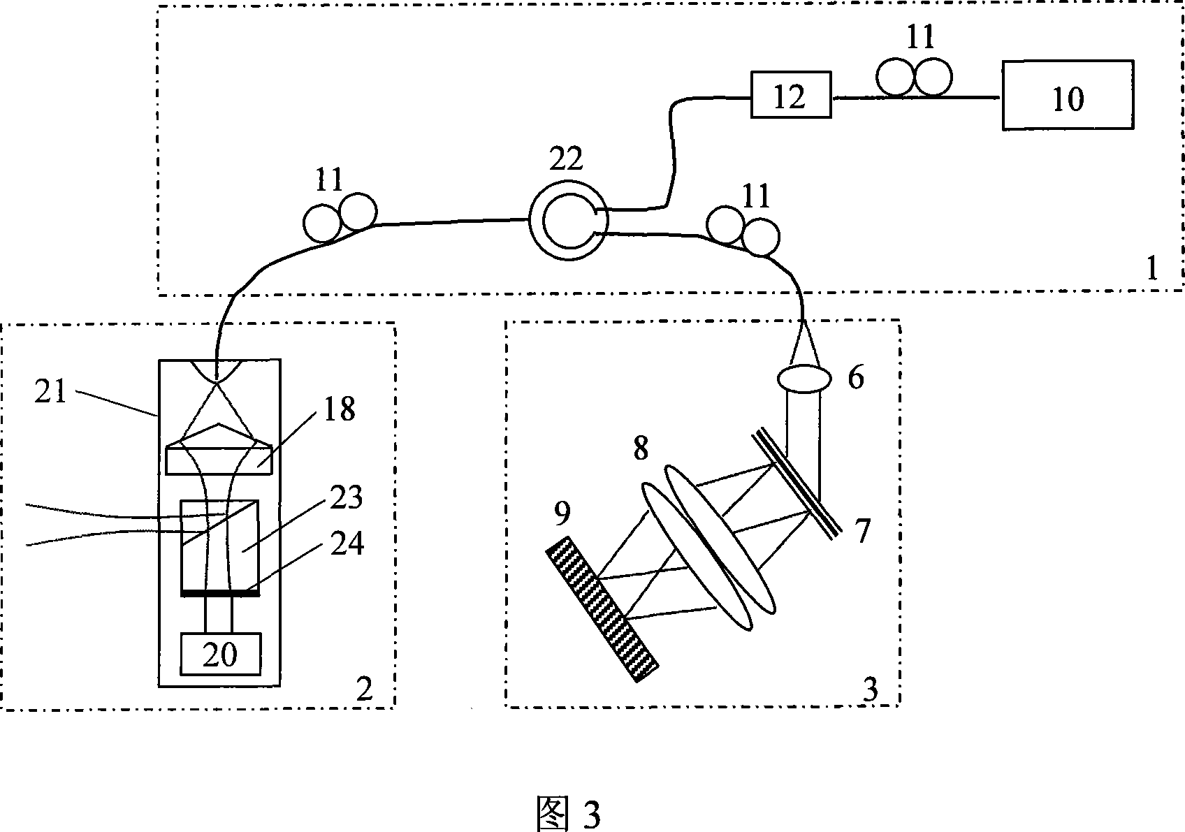
[0025] As Embodiment 1, FIG. 2 shows a composition structure of a spectral domain OCT endoscopic imaging system, including a broadband light source 10, four polarizat...


PUM


Abstract

The present invention discloses one kind of spectral-domain optical coherent tomography endoscopic image system for in vivo optical biopsy, and the system includes one fiber optical interferometer, one imaging probe, one detection unit, one image acquiring card and one computer. The detection unit has grating spectrograph for high imaging speed, and the imaging probe has axial axicon lens and inside rotating right angle prism or circularly symmetric beam splitter combined to ensure high transverse resolution in the whole depth range, so as to realize circularly scanning endoscopic imaging. The present invention proposes two embodiments of circularly scanning probe and their corresponding system structures. The present invention may be applied in the optical endoscopic biopsy and analytic study of oral cavity, respiratory tract, gastrointestinal tract, etc.

Description

technical field [0001] The invention relates to optical coherence tomography (OCT) technology, in particular to a spectral domain OCT endoscopic imaging system for optical biopsy in vivo. Background technique [0002] Optical coherence tomography (OCT) is an emerging optical imaging technology. Compared with traditional clinical imaging methods, it can achieve non-contact, non-invasive, High-resolution imaging is an important potential tool for basic medical research and clinical diagnostic applications, and can be used for early diagnosis of diseases such as ophthalmology, skin, and tumors. [0003] Rapid imaging technology has always been the core of OCT technology. Time-domain OCT obtains depth information through mechanical scanning of the reference arm or other optical path scanning methods, and the imaging speed is limited to a certain extent. Based on the grating-type fast-scanning optical delay line, generally only the axial scanning speed of 1K can be achieved. I...

Claims


Application Information

Patent Timeline
IPC IPC(8): A61B1/04A61B5/00
Inventor 丁志华孟婕王凯朱瑛吴彤
Owner ZHEJIANG UNIV
Learn More